

Sharaf Ali Shah ( Bridge Consultants Foundation, Karachi. )
Abdul Aziz Memon ( Department of Dermatology, Civil Hospital, Sukkur. )
Auwj-e-Shamim ( Department of Dermatology, Civil Hospital, Sukkur. )
Shehla Baqi ( Department of Infectious Diseases, Sindh Institute of Urology and Transplantation, Karachi. )
Richard Witzig ( Section of Infectious Diseases, Tulane University School of Medicine; Social Vaccine Strategies, New Orleans, USA. )
December 2014, Volume 64, Issue 12
Original Article

Abstract
Objective: To study the efficacy of a low-cost heating pack device used for thermotherapy in the treatment of cutaneous leishmaniasis.
Methods: The study was conducted at the Department of Dermatology, Civil Hospital Sukkur, Pakistan, from April 20, 2012, to January 3, 2013. Thermotherapy with Hand-Held Exothermic Crystallisation Therapy for cutaneous leishmaniasis was performed on each lesion of the participating subjects at an average initial temperature of 51.6°C for 3 minutes daily for 7 days. Patients were followed regularly for 6 months after the therapy. SPSS 20 was used for statistical analysis.
Results: Even though all 27 patients completed 1 week of thermotherapy, only 23(85.2%) patients could be evaluated for full treatment response since 4(14.8%) were lost to complete follow-up. By the final 180-day evaluation, 19 (83%) patients had been cured. Applications were well tolerated with no side effects.
Conclusion: The devise was a convenient, safe, non-toxic and effective treatment for cutaneous leishmaniasis at a fraction of the cost of standard antimonial treatment. Further studies are needed to certify its safety and efficacy as monotherapy for the condition.
Keywords: Old world, Cutaneous leishmaniasis, Thermotherapy, Low cost, Pakistan. (JPMA 64: 1398; 2014).
Introduction
Cutaneous Leishmaniasis (CL) is a sand fly-transmitted protozoan disease on the World Health Organisation (WHO) list of Neglected Tropical Diseases, with an estimated 1 to 1.5 million new cases annually.1 More than 90% of the world\'s CL cases occur in only seven countries: Afghanistan, Brazil, Iran, Pakistan, Peru, Saudi Arabia and Syria.1,2
The incidence of CL is rising in Pakistan; the situation exacerbated by poverty, poor healthcare, limited access to drugs, inadequate vector control, regional warfare, and refugee migration.3-5 In Pakistan, L. tropica and L. major are the causative CL organisms, and two epidemiologic types of CL are prevalent - zoonotic CL is present mainly in the south-western province of Balochistan, and anthroponotic CL is more prevalent in the central regions of Sindh and Punjab.6,7 CL is generally considered endemic in affected areas, but epidemic CL has been sporadic in the last decade, with some villages reporting >50% of individuals with active CL lesions.
Treatment of Old World CL has been recently reviewed with sodium stibogluconate (SC) chemotherapy still the current first-line treatment worldwide,8 but Leishmania species are developing SC resistance (in some areas up to 65% resistance).9 In addition, SC has potential systemic toxicities, is expensive, and the 20-day SC regimen under medical supervision places a considerable burden on impoverished patients and their families.10,11 Second-line treatments with intravenous amphotericin B deoxycholate (AB) and pentamidine are expensive and potentially toxic; paromomycin and oral miltefosine are not yet available in Pakistan. Despite advances in basic scientific research, there has been little progress in vaccine or new drug development for what remains a neglected tropical disease (NTD).12
Leishmania parasites do not readily multiply in macrophages above 39°C, leading to thermotherapy (TT) as a possible local treatment strategy.8,13,14 Several TT modalities over the last two decades have demonstrated CL treatment efficacy without the potential toxicities of chemotherapy, with some methods demonstrating shorter healing times.15-18 The best-studied and the only thermotherapy device approved by the Food and Drug Administration (FDA) is the radio-wave modality ThermoMed®.15,16,19-21 However, to date its initial cost has been prohibitively high, making it impractical for global CL treatment and control.
We present the first clinical evaluation of Old World CL using a new TT device called the Hand-Held Exothermic Crystallisation Therapy for CL (HECT-CL), a customised heating pack that delivers controlled conduction heat starting at 52°C and decreasing to 48°C over a 3-minute period when applied over a CL lesion. The HECT-CL device is a vinyl pouch containing a super-saturated sodium acetate salt solution and a flexible 2cm stainless-steel disc at room temperature. Flexing the disc triggers an exothermic liquid-to-solid phase crystallization reaction that in 10 seconds achieves an initial predictable 52°C (Figure-1).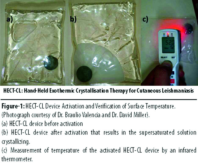
The HECT-CL is safe, low-cost (
Patients and Methods
The pilot study was conducted at the Department of Dermatology, Civil Hospital Sukkur, Pakistan, which is a CL referral facility from Sindh and neighbouring provinces. Two physicians, of whom one was a consultant dermatologist, conducted the study from April 20, 2012 to January 3, 2013, after receiving Institutional Review Board approval from the Tulane University School of Medicine, New Orleans, and Bridge Consultants Foundation, Karachi.
Patients presenting to the outpatient clinic with cutaneous lesions suggestive of CL were skin smear tested for Leishmania Donovan (LD) bodies. If positive, the patients were offered enrolment in the study after a thorough history and physical examination and if they met further inclusion criteria: age 10-65 with one, two, or three LD-proven lesions on the trunk or limbs, each with size of < 6cm diameter. Cranial and facial lesions were also included. Patients below the age of 10 were enrolled in the study for thermotherapy only on a compassionate-use basis on the request of parents who wanted to avoid antimonial injections either due to cost, pain of administration to their child, and/or time constraints.
Patients with lesions > 6cm diameter, and/or lesions on eyelids or mucosal surfaces were excluded. Also excluded were pregnant and lactating women, patients with uncontrolled medical illnesses and patients with immune-compromising conditions such as Human Immunodeficiency Virus/Acquired Immunodeficiency Syndrome (HIV/AIDS), solid organ transplantation, or chronic steroid use.
CL was confirmed by direct microscopic identification of amastigotes (LD bodies) on a Giemsa-stained slide from a skin slit smear sample. Patients who tested positive for LD bodies and met the inclusion criteria were counselled and consent was taken in the language of the patient either by signature, or, if illiterate, by thumb impression. Children under the age of 18 were included after obtaining informed consent from the parent or guardian as well as the participant. Patients were offered reimbursement for their clinic visit transportation costs if they lived outside of Sukkur city.
On Day 1 of treatment, a data collection form was filled that included demographics, medical history and physical examination. For each cutaneous lesion, information was documented regarding the site of location, diameter in centimetres, and type of lesion (ulcerative, verrucous, or nodular). Measurement was performed by holding two centigrade scales at right angles to each other, and a photograph was taken with the scales in position. The total area of the lesion in cm2 was calculated and documented.
Thermotherapy was performed using HECT-CL for 3 minutes daily for 7 consecutive days. For each lesion, the HECT-CL was first activated by flexing the disc. An infrared thermometer (accurate to ±1°C) was used to determine when the device reached 52°C. Simultaneously, the skin temperature at the lesion was also recorded. The HECT-CL, once at a temperature of 52°C, was pressed down directly over the cutaneous lesion. The investigator used a clean paper towel to hold down the device. The device was applied for a total of 90 seconds, either continuously if tolerated, or divided into 2 fractions of 60 and 30-second applications, not more than 5 seconds apart. After removal of the device, skin temperature over the lesion and of the HECT-CL was recorded. Any pain reported during application was documented. The lesion was examined for oedema, erythema and blistering after the completion of application. The HECT-CL treatment was repeated for another 90 seconds 1-2 hours later. Thus, every patient received 2 applications of thermotherapy of 90 seconds each (total of 3 minutes daily) for each cutaneous lesion (1-3) daily for 7 days. At the end of treatment on the 7th day, lesions were again measured, photographed and evaluated.
The patients were scheduled for five follow-up visits at Day 15, 30, 60, 90 and 180. At each follow-up visit, the lesions were examined for signs of healing. The diameter was recorded as well as presence of elevated borders, infiltration, exudation and inflammation. The appearance of new satellite lesions, nodules and/or lymphadenopathy and superinfection was also recorded. Scaled digital photographs were taken on each visit to evaluate treatment progress.
Treatment response was staged at each visit using uniform clinical criteria: No improvement: lesion remained active, having the same characteristics or becoming larger than at prior visit; Improvement: size of the lesion decreased with fewer inflammatory signs compared to the prior visit.
Final outcome was determined as either
\'Clinical Cure\' (complete re-epithelialisation with scar formation by Day 180) or ;Failure\' (increase in size by Day 30, new lesions, or recurrence by Day 180).
Patients who failed the treatment were offered glucantime intralesional therapy, but were not followed further for the purpose of the study.
Secondary patient outcomes included pain reported during HECT-CL application, and occurrence of burn, which was graded as first degree by the presence of erythema and as second degree by the occurrence of painful blistering.
Statistical analysis was performed using SPSS 20. Analysis was performed by calculating mean ± standard deviation (SD) for continuous, and proportions for categorical variables, as well as descriptive frequencies.
Results
During the study period, 60 patients were evaluated and 27(45%) were enrolled in five batches of 5-6 patients each. Of them 24(88.8%) were treatment naive and only 3(11.1%) had received and failed prior antimonial therapy. A one-year-old child was enrolled on compassionate grounds on the request of the parents.
All 27 patients completed 1 week of thermotherapy, but only 23(85.2%) could be evaluated for treatment response since 4(14.8%) were lost to follow-up; 2(7.4%) after the 7th and final day of treatment, and 2(7.4%) more after their 15-day visit.
The overall median age was 27 (range 1-60), and there were 14(51.8%) males and 13(48.2%) females (Table-1).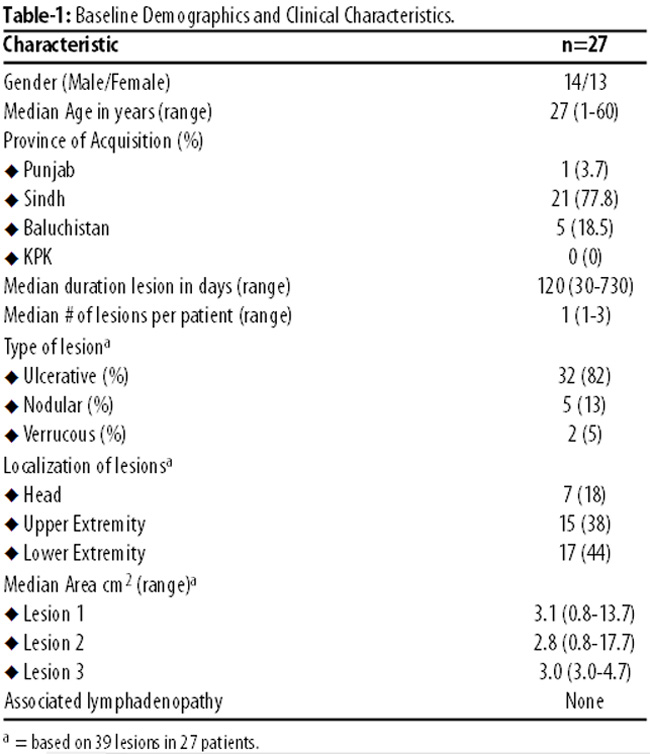
There were a total of 39 lesions in 27 patients; 6(22%) patients had 2 lesions each, and 3(11.1%) had 3 lesions each. The median lesion duration was 4 months. The majority of patients 21(77%) had acquired the infection in Sindh province.
There were a total of 546 applications of the HECT-CL device over 1 week of thermotherapy, and the average temperature of the device at initial application was 51.6±0.8°C (range: 50.1-52.0°C). Of the 23 patients who were followed up, by the 180-day evaluation, overall 19(83%) had been cured (Table-2);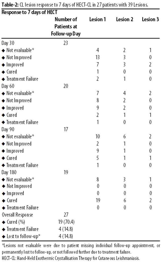
1(5.2%) at 30 days, 2(10.4%) by 60 days, 5(26%) by 90 and 11(57.8%) stood cured at some point between the visits on the 90th and 180th day (Figure-2).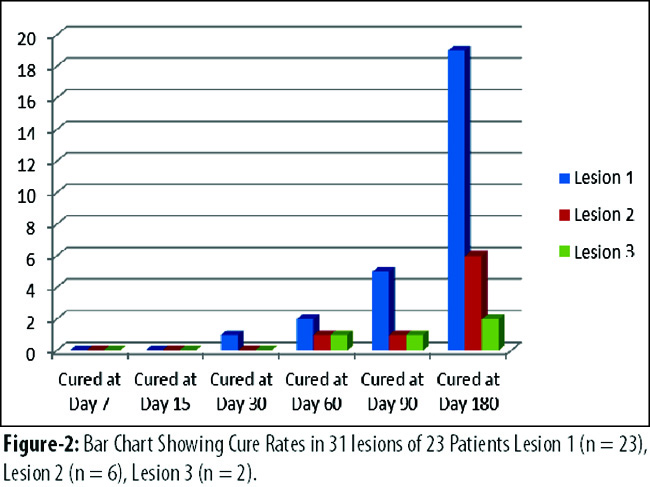
Of the 23 patients with complete data, there were 4(17%) treatment failures; 2(8.5%) at 30 days, 1(4.2%) at 60 and 1(4.2%) at 90 days. Photographic documentation of response to thermotherapy was done (Figure-3).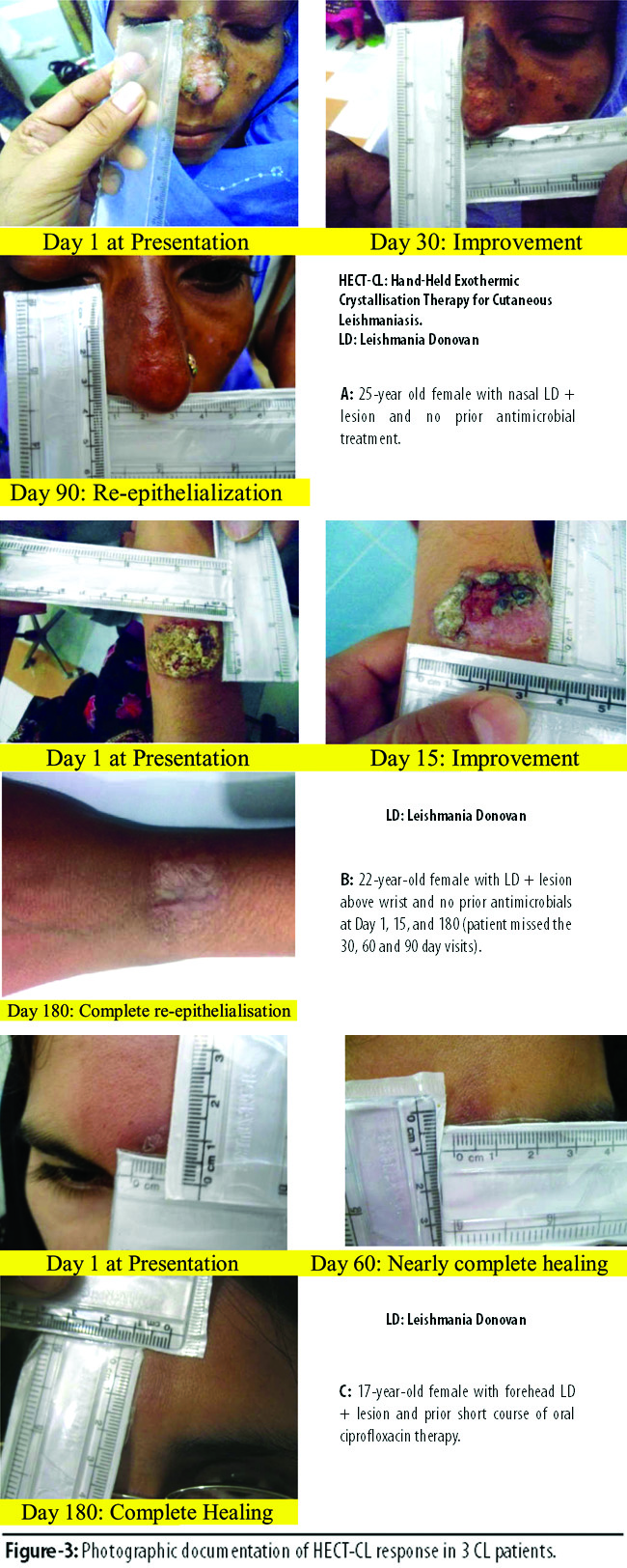
The average temperature of the HECT-CL at initial application and the temperature elevation of the skin achieved after application for each lesion were calculated. Days 1, 4 and 7 of thermotherapy were compared and temperatures were found to be fairly uniform on all days. However, applications were increasingly better tolerated with less adverse effects documented on Day 7 compared with Day 1 (Table-3).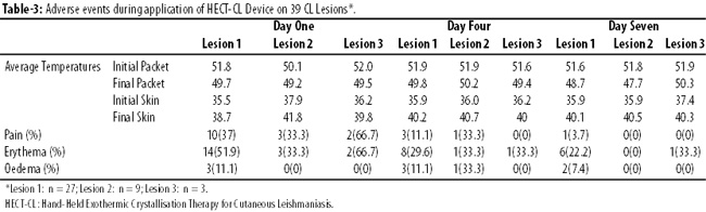
None of the patients had blistering at the site of HECT-CL application.
Discussion
This pilot study provides first proof of concept that 7 days of HECT-CL conduction thermotherapy for Old World CL is safe, well-tolerated, and efficacious with no significant adverse events.
A 2013 pilot HECT-CL study of New World CL in Peru demonstrated similar study goals on 38 patients.22 They describe a clinical cure rate of 60% (15/25), although when 13 compassionate use patients (11 of which had failed SC before) were added in a meta-analysis, the cure rate rose to 68.4%. Though lower than our cure rate of 83%, the Peruvian study reports earlier improvement with re-epithelialisation and resolution of inflammation occurring by one-month follow-up in over two-third of the lesions. Of 25 prospective patients, 24 (96%) subjects had already improved, with one clinical cure documented as early as Day 7 of HECT-CL thermotherapy, and 15 (60%) were cured by 2 months (all 15 remained clinically cured at the 90 and 180 day follow-up visits). However, our study was affected by non-attendance of clinic follow-up at 60 (5 no-shows) and 90 (7 no-shows) days, which obscured the exact time that patients had their lesions cured. Future studies with more rigorous patient follow-up should show earlier healing times.
The earlier cure rates seen in the Peruvian study could also be because the temperature of the HECT-CL device at initial application ranged between 51°C-53°C, which was on average 1°C higher than the temperatures employed in our study. Another possibility could be that the Peruvian trial used continual HECT-CL heat over three minutes, while our trial broke up the daily 3-minute treatment into 2-4 fractions. A final contribution to earlier cure rates in Peru for the HECT-CL could be regional variation in virulence and heat-susceptibility within species. Variation in clinical cure rate by species was seen in the Peruvian study; 56.2% in L. (V) peruviana, 50% in L. (V) braziliensis and 100% in L. (V). guyanensis.22 A study of thermosensitivity of 8 CL strains suggested, albeit in vitro in a murine cell line, that Old World strains such as L. tropica might be less responsive to heat therapy than New World CL strains.14
Minimal adverse events to thermotherapy with HECT-CL device were noted in our study, with a good safety profile, similar to findings in the Peruvian study. However, in the latter, 2 cases of blistering occurred, in both of which the heat pack temperature at application was 53°C, suggesting limiting initial treatment temperatures to 52±1°C. Indeed, we employed temperatures lower than 53°C and did not document any case of blistering.
When our HECT-CL cure rate of 83% is compared to the cure rates using other modalities of thermotherapy in Old World CL, a randomised controlled trial in Kabul, Afghanistan, reported clinical cure in 75 of 108 patients (69.4%) with a radiofrequency TT device.15 The time to cure was shorter in the Kabul study (median 53 days) compared to our study.
Local infiltration with antimonials (sodium stibogluconate [SSG]100 mg/mL 0.3-3 mL/lesion or meglumine antimonate 0.2-0.8 mL/lesion) was studied in Saudi Arabia in regions where L. major and Leishmania tropica are endemic. Two to 15 infiltrations were needed to achieve cure rates of 72-99%.23,24 According to 2002 WHO estimates, 90% of L. tropica lesions in Pakistan can be healed by intralesional pentavalent antimonials.25 Our cure rate with the HECT-CL device is comparable and avoids intralesional infiltration, a painful and technically difficult procedure.
Systemic therapy with intramuscular SSG therapy daily for 20 days is an alternative if intralesional therapy fails, but clinical trials demonstrating efficacy of parenteral SSG in L. major and L. tropica are limited. A cure rate of 44.8% with intramuscular SSG was reported in the study from Kabul.15 Oral systemic therapy with fluconazole has demonstrated a cure rate of 59% after 3 months, and itraconazole has also demonstrated only modest beneficial effects. Therefore, overall, our cure rate with HECT-CL appears to be comparable to, or better than, other local and systemic treatment modalities, but with no toxicity and at a fraction of the cost.
Our study used a 7-day extended HECT-CL intervention (like the Old World Peru HECT-CL study) contrasting with a single application of radiowave thermotherapy, due to concerns about the efficacy of a single application with HECT-CL. As CL protozoans are unlikely to develop thermo-resistance, we could consider elongated or recurrent treatment courses in unsatisfactory lesion responses. Therefore, future controlled clinical trials considering modifications of our current HECT-CL thermotherapy procedures should be designed to identify whether prolongation or reduction of applications offers the best therapeutic outcomes. We concur that the optimal temperature of HECT-CL at initial application should be 52±1°C, as was recommended by the Peruvian study.22
It has been seen that in patients with multiple lesions, TT of one lesion can induce complete simultaneous remission of other lesions. A study demonstrated that local TT in CL lesions leads to systemic cytokine responses similar to that induced by systemic glucantime therapy and found that healing occurred in untreated lesions as well.16 Therefore, future trials should also incorporate immunological studies to investigate molecular and cellular phenomena during HECT-CL therapy.
Conclusion
Further HECT-CL studies should be conducted on Old World CL to verify our results. If future results confirm similar efficacy, treatment of CL could move from the tertiary care hospital to the community/primary health centre, thereby lowering patient costs and simplifying logistics. HECT-CL thermotherapy utilises low-cost and safe technology that village health workers can easily learn, and the increased access to treatment could be a breakthrough for CL treatment and control, with an end-goal of CL elimination from communities.
Acknowledgement
Mr. Hafeez Siddiqui, Bridge Consultants Foundation, for data analysis.
References
1. World Health Organization. Leishmanisis: burden of disease. (Online) (Cited 2014 Jan 1) Available from URL: http:// www.who.int/leishmaniasis/burden/en.
2. Desjeux P. Leishmaniasis: current situation and new perspectives. Comp Immunol Microbiol Infect Dis 2004; 27: 305-18.
3. Murray HW, Berman JD, Davies CR, Saravia NG. Advances in leishmaniasis. Lancet 2005; 366: 1561-77.
4. Brooker S, Mohammed N, Adil K, Agha S, Reithinger R, Rowland M, et al. Leishmaniasis in refugee and local Pakistani populations. Emerg Infect Dis 2004; 10: 1681-4.
5. ul Bari A, Hasshim R, Mahmood K, Muhammad I, Shahbaz N, Tariq KM. Clinico-epidemiological pattern of cutaneous leishmaniasis in armed forces personnel fighting war against terrorism in Khyber Pakhtunkhwa Province and Fata regions. J Pak Assoc Dermatol 2011; 21: 10-5.
6. Bhutto AM, Soomro RA, Nonaka S, Hashiguchi Y. Detection of new endemic areas of cutaneous leishmaniasis in Pakistan: a 6-year study. Int J Dermatol 2003; 42: 543-8.
7. Marco JD, Bhutto AM, Soomro FR, Baloch JH, Barroso PA, Kato H, et al. Multilocus enzyme electrophoresis and cytochrome B gene sequencing-based identification of Leishmania isolates from different foci of cutaneous leishmaniasis in Pakistan. Am J Trop Med Hyg 2006; 75: 261-6.
8. Blum J, Desjeux P, Schwartz E, Beck B, Hatz C. Treatment of cutaneous leishmaniasis among travellers. J Antimicrob Chemother 2004; 53: 158-66.
9. Lira R, Sundar S, Makharia A, Kenney R, Gam A, Saraiva E, et al. Evidence that the high incidence of treatment failures in Indian kala-azar is due to the emergence of antimony-resistant strains of Leishmania donovani. J Infect Dis 1999; 180: 564-7.
10. Saldanha AC, Romero GA, Merchan-Hamann E, Magalhaes AV, Macedo Vde O. A comparative study between sodium stibogluconate BP 88R and meglumine antimoniate in the treatment of cutaneous leishmaniasis. The efficacy and safety. Rev Soc Bras Med Trop 1999; 32: 383-7.
11. Saldanha AC, Romero GA, Guerra C, Merchan-Hamann E, Macedo Vde O. Comparative study between sodium stibogluconate BP 88 and meglumine antimoniate in cutaneous leishmaniasis treatment. II. Biochemical and cardiac toxicity. Rev Soc Bras Med Trop 2000; 33: 383-8.
12. Ameen M. Cutaneous and mucocutaneous leishmaniasis: emerging therapies and progress in disease management. Expert Opin Pharmacother 2010; 11: 557-69.
13. Berman JD, Neva FA. Effect of temperature on multiplication of Leishmania amastigotes within human monocyte-derived macrophages in vitro. Am J Trop Med Hyg 1981; 30: 318-21.
14. Sacks DL, Barral A, Neva FA. Thermosensitivity patterns of Old vs. New World cutaneous strains of Leishmania growing within mouse peritoneal macrophages in vitro. Am J Trop Med Hyg 1983; 32: 300-4.
15. Reithinger R, Mohsen M, Wahid M, Bismullah M, Quinnell RJ, Davies CR, et al. Efficacy of thermotherapy to treat cutaneous leishmaniasis caused by Leishmania tropica in Kabul, Afghanistan: a randomized, controlled trial. Clin Infect Dis 2005; 40: 1148-55.
16. Lobo IM, Soares MB, Correia TM, de Freitas LA, Oliveira MI, Nakatani M, et al. Heat therapy for cutaneous leishmaniasis elicits a systemic cytokine response similar to that of antimonial (Glucantime) therapy. Trans R Soc Trop Med Hyg 2006; 100: 642-9.
17. Sadeghian G, Nilfroushzadeh MA, Iraji F. Efficacy of local heat therapy by radiofrequency in the treatment of cutaneous leishmaniasis, compared with intralesional injection of meglumine antimoniate. Clin Exp Dermatol 2007; 32: 371-4.
18. Aronson NE, Wortmann GW, Byrne WR, Howard RS, Bernstein WB, Marovich MA, et al. A randomized controlled trial of local heat therapy versus intravenous sodium stibogluconate for the treatment of cutaneous Leishmania major infection. PLoS Negl Trop Dis 2010; 4: e628.
19. Navin TR, Arana BA, Arana FE, de Merida AM, Castillo AL, Pozuelos JL. Placebo-controlled clinical trial of meglumine antimonate (glucantime) vs. localized controlled heat in the treatment of cutaneous leishmaniasis in Guatemala. Am J Trop Med Hyg 1990; 42: 43-50.
20. Velasco-Castrejon O, Walton BC, Rivas-Sanchez B, Garcia MF, Lazaro GJ, Hobart O, et al. Treatment of cutaneous leishmaniasis with localized current field (radio frequency) in Tabasco, Mexico. Am J Trop Med Hyg 1997; 57: 309-12.
21. Willard RJ, Jeffcoat AM, Benson PM, Walsh DS. Cutaneous leishmaniasis in soldiers from Fort Campbell, Kentucky returning from Operation Iraqi Freedom highlights diagnostic and therapeutic options. J Am Acad Dermatol 2005; 52: 977-87.
22. Valencia BM, Miller D, Witzig RS, Boggild AK, Llanos-Cuentas A. Novel low-cost thermotherapy for cutaneous leishmaniasis in Peru. PLoS Negl Trop Dis 2013; 7: e2196.
23. Alkhawajah AM, Larbi E, al-Gindan Y, Abahussein A, Jain S. Treatment of cutaneous leishmaniasis with antimony: intramuscular versus intralesional administration. Ann Trop Med Parasitol 1997; 91: 899-905.
24. Tallab TM, Bahamdam KA, Mirdad S, Johargi H, Mourad MM, Ibrahim K, et al. Cutaneous leishmaniasis: schedules for intralesional treatment with sodium stibogluconate. Int J Dermatol 1996; 35: 594-7.
25. Munir M, Mohammed K, Babkerhyl M. Guidelines for the Treatment and Prevention of Cutaneous Leishmaniasis in Pakistan. Ministry of Health Pakistan. WHO, Health Net International 2002.
Journal of the Pakistan Medical Association has agreed to receive and publish manuscripts in accordance with the principles of the following committees:


