

Abdul Razaque Mari ( Department of Neurosurgery, DUHS & Civil Hospital, Karachi. )
Irfanullah Shah ( Spine Fellow, National Neurosciences Institute, King Fahad Medical City Riyadh, KSA. )
Muhammed Imran ( Department of Neurosurgery, DUHS & Civil Hospital, Karachi. )
Junaid Ashraf ( Department of Neurosurgery, DUHS & Civil Hospital, Karachi. )
December 2014, Volume 64, Issue 12
Original Article
Role of intraoperative ultrasound in achieving complete resection of intra-axial solid brain tumours

Abstract
Objectives: To determine the frequency of completeness of resection for intra-axial solid brain tumours with the help of intra-operative ultrasound to detect residual brain tumour.
Methods: The cross-sectional study was conducted at the Department of Neurosurgery, Dow University of Health Sciences and Civil Hospital Karachi, from September 2009 to June 2010 and comprised patients with intra-axial solid brain lesion. During operation following standard craniotomy, multi-plane sonographic examination was performed using intra-operative ultrasound for tumour localisation and calculation of dimension, followed by tumour resection in the standard fashion. At the end of tumour resection ultrasound was again used for the detection of any residual tumour. Results of intra-operative ultrasound were compared with post-operative contrast magnetic resonance imaging.
Results: Of the 39 cases in which intra-operative ultrasound was performed, 32(82.1%) were males and 7(17.9%) were females, with an overall mean age of 42.6±19.7 years. Intra-operative ultrasonography was able to localise and delineate the tumour in all 39 (100%) cases. It showed no residual tumour in 36 (92.3%) cases, but in 3(7.7%) cases residual tumour was detected. Post-operative contrast enhancing magnetic resonance imaging showed no residual tumour in 35(89.7%) cases and in 4(10.3%) cases residual tumour was detected. The frequency of completely resected intra-axial solid brain tumour was 35(89.7%), while in 4(10.3%) cases incomplete resection was observed.
Conclusion: The study concluded that intra-operative ultrasonography has an important role in achieving increased frequency of completely resected intra-axial solid brain tumours.
Keywords: Brain tumour, Intra-operative ultrasonography (IOUS), Extent of resection. (JPMA 64: 1343; 2014).
Introduction
It is well documented that maximum resection of brain tumours increases the survival rate, but on the other hand it also increases the risk of damaging the normal peritumoural neural tissues.1,2 In the quest for equilibrium on tumour resection, many parallel fields were focussed and improvements in neuroanaesthesiology, neurophysiology and neuroimaging have opened new horizons for researchers during the last few decades3,4 resulting in the introduction of neuronavigation system. Although it has revolutionised the operative technique, but brain shifting with cerebrospinal fluid (CSF) drainage and tumour debulking is a well-documented drawback.5 Real-time peroperative imaging has eventually solved this problem to a large extent. Magnetic resonance imaging (MRI) was available for utilisation which led to the development of MRI-compatible operation rooms. But it was not practical in countries with limited financial resources for human health. The alternative modality available was ultrasound, which is being used since the early 1970s.6 It is a dynamic, highly interactive, efficacious and cost-effective imaging study that is easily available. The main obstacle in usage of ultrasound during neurosurgery is the thick human skull, which does not let the sound waves to pass through. After craniotomy, it is as useful as anywhere else on human body.
Intra-operative ultrasound (IOUS) is the modality which picks up real-time changes. After craniotomy it localises the mass, and helps in appropriate dural opening. It also facilitates cortical incision in deep-seated lesions which minimises damage to neural tissue.7 Due to the increased echogenicity of the solid brain tumours compared to normal echoic brain tissue, ultrasound can be used to delineate the residual tumour from normal brain tissue during operation, thereby achieving complete tumour resection. It also provides a chance to confirm the final picture of operative field for residual tumour before completing the surgery.8-10 Its applicability was limited previously in neurosurgery because of poor image quality, but recent improvements in ultrasonography equipment and image quality, small sized sensitive probes, ability to freeze real-time display, printing facility of images, built-in cursors for measurement and Doppler technology, have remarkably renewed the concept for its use as a neuronavigation tool worldwide.11
Patients and Methods
The cross-sectional study was conducted at the Department of Neurosurgery, Dow University of Health Sciences and Civil Hospital Karachi, from September 2009 to June 2010 and comprised patients with intra-axial solid brain lesion. All patients fulfilling the inclusion criteria were assessed through September 2009 to August 2010. During surgery following standard craniotomy, before opening of dura, multi-plane sonographic examination with Aloka (SSO-500 Echo Camera, Japan) ultrasound device with a 5 MHz convex probe was performed for tumour localisation. A particular consultant radiologist was involved right through the study. At the end of tumour resection ultrasound was used for the detection of any residual tumour. Any hyperechoic margin extending from surgical cavity into normal brain parenchyma >5mm was taken as residual, and removed with the same microsurgical technique by the same surgeon. Hyperechoic margin <5mm was taken as complete tumour excision. All surgeries were performed by the same consultant neurosurgeon. A post-operative contrast MRI was obtained 10 days after surgery12 for confirmation of ultrasound findings and completeness of tumour resection.
Patients with recurrent intra-axial brain tumours or with history of radiation therapy were excluded from the study as both recurrence and irradiation alters the sonographic findings.13 Secondly, intra-axial solid brain tumours were studied as they have similar sonographic findings of hyperechoic mass lesion. A pre-designed proforma was used for the collection of data. It included information about patient's age, gender, hospital registration number, intra-operative ultrasound findings, post-operative contrast MRI and level of tumour resection (complete/incomplete).
Results
Of the 39 patients fulfilling the inclusion criteria, 32(82.1%) were males. The overall age ranged from 6 to74 years with a mean of 42.6±19.7 years (Table-1).
As in the case of brain tumours, clinical presentation was mainly according to the location of the tumour. Of all the patients, 14(35.8%) had parietal lobe tumours, and presented with new onset seizures, focal neurological deficit like, hemiplegia/hemiparesis along with signs and symptoms of raised intracranial pressure (headache, nausea, vomiting and papilledema), 9 (23%) patients had frontal lobe lesions and were admitted for symptoms of raised intracranial pressure, visual problems and focal neurological deficit which could be aphasia or hemiplegia. Three (7.6%) patients having temporal lobe mass came with partial seizure. Occipital lobe tumours were diagnosed in 2 (5.1%) patients and presented mainly with visual focal neurological deficits. Besides, 11 (28.2%) patients with posterior fossa tumour presented with symptoms of hydrocephalus (raised intracranial pressure) and cerebellar signs.
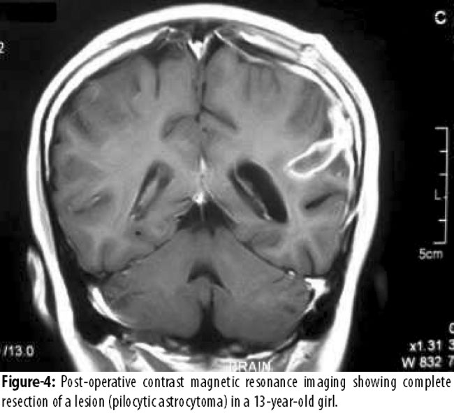
In 14 (35.8%) patients after apparent gross total excision of tumour, ultrasound showed residual tumour (margins >5mm into brain parenchyma) and surgery continued. In 3 (7.6%) patients with residual tumour further excision was not possible due to anatomical limitations during surgery. In 36 (92.3%) patients ultrasound obtained at the end of surgery before dural closure revealed no residual tumour, and routine surgical closure was carried out. In all patients, post-operative contrast MRI was performed 10 days after surgery for confirmation of IOUS findings and detection of residual tumour if any. MRI showed residual tumour in 4(10.3%) patients. Three (7.7%) of these patients were those where further resection was not possible, while in 35 (97.2%) patients with ultrasound-confirmed total resection MRI confirmed the IOUS findings, and showed no residual tumour (Figures 1-4).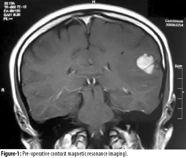
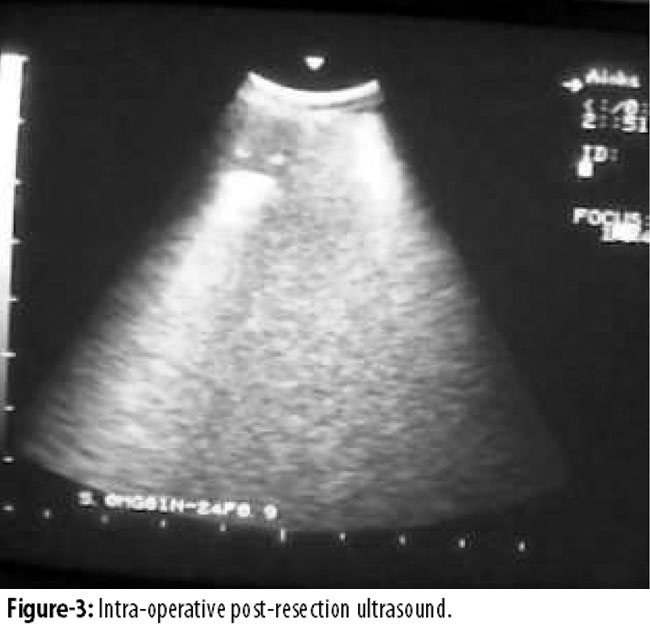
In only 1(2.56%) case there was disagreement between IOUS and MRI findings, as ultrasound showed no residual tumour while on MRI there was residual tumour. In final outcome, the frequency of completely resected intra-axial solid brain tumours in our study was 35(89.7%) (Table-2).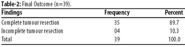
Discussion
Usage of ultrasound in neurosurgery was reported by a 1970 study.14 Use of IOUS does not require any extra investment. Routine scanners available in hospitals are sufficient, even if the probe happens to be small and specific. Frequency of the probe usually ranges from 7.5 to 10 MHz, but for deep-seated lesions 3 to 5 MHz frequency is usually required.15 Due to hyper-echogenicity of the solid brain tumours compared to normal echoic brain tissue, IOUS can be used to localise and delineate the residual tumour from normal brain tissue, thus achieving complete tumour resection and avoiding damage to normal brain parenchyma. IOUS has proved good for determining residual glioblastoma multiforme, metastases or other diffuse glial tumours during surgery where naked eye may not differentiate.16 IOUS is not good for recurrent lesions, where post-treatment changes may not be differentiated.17 It may also not identify the histopathological pattern of different brain tumours. IOUS can be used at various times during surgery, from localisation of lesion up to assessing the residual tumour after apparent completion of resection.
A study18 conducted on 70 patients with intra-axial brain tumours (36 gliomas and 34 metastasis had 38 males and 32 females with a median age of 49 years (range: 20-90 years). In that study recurrent brain tumours, which received radiation therapy, were also included. It evaluated the efficacy of IOUS in localising the brain tumour, defining its margins and to determine the extent of resection during surgery. Post-excision IOUS and MRI findings were compared to measure the residual tumour volume. Post-operative MRI in 9 patients revealed no residual tumour while IOUS showed no residual tumour in only 6 patients. This difference was not statistically significant (p=0.97).
Researchers in a prospective clinical study19 assessing supratentorial gliomas mentioned that IOUS proved extremely helpful in easily localising a small, deep-seated supratentorial lesion without taking additional operating time. The study further added that the learning curve was with the use of the IOUS was not long.
Another set of researchers20 in 2005 used intraoperative ultrasound in 32 patients of intracranial brain tumours. There were 18 female patients and 14 were males with a mean age of 49.5 years (range: 11-71 years). It excluded recurrent tumours. Both intra-axial and extra-axial tumours were assessed; and both cystic and solid tumours were included. It determined the method of agreement between IOUS and post-operative contrast MRI for the detection of residual tumour. After resection of tumour, resection cavity borders were examined with IOUS to assess hyperechoic shadow which highlighted residual tumour. It compared the findings of ultrasound with contrast enhancing MRI, obtained within 48 hours after surgery. It concluded with fair to good kappa level (k=0.72) for inter-method agreement, a level showing that IOUS was comparable to MRI in detecting the residual tumour and maximising tumour resection. There was disagreement in 4 cases, in which ultrasound showed no residual tumour but on post-operative contrast enhancing MRI residual tumour was detected.
Our study was different from this study as we performed post-operative contrast MRI after 10 days of surgery after post-operative changes had abated to get more accurate MRI findings. A study21 in 2006 used intra-operative ultrasound in 35 patients with intracranial brain tumours. It revealed that IOUS could detect location, size and depth of cerebral tumours. IOUS localised the tumour in all 35 patients (100%), in 3(9%) patients residual tumour was detected on ultrasound, which was confirmed by post-operative MRI scan. In 32 (91%) patients the study achieved complete tumour resection. The frequency of completely resected tumours was 91%.
In our study majority of the patients were males 32(82.1%) and only 7(17.9%) were females. Mean age of the patients was 42.62 years (range: 6-74 years). We included in our study those patients who were diagnosed pre-operatively on MRI as intra-axial solid brain tumours. This was different from the earlier study20 as it included intra-axial as well extra-axial brain tumours and also both cystic and solid tumours, while they excluded previously-treated cases. Researchers in a study included patients with recurrent tumours and/or those who received radiation therapy. This was different from our study as we excluded those patients having history of recurrent tumour and those who had received radiation therapy, as IOUS could not differentiate the radiation-induced changes accurately as the study mentioned later in its results.17,22
In recent years 3D ultrasound is being used as navigational tool. One of the case report for temporal glioma, where optic radiation was involved. Awake craniotomy with electrical stimulation and IOUS with miniature probe, could achieve 97% of tumour, rest of the tumour was adhered with optic radiation and hence was left intentionally.23
In our study IOUS was able to localise the tumour in all 39 (100%) patients and in 36 (92.3%) patients showed complete resection of tumour, while post-operative contrast MRI confirmed complete tumour resection in 35(89.7%) of these 36 patients. IOUS was able to detect residual tumour in 3 (7.7%) patients during surgery, while post-operative contrast MRI detected residual tumour in 4 (10.3%) patients.
In 1(2.54%) patient MRI did not confirm the IOUS findings. as ultrasound was not able to detect residual tumour but post-operative contrast MRI showed residual tumour but this was not statistically significant. In our study the frequency of intra-operative ultrasound guided complete resection of solid intra-axial brain tumours was 89.7 %, which is comparable to other studies.
Results from our study offer a greater understanding potential benefit of ultrasonography as the real-time imaging modality in neurosurgical field.
In current clinical practice, intra-operative MRI guided tumour resection represents state-of-the-art technology. However, this requires an expensive MRI scanner dedicated to this procedure alone. Moreover the feedback provided is not in real time. On the other hand, intra-operative ultrasound is a safe and inexpensive modality of obtaining an intra-operative feedback. Although the resolution of images is not as high as that in the intra-operative MRI the feedback provided is in real time and imaging can be safely and quickly repeated during surgery. Hence, it appears reasonable to infer that this is a reasonable form of surgery in our part of the world.
Conclusion
Intra-operative ultrasonography has turned out to be a good substitute for intra-operative MRI (iMRI) in neurosurgery operative rooms, especially in countries like ours where iMRI is not available due to cost constraints. Intra-operative ultrasonography is a non-invasive, safe, cost-effective and easily available technique that may easily be used in neurosurgery and appears a reasonable form of surgery in our part of the world.
References
1. Smith JS , Chang EF, Lamborn KR, Chang SM, Prados MD, Cha S, et al. Role of extent of resection in long-term outcome of low- Grade Hemispheric gliomas. J clin Oncol 2008; 26: 1338-45.
2. Quigley MR, Maroon JC. The relationship between survival and the extent of the resection in patients with supratentorial malignant gliomas. Neurosurgery 1991; 29: 385-8.
3. Duffau H, Moritz-Gasser S, Gatignol P. Functional outcome after language mapping for insular World Health Organization Grade II gliomas in the dominant hemisphere: experience with 24 patients. Neurosurg Focus 2009; 27: E7.
4. Cabrera HN, Almeida AN, Silva CC, Fonoff ET, Martin Md, Leite Cda C, et al Use of intraoperative MRI for resection of gliomas. Arq Neuropsiquiatr 2011; 69: 949-53.
5. Wadley J, Dorward N, Kitchen N, Thomas D. Pre-operative planning and intra-operative guidance in modern neurosurgery: a review of 300 cases. Ann R Coll Surg Engl 1999; 81: 217-25.
6. Tronnier V, Wirtz C, Knauth M. Intraoperative diagnostic and interventional magnetic resonance imaging in Neurosurgery. Neurosurgery 1997; 40: 891-902.
7. Hayashi K, Matsuo T, Suyama K, Nagata I. Usefulness of ultrasonography with a burr-hole transducer during surgery through a burr hole. Neurol Med Chir (Tokyo) 2012; 52: 165-8.
8. Rubin JM, Doharmann GJ, Duda EF, Beezhold C. Intraoperative meningioma. Am J Neuroradiol 1982; 3: 305-8.
9. Zhang Y, Lin S, Zhao JZ, Zhao YL, Wang S, Jiang ZL, et al Application of intraoperative ultrasound combined with neuronavigation for resection of intracranial cavernous malformations in minimally invasive neurosurgery Zhonghua Wai Ke Za Zhi 2011; 49: 716-9.
10. Zhao P, Zhao Y, Zhang W, Zhang MZ, Zhao JZ. Ultrasound guided minimally invasive neurosurgery in treatment of cranial tumours: clinical study. Zhonghoa Yixue Za Zhi 2006; 86: 1600-3.
11. Moiyadi A, Shetty P. Objective assessment of intraoperative ultrasound in resection of central nervous system tumour: A cost effective tool for intraoperative navigation in neurosurgery. 2011; 2: 4-11.
12. Campbell JW, Pollack IF, Martinez AJ. High grade astrocytomas in children:radiologically complete resection is associated with an excellent long term prognosis. Neurosurgery 1994; 38: 258-64.
13. Becker G,Hofman E,Woydt M. Postoperative neuroimaging of high grade gliomas: comparison of transcranial sonography, magnetic resonance imaging and computed tomography. Neurosurgery 1999; 44: 469-78.
14. Ram Z, Bruck B, Hadani M. Ultrasound in pituitary tumour surgery. Pituitary. 1999; 2: 133-8.
15. Sawaya R. Radical resection of glioblastoma: techniques and benefits. Contemp Neurosurg 2002; 24: 1-5.
16. Lang FF, Gilbert MR. Diffusely infiltrative low grade gliomas in adults. J clin Oncol 2006; 24: 1236-45.
17. Mahmood T. Experience of intraoperative ultrasound compared to CT, MR & percutaneous ultrasound. J Surg Pakistan 2003; 8: 35-8.
18. Hammoud MA, Ligon BL, ElSouki R. Use of intraoperative ultrasound for localizing tumours and determining the amount of resection: a comparative study with magnetic resonance imaging. J Neurosurg 1996; 84: 737-41.
19. Koivukangas J, Louhisalmi Y, Alakuijala J. Ultrasound controlled neuronavigator guided brain surgery. J Neurosurg 1993; 79: 36-42.
20. Erdogan N, Tucer B, Mavili E, Menku A, Kurtosy A. Ultrasound guidance in intracranial tumour resection: Correlation with postoperative magnetic resonance findings. Acta Radiol 2005; 46: 743-9.
21. Zhao P, Zhao Y, Zhang W, Zhang MZ, Zhao JZ. Ultrasound guided minimally invasive neurosurgery in treatment of cranial tumors: clinical study. Zhonghoa Yixue Za Zhi 2006; 86: 1600-3.
22. Keles GE, Chang EF, Kethleen R. Volumetric extent of resection and residual contrast enhancement on initial surgery as predictors of outcome in adult patients with hemispheric anaplastic astrocytoma. J Neurosurg 2006; 105: 34-40.
23. Steno A, Karlík M, Mendel P, Cik M, Steno J. Navigated three-dimensional intraoperative ultrasound-guided awake resection of low-grade glioma partially infiltrating optic radiation Acta Neurochir 2012; 154: 1255-62.
Journal of the Pakistan Medical Association has agreed to receive and publish manuscripts in accordance with the principles of the following committees:


