

Hamza Karabag ( Department of Clinical Neurosurgery, University of Harran, Sanliurfa, Turkey. )
Erkan Cakmak ( Department of Clinical Neurosurgery, University of Harran, Sanliurfa, Turkey. )
Bahattin Celik ( Department of Clinical Neurosurgery, University of Harran, Sanliurfa, Turkey. )
Ahmet Celal Iplikcioglu ( Department of Clinical Neurosurgery, University of Harran, Sanliurfa, Turkey. )
Ahmet Faruk Soran ( Department of Clinical Neurosurgery, University of Harran, Sanliurfa, Turkey. )
December 2014, Volume 64, Issue 12
Original Article

Abstract
Objective: To evaluate the effectiveness and safety of cervical arthroplasty and anterior cervical discectomy fusion methods.
Methods: The randomised clinical trial was conducted at the neurosurgical clinic of University of Harran, Turkey, between February 2009 and January 2010. The patients had single level disc disorder between C4-C7 levels. Before surgery, all of the patients had taken medical treatment with no improvement. Surgery was conducted with anterior approach, and disc prosthesis or polyetheretherketone cage for fusion were applied after patients were randomly divided into two groups. For preoperative and postoperative clinical evaluations Neck Disability Index and Visual Analogue Scale were used. Surgical results were evaluated according to Odom\'s criterion, and \'excellent\' and \'good\' results were accepted as successful. P<0.05 was taken as statistically significant.
Results: Of the 42 patients in the study, 23(54.76%) were treated with Anterior Cervical Discectomy and Fusion, and 19(45.23%) with Cervical Disc Arthroplasty. There were no statistical differences between postoperative mean Visual Analogue Scale score (p<0.86) and Neck Disability Index scores (p<0.11) in the two groups. Average decrease in lordosis angle was 1.2 degree in Arthroplsty group, while it was 1 degree in the Fusion group. Postoperative adjacent segment degeneration was not detected in either group.
Conclusion: Anterior Cervical Discectomy and Fusion, and Cervical Disc Arthroplasty are safe and successful methods for the treatment of single level cervical disc disease. Although the latter is a relatively new technique performed with increased frequency, but its superiority is still uncertain.
Keywords: Cervical disc, Arthroplasty, Fusion/stabilisation, PEEK cage, Disc prosthesis, Radiography. (JPMA 64: 1348; 2014).
Introduction
The standard conservative treatment for single level cervical disorder which is non-responsive is Cervical Disc Arthroplasty (CDA) or Anterior Cervical Discectomy and Fusion (ACDF).1,2 Adjacent segment disease and dysmotility in fusion distance were seen in the patients who were treated with ACDF. As an alternative, in order to preserve movement in distance and disc height, to avoid fusion development and to conserve normal segmental lordosis, cervical disk prothesis were designed.3,4 However, randomised clinical controlled trials comparing the surgical results of two different methods have remained limited and controversial. The aim of the study was to evaluate the effectiveness and safety of cervical arthroplasty and ACDF methods by a randomised clinical trial.
Patients and Methods
The randomised clinical trial was conducted at the neurosurgical clinic of University of Harran, Turkey, between February 2009 and January 2010. The patients had single level disc disorder between C4-C7 levels. Before surgery, all of the patients had taken medical treatment with no improvement. Surgery was conducted with anterior approach, and disc prosthesis or polyetheretherketone (PEEK) cage for fusion were applied after patients were randomly divided into two groups. For preoperative and postoperative clinical evaluations Neck Disability Index (NDI) and Visual Analogue Scale (VAS) were used. Surgical results were evaluated according to Odom\'s criterion as \'excellent\', \'good\', \'mean\' and \'bad\' depending on the resolution, improvement, persistence of preoperative symptoms and findings (Table-1);5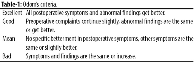
and \'excellent\' and \'good\' results were accepted as successful. P<0.05 was taken as statistically significant.
Radiological examination was done by preoperative and postoperative plain and functional radiographies. Measurements were taken from various perspectives: cervical lordosis in neutral position and in flexion and extension cervical lordosis was measured between C2 and C7 according to Cobb.6 In the follow-up, new bone formation in anterior and posterior of vertebral corpus and collapse in operation spacing (>2mm)7 were evaluated. In flexion-extension position, >2° movement in lateral graphy was accepted as pseudoarthrosis.8,9 Adjacent segment disease was considered as developed in patients who came with upper level or level-of-disc disorder established clinically or radiologically in two years.
Clinical and radiological (NDI, VAS, cervical lordosis) parameters were analysed by Mann-Whitney U test. Chi square test was used for the evaluation of surgical success.
Results
Of the 42 patients in the study, 23(54.76%) were treated with ACDF, and 19(45.23%) with Cervical Disc Arthroplasty (CDA). The mean age of ACDF patients was 46.2±4.7 years while that of CDA patients was 43.1±6.1 years. In none of the patients any major surgical complications such as oesophageal perforation, neural or vascular injuries occurred.
The operation level was C4-C5 in 3(13%) ACDF patients and 3(15.7%) CDA patients. It was C5-C6 in 11(47.8%) patients and 10(52.6%) CDA patients; C6-C7 in 9(39%) ACDF patients and 6(31.5%) CDA patients (Table-2).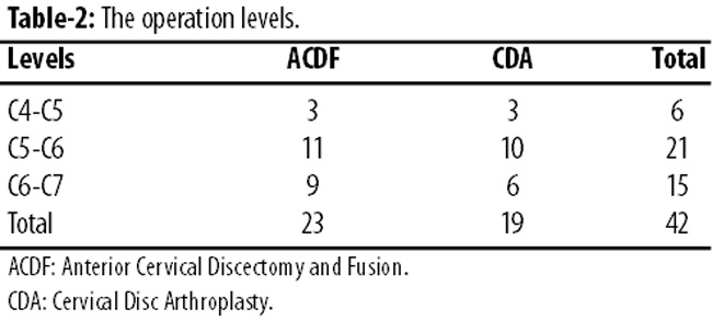
In the 23rd month of the postoperative phase, in 1(10%) C5-C6 patient of the CDA group, the implant on vertebra corpus skidded into the posterior (Figure-1).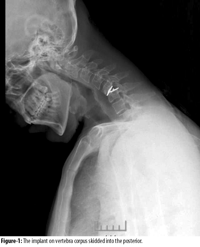
There was no neurological deficit, but there was pain on the right side starting from shoulder towards forearm medialis. The patient was shifted to PEEK cage instead of disc prosthesis (Figure-2).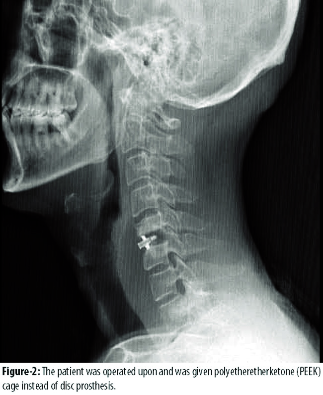
Two-year follow-up evaluated the preoperative-postoperative VAS and NDI averages (Table-3).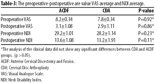
There were no statistical differences between postoperative mean VAS score (p<0.86) and NDI scores (p<0.11) in the two groups. Average decrease in lordosis angle was 1.2 degree in Arthroplsty group, while it was 1 degree in the Fusion group. Postoperative adjacent segment degeneration was not detected in either group.\'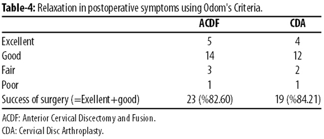
On Odom\'s criterion, the surgery in 19(82.6%) ACDF patients and 16(84.2%) CDA patients was considered a success.
Discussion
There are two surgical treatments of cervical disc disorder: ACDF and CDA. The latter was developed in recent years to protect movement and to prevent adjacent segment disease. But no clear results are available about the superiority of one method over the other in randomised clinical studies. For ACDF, systems like allograft, autograft, cage (titanium, PEEK), graft+plaque+screw are used. Today for fusion, mostly PEEK anatomic cervical cage is used. Also, some in vitro studies were done about PEEK cages for biological accordance, the absence of cytotoxic and mutagenic effects,10 non-absorbable property and corrosion situation against resistance. In these studies, it was proved that PEEK cages are secure biomaterial for intermediate ring11 and the flexibility is similar with bones.12 We also used PEEK cage in the study.
The most important disadvantage of ACDF is the loss of movement in fusion segment and it was thought that there was a development of adjacent segment degeneration and instability due to this. Because of this, using of disc arthroplasty as an alternative treatment is increasing. Some authors suggested that adjacent segment was found higher in ACDF. Two reasons were reported for this. The first reason was the protection of disc height in CDA and spinal dynamism reducing movement difficulty in adjacent segment.13 The second reason was fusion providing higher stress in adjacent segment and speeding up disc degeneration.14,15 Recent studies show that CDA is a secure alternative.16,17 However, the results of comparative studies with ACDF should be discussed since the accepted superiority of CDA in theoretical base cannot be established. A meta-analysis18 showed that CDA group has lower risks of reoperation related to adjacent-segment degeneration although there was no statistically significant difference in NDI neck and arm pain scores. In three systematic reviews comparing this to surgical methods produced controversial results. In these studies, fusions were made of different grafts and different cages, plaques and plaque-screw systems were used. The compared disc prostheses also were of different systems.19-21 In our study, PEEK cage for fusion, osimplant Byrain disc prosthesis was used for arthroplasty.
In literature, dysphagia was found higher in ACDF.22 The reason is believed to be related to over-retraction and dissection.23 Since we used PEEK cage, there was no such complication seen in either group.
Although there are fewer results of neck pain scores, in these studies arthroplasty results are better.19 Again in some of these studies the success rate is higher.19 The high percentage of success of cervical arthroplasty was connected to the rareness of overloading and the stability of intervertebral pressure due to this. However, the patients having serious facet degeneration generally are not put into arthroplasty group. Most of the studies in literature are related to adjacent segment surgery. One study reported that 3 out of multi-centred 276 patients in arthroplasty group and 9 out of 265 patients in fusion group were operated for adjacent segment disease.24 On the other hand, another study25 detected adjacent segment disease in the same ratio in a multi-centred group of 269 patients. In our study, no operation was done in both groups for adjacent segment disease. The most important reason of this was the single-centre nature of the study. However, it was reported that the fusion done with PEEK cage is better than the other fusion methods.26
Some clinical studies reported that the results are equal or better than ACDF in cervical arthroplasty.19 However, such results are limited because of the usage of different fusion methods, different disk prostheses and different surgical indications. Yet there are no studies about the life of prostheses used in arthroplasty. There was no differences in surgical results in our two groups.
There are several limitations in our study. First, the number of patients was small. Second, there was no extended follow-up to evaluate the differences between the two methods, larger randomised controlled trials and long-term follow-up are recommended.
Conclusion
The existing standard treatment of surgery for symptomatic cervical disc disorder is anterior cervical decompression. There was no statistical difference in surgical and radiological results between ACDF and CDA groups in the study. However, it is still uncertain whether CDA is more effective and safer than ACDF.
References
1. Clements DH, O\'Leary PF. Anterior cervical discectomy and fusion. Spine (Phila Pa 1976) 1990; 15: 1023-5.
2. Walker AE, Robinson RA. Anterior cervical fusion. Dia Med 1962; 34: 894-900.
3. Cummins BH, Robertson JT, Gill SS. Surgical experience with an implanted artificial cervical joint. J Neurosurg 1998; 88: 943-8.
4. Wigfield CC, Gill SS, Nelson RJ, Metcalf NH, Robertson JTL. The new Frenchay artificial cervical joint: results from a two-year pilot study. Spine (Phila Pa 1976) 2002; 27: 2446-52.
5. Odom GL, Finney W, Woodhall B. Cervical disc lesions. J Am Med Assoc 1958; 166: 23-8.
6. Gwinn De, ?annOtti CA, Benzel EC, Steinmetz MP. Effective lordosis: Analysis of sagittal spinal canal alignment in cervical spondylotic myelopathy. J.Neurosurg spine 2009; 11: 667-2.
7. Barsa P, Suchomel P. Factors affecting sagittal malalignment due to cage subsidence in standalone cage assisted anterior cervical fusion. Eur Spine J 2007; 16: 1395-400.
8. Hacker RJ. A randomized prospective study of an anterior cervical interbody fusion device with a minimum of 2 years of follow-up results. J Neurosurg 2000; 93(2 Suppl): 222-26.
9. Hacker RJ, Cauthen JC, Gilbert TJ, Griffith SL. A prospective randomized multicenter clinical evaluation of an anterior cervical fusion cage. Spine (Phila Pa 1976) 2000; 25: 2646-55.
10. Katzer A, Marquardt H, Westendorf J, Wening JV, von Foerster G. Polyetheretherketone-cytotoxicity and mutagenicity in vitro. Biomaterials 2002; 23: 1749-59.
11. Toth JM, Wang M, Estes BT, Scifert JL, Seim HB III, Turner AS Polyetheretherketone as a biomaterial for spinal applications. Biomaterials 2006; 7: 324-34.
12. Wenz LM, Merritt K, Brown SA, Moet A, Steffee AD. In vitro biocompatibility of polyetheretherketone and polysulfone composites. J Biomed Mater Res. 1990; 4: 207-15.
13. Nabhan A, Steudel WI, Pape D, Ishak B. Segmental kinematics and adjacent level degeneration following disc replacement versus fusion: RCT with three years of follow-up. J Long Term Eff Med Implants 2007; 17: 229-36.
14. Rabin D, Pickett GE, Bisnaire L, Duggal N. The kinematics of anterior cervical discectomy and fusion versus artificial cervical disc: a pilot study. Neurosurgery 2007; 61(3 Suppl): 100-4 (Discussion 104-105).
15. Chang UK, Kim DH, Lee MC, Willenberg R, Kim SH, Lim J. Changes in adjacent-level disc pressure and facet joint force after cervical arthroplasty compared with cervical discectomy and fusion. J Neurosurg Spine 2007; 7: 33-9.
16. Phillips FM, Garfin SR. Cervical disc replacement. Spine (Phila Pa 1976) 2005; 30(17 Suppl): S27-S33.
17. Anderson PA, Sasso RC, Riew KD. Update on cervical artificial disk replacement. Instr Course Lect. 2007; 56: 237-45.
18. Jiang H, Zhu Z, Qiu Y, Qian B, Qiu X, Ji M. Cervical disc arthroplasty versus fusion for single-level symptomatic cervical disc disease: a meta-analysis of randomized controlled trials. Arch Orthop Trauma Surg 2012; 132: 141-51.
19. Xing D, Ma XL, Ma JX, Wang J, Ma T, Chen Y. A meta-analysis of cervical arthroplasty compared to anterior cervical discectomy and fusion for single-level cervical disc disease. J Clin Neurosci 2013; 20: 970-8.
20. Pickett GE, Sekhon LHS, Sears WR, Duggal N. Complications with cervical arthroplasty. J Neurosurg Spine. 2006; 4: 98-105.
21. Sears WR, Duggal N, Sekhon LH, Williamson OD. Segmental malalignment with the Bryan cervical disc prosthesis-contributing factors. J Spinal Disord Tech. 2007; 20: 111-7.
22. Baron EM, Young WF. Cervical spondylotic myelopathy: a brief review of its pathophysiology, clinical course, and diagnosis. Neurosurgery. 2007; 60(1 Supp11): 35-41.
23. McAfee PC, Cappuccino A, Cunningham BW, Devine JG, Phillips FM, Regan JJ, et al. Lower incidence of dysphagia with cervical arthroplasty compared with ACDF in a prospective randomized clinical trial. J Spinal Disord Tech 2010; 23: 1-8.
24. Mummaneni PV, Burkus JK, Haid RW, Traynelis VC, Zdeblick TA. Clinical and radiographic analysis of cervical disc arthroplasty compared with allograft fusion: a randomized controlled clinical trial. J Neurosurg Spine 2007; 6: 198-209.
25. Coric D, Nunley PD, Guyer RD, Musante D, Carmody CN, Gordon CR, et al. Prospective, randomized, multicenter study of cervical arthroplasty: 269 patients from the Kineflex C artificial disc investigational device exemption study with a minimum 2-year follow-up: clinical article. J Neurosurg Spine 2011; 15: 348-58.
26. Faldini C, Chehrassan M, Miscione MT, Acri F, d\'Amato M, Pungetti C, et al. Single-level anterior cervical discectomy and interbody fusion using PEEK anatomical cervical cage and allograft bone. J Orthop Traumatol 2011; 12: 201-5.
Journal of the Pakistan Medical Association has agreed to receive and publish manuscripts in accordance with the principles of the following committees:


