

Aneela Altaf Kidwai ( Department of Medicine, Abbasi Shaheed Hospital, Karachi. )
Syeda Urooj Riaz ( Department of Medicine, Abbasi Shaheed Hospital, Karachi. )
Sabeen Aatif ( Department of Medicine, Abbasi Shaheed Hospital, Karachi. )
Saleemullah Paracha ( Department of Medicine, Abbasi Shaheed Hospital, Karachi. )
December 2014, Volume 64, Issue 12
Original Article

Abstract
Objective: To determine the spontaneous platelet recovery time in primary and secondary dengue infection in a tertiary care hospital.
Method: The cross-sectional observational study was conducted at Abbasi Shaheed Hospital, Karachi, from July 2010 to January 2011, and comprised 138 seropositive patients with ages 13 years and above who fulfilled the World Health Organisation criteria of probable dengue infection, and presented with platelet count of <50,000/mm3 were enrolled. Serology was performed using rapid immunochromatographic assay and enzyme-linked immunosorbent assay with differential detection of immunoglobulin M and G. Spontaneous platelet recovery time (days) in both primary and secondary dengue infection was recorded. SPSS 20 was used for statistical analysis.
Result: Of the total 138, patients, 38(27.5%) had primary infection and 100(72.5%) had secondary infection. Male-to-female ratio was 2.3:1. Among primary and secondary infections, platelet count on presentation was not significantly different (p<0.64). Mean spontaneous platelet recovery time was 3±2.6 days and 3±1.87 days in primary and secondary infection respectively. Higher platelet count at presentation was associated with early recovery time (p<0.033). Of 108(78%) patients who presented with platelet count of 20,000-<50,000/mm3, platelet count of 36(33.33%) rose to >50,000/mm3 within 2 days, and 62(57.4%) rose to >50,000 in 3-5 days. . In primary and secondary dengue infections, no statistically significant difference was observed in spontaneous platelet recovery time (-=0.87).
Conclusion: Platelet count at presentation and spontaneous platelet recovery time do not significantly differ in primary and secondary dengue infection.
Keywords: Platelet recovery time, Primary dengue infection, Secondary dengue infection. (JPMA 64: 1380; 2014).
Introduction
Dengue infection has emerged as the most rapidly spreading mosquito-borne viral disease of public health concern affecting tropical and subtropical countries across the globe. In Southeast Asia, including Pakistan, multiple outbreaks of dengue infection have occurred over the past few decades. Clinical manifestation caused by dengue virus ranges from an asymptomatic infection to a self-limiting febrile illness to life-threatening infection characterised by increased capillary permeability and shock.
Higher prevalence of secondary dengue infection is reported in dengue endemic areas and has been shown to be associated with more severe disease areas.1,2 Thrombocytopenia is a constant laboratory finding in dengue infection. The platelets usually drop to below 100,000/mm3 in febrile phase or around defervescence and may remain low for the first few days of recovery.3 Variable degree of thrombocytopenia is observed in both primary and secondary dengue infection. Thrombocytopenia i.e. platelet count <100,000/mm3 is found to be one of the major reasons for tertiary care referred in both public and private sectors even in uncomplicated dengue. The aim of the present study was to determine the spontaneous platelet recovery (SPR) time in primary and secondary dengue infection. Declining pattern of platelet count often creates an overwhelming response on behalf of the patients and their relatives and results in unnecessary hospitalization and platelet transfusion. Understanding the natural history of SPR time would not only help in clinical management, but also decrease the economic burden in terms of hospitalisation and inappropriate platelet transfusions.
Patients and Methods
This cross-sectional observational analytical inferential study was conducted at the Abbasi Shaheed Hospital, Karachi, from July 2010 to January 2011. According to the inclusion criteria, all patients aged 13 years and above presenting with acute febrile illness (fever for 2-7 days) and fulfilling the World Health Organisation (WHO) case definition criteria of Dengue Fever (DF) and Dengue Haemorrhagic Fever (DHF) were included with random purposive sampling. We did not have any study that had SPR time as the outcome, so we enrolled as many patients as we could during our study period.
Patients were enrolled as probable DF cases as per the WHO criteria of probable DF i.e. acute febrile illness with two or more of the following manifestations: headache, retro-orbital pain, myalgia, arthralgia, rash, haemorrhagic manifestations, leukopenia and supportive serology. A positive immunoglobulin M (IgM) antibody test on serum samples collected five or more days after the onset of fever, or occurrence at the same location and time as other confirmed cases of DF. DHF is defined as probable cases of dengue and haemorrhagic tendencies, thrombocytopenia and evidence of plasma leakage i.e. >20% rise in haematocrit or signs of plasma leakage (pleural effusion, ascites or hypo-proteinemia).
Patients with bleeding diathesis, haematological malignancies, cirrhosis, malaria, sepsis and enteric fever were excluded. Patients who received platelet transfusion were also excluded.
As per the admission criteria, during dengue outbreak only those suspected cases of DF were hospitalised who either presented with platelet count of <50,000/mm3 or who had haemorrhagic magnification at any platelet count. Likewise, all those haemodynamically stable patients whose platelet rose to >50,000/mm3 were discharged. We, therefore, recorded SPR time till platelet counts had risen to 50,000/mm3.
Serological diagnosis of dengue infection was performed using rapid immunochromatography test (ICT) with qualitative detection of dengue IgM and IgG. Acute plasma sera of all the enrolled patients were collected after 5 days of fever onset. In non-reactive cases, repeat ICT was performed after 3-5 days of first sera (within 12 days of fever onset). Dengue IgM capture enzyme-linked immunosorbent assay (ELISA) was done in some suspected patients to confirm the status. In ICT assay, specimen reacts with dengue antigen coated particles (type 1-4) in the test strip and the mixture is captured by ligand anti-IgM or anti-IgM, forming a visible band. The IgG cut-off in the test strip has been set to detect the high IgG levels characteristic of secondary infection, therefore the primary dengue infection was defined by a visible IgM band without a visible IgG band, whereas, secondary infection was defined by a positive IgG band with or without positive IgM band.
Data was analysed using SPSS 20.0. Mean and standard deviations (SD) were calculated for continuous variables and frequencies were done to determine relationship between different variables. Statistical significance was considered at p <0.05.
Results
Of the patients initially enrolled, 3(2%) had concomitant plasmodium vivax infection and were excluded. The final sample, as such, comprised 138(98%) patients. The maximum number of patients were admitted in September 46(33.3%), October 72(52%), November 9(6.5%) and December 4(2.9%). Overall male-to-female ratio was 2.3:1 with 97 (70%) males and 41 (30%) females. In terms of age, 110 (79.7%) patients belonged to 13-33 group. Among the cases, 38(27.5%) had primary dengue infection and 100(72.5%) had secondary dengue infection (Table-1). 
The distribution of thrombocytopenia on presentation among cases of primary and secondary dengue infection was also noted (Figure-1).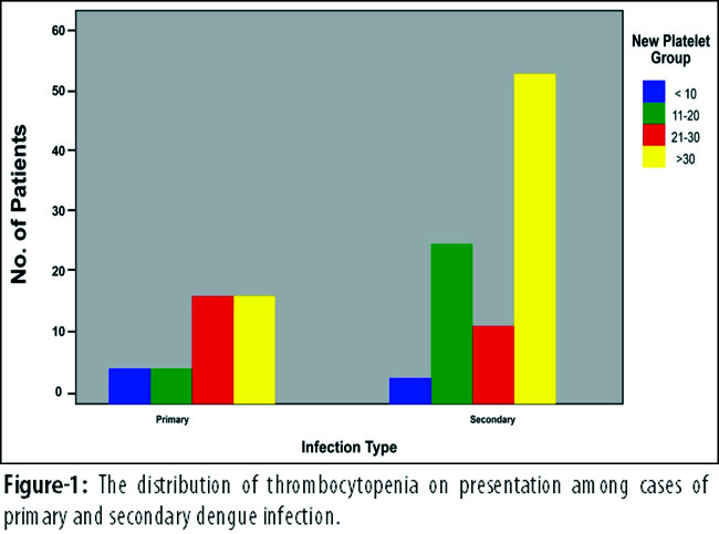
Platelet count was recorded daily in all patients till the count had risen to 50,000/mm3. The count of 83 (60%) patients rose to 50,000/mm3 in 3-5 days\' time (Table-2).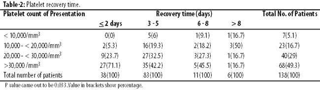
Higher platelet count at presentation was associated with early recovery time (p<0.03).
Mean platelet count in cohort of -primary dengue infection was 28±13/mm3 and secondary dengue infection was 31±11/mm3. Mean SPR time (days) was 3±2.6 days in primary dengue infection and 3±1.87 days in secondary dengue infection (Figure-2).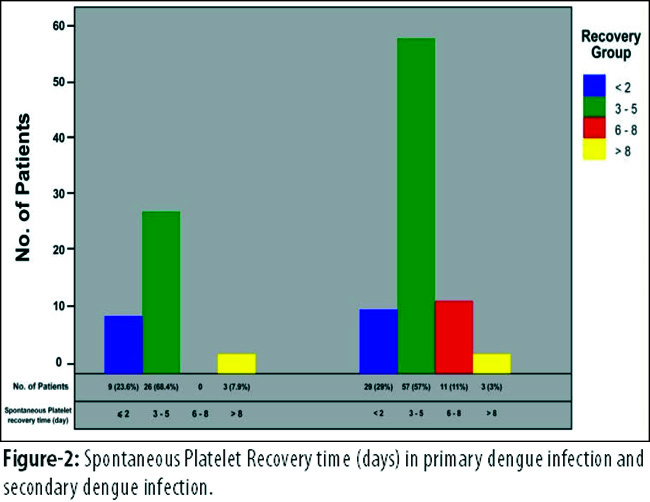
No statistically significant difference was observed in SPR time between primary and secondary dengue infections (p<0.87).
Discussion
In Pakistan significant dengue outbreaks have been witnessed over past the few years. Post-monsoon seasonal trend of dengue outbreaks have been reported in Southeast Asia.4,5 Our study also supports this seasonal trend, as peak incidence was observed in September and October. The most affected age group with both primary and secondary dengue infection belonged to 13-33 age group (p=0.871). Similar to our findings another study6 also found no significant statistical difference in age group among primary and secondary dengue infections. In contrast to our findings, one study7 showed higher percentage of secondary infection in patients >50 years of age.
Serological studies conducted in four provinces of Pakistan identified all four dengue serotypes (DEN1 to DEN4) during multiple outbreaks. In 1994 outbreak of dengue in Karachi, DEN1 and DEN2 were identified, DEN3 was isolated in 2005 outbreak and co-circulation of DEN2 and DEN3 was found in the 2006 outbreak.8-10 In the 2008 outbreak in Lahore, DEN4, DEN2 and DEN3 were identified, whereas in the 2010 outbreak DEN2 and DEN1 were found.11,12 This sequential circulation of different serotypes during regular outbreaks in dengue-endemic areas results in an increased rate of secondary dengue infection. The current study also reports high percentage of secondary infection 72% (n=100) in138 patients. Our pervious study from the same tertiary care hospital in Karachi also identified high incidence of secondary infection 83% (n=209).13 Our findings are consistent with reports from other parts of Southeast Asia. One study1 found 92% cases of secondary dengue infection, while another14 reported 79% cases of secondary dengue infection.
Secondary dengue infection has been shown to be associated with more severe disease,15 but pathogenesis that lead to severe dengue is not clearly understood. Lack of significant association between disease severity and secondary dengue infection has also been reported.16,17 We studied clinical manifestations among primary and secondary dengue infection at presentation. No statistically significant difference in clinical presentation was observed among primary and secondary dengue infection. Thrombocytopenia is a common presenting feature is dengue infection. In our cohort of patients, we did not find any significant difference in platelet count on presentation (p<0.64) among primary and secondary dengue cases.
Despite the poor correlation between dengue thrombocytopenia and bleeding manifestation,18,19 most clinicians show an interest in chasing platelet count very closely. Studies have shown inappropriate platelet transfusions in hospitalised patients during dengue outbreaks. A study20 reported 51% inappropriate platelet transfusion in hospitalised patients, whereas another21 reported 56% during dengue epidemic in Dehli. We evaluated SPR time in relation to presenting platelet/mm3. Our study showed that high platelet count at presentation was significantly associated with early recovery time (p<0.033). Of 78% (n=108) patients who presented with platelet count of 20,000-<50,000/mm3, platelet count of 36(33.33%) rose to >50,000/mm3 within 2 days, and 62(57.4%) rose to >50,000 in 3-5 days. In patinets of primary and secondary dengue infection we did not observe statistically significant difference in SPR time (p<0.87).
Conclusion
Out experience of managing patients of uncomplicated primary and secondary dengue cases should be considered to reduce the hospital cost and unnecessary transfusions.
Acknowledgement
We are grateful to Rehan Malick for statistical help and Dr. Saad Rehman and Dr. Zohaib Ahmed for help in data collection.
References
1. Atul G, Garg J, RaoY, Upadhyay, Sakhuja S. Prevalence of dengue among clinically suspected febrile episodes at a teaching hospital in North India. J. Infect.Dis.Immun 2011; 3: 85-9.
2. Guzmán MG., Kourí G, Valdés L, Bravo J, Vázquez S, Halstead SB. Enhanced severity of secondary dengue-2 infections: death rates in 1981 and 1997 Cuban outbreaks. Rev Panam Salud Publica 2002; 11: 223-7.
3. World Health Organization. Guidelines for dengue clinical case management of dengue fever/ dengue hemorrhagic fever/ DSS 2011 in Pakistan context. 1st ed. WHO, Pakistan.
4. Mumtaz K, Khan MS, Patel J, TariqM, Hilal H, Ahmed S etal. Out break of dengue fever in Karachi 2006: a clinical perspective. J Pak Med Assoc 2009; 59: 339.
5. Ukey PM, Bondade SA, Paunipagar PV, Powar RM, Akulwar SL .Study of seroprevalence of dengue fever in central India. Indian J Comm Med 2010, 35: 517-9.
6. Lye D C, Chan M, Lee V J, Leo Y S. Do young adults with uncomplicated dengue fever need hospitalisation? A retrospective analysis of clinical and laboratory features. Singapore Med J 2008; 49: 476.
7. Guilarde AO, Turchi MD, Siqueira JB Jr., Feres VC, Rocha B, Levi JE, et al. Dengue and dengue hemorrhagic fever among adults: Clinical outcomes related to viremia, serotypes, and antibody response. J Infect Dis 2008; 197: 817-24.
8. Chan YC, Salahuddin NI, Khan J, Tan HC, Seah CL, Li J, et al. Dengue haemorrhagic fever outbreak in Karachi, Pakistan, 1994. Trans R Soc Trop Med Hyg 1995; 89: 619-20.
9. Jamil B, Hasan R, Zafar A, Bewley K, Chamberlain J, Mioulet V, et al. Dengue virus serotype 3, Karachi, Pakistan. Emerg Infect Dis 2007, 13: 182-3.
10. Khan E, Hasan R, Mehraj V, Nasir A, Siddiqe J, Hewson R. Co-circulations of two genotypes of dengue virus in 2006 out-break of dengue hemarrhogic fever in Karachi, Pakistan. J ClinVirol 2008; 43: 176-9.
11. Humayoun MA, Waseem T, Jawa AA, Hashmi MS, Akram J. Multiple dengue serotypes and high frequency of dengue hemorrhagic fever at two tertiary care hospitals in Lahore during the 2008 dengue virus outbreak in Punjab, Pakistan. Int J Infect Dis 2010; 14: 54-9.
12. Mahmood N, Rana MY, Qureshi Z, Mujtaba G, Shaukat U. Prevalence and molecular characterization of dengue viruses serotypes in 2010 epidemic. Am J Med Sci 2010; 343: 61-4.
13. Kidwai AA, Jamal Q, Saher, Mehrunnisa, Farooqi FR, Ullah S. Serodiagnosis of dengue infection using rapid immunochromatography test in patients with probable dengue infection. J Pak Med Associ 2010; 60: 936-9.
14. Neeraja M, Lakshmi V, Umabala P, Subbalakshmi MV. Serodiagnosis of dengue virus infection in patients presenting to a tertiary care hospital .Indian J Med Microbiol 2006; 11: 280-2.
15. Guzmán MG, Kouri G, Valdes L, Bravo J, Alvarez M, Vasques S, et al. Epidemiologic studies on Dengue in Santiago de Cuba, 1997. Am J Epidemiol 2000; 152: 793-9.
16. Harris E, Videa E, Perez L, Sandoval E, Tellez Y, Perez ML, et al: Clinical, epidemiologic, and virologic features of dengue in the 1998 epidemic in Nicaragua. Am J Trop Med Hyg 2000; 63: 5-11.
17. Morens DM, Rigau-Perez JG, Lopez-Correa RH, Moore CG, Ruiz-Tiben EE, Sather GE, et al: Dengue in Puerto Rico, 1977: public health response to characterize and control an epidemic of multiple serotypes. Am J Trop Med Hyg 1986, 35: 197-211.
18. Lum LC, Goh AY, Chan PW, Abdel-Latif ME, Lam SK. Risk factors for severe hemorrhage in severe dengue infections. J Pediatr 2002; 140: 629-31.
19. Lye CD, Lee VJ, Sun Y, Leo YS.. Lack of efficacy of prophylactic platelet transfusion for severe thrombocytopenia in adults with acute uncomplicated dengue infection. Clin Infect Dis 2009; 48: 1262-5.
20. Kulkarni N. Study on the effectiveness of transfusion program in dengue patients receiving platelet transfusion. Int J Blood Transfus Immunohematol 2012; 2: 11-5.
21. Kumar ND, Tomar V, Singh B, Kela K. Platelet transfusion practice during dengue fever epidemic. Indian J Patho lMicrobiol 2000; 43: 55-60.
Journal of the Pakistan Medical Association has agreed to receive and publish manuscripts in accordance with the principles of the following committees:


