

A. H. Haquani ( Dept. of Pediatrics National Institute of Child Health, Jinnah Postgraduate Medical Centre, Karachi, Pakistan. )
Shanaz Shams ( Dept. of Pediatrics National Institute of Child Health, Jinnah Postgraduate Medical Centre, Karachi, Pakistan. )
August 1985, Volume 35, Issue 8
Original Article

Abstract
Oral rehydration therapy with electrolytes and glucose has been generally accepted in the treatment of cholera and non-cholera diarrhoeas with less than 10% dehydration. Oral fluid for cholera patients contained 90 mmol/L of Sodium because of high facal excretion rate of sodium in cholera. The same fomula has been introduced by the World Health Organisation in developing countries for non-cholera diarrhoeas where fecal excretion rates of sodium are much less compared to cholera. There has been some concern about its use in children specially infants and malnourished children. We conducted a comparative study of Orolyte (WHO formula) and Lactade, a low solute formula with glycine, the latter was found to be more effective. Reasons are discussed for recommending a low solute formula in this country.(JPMA: 35, 255, 1985).
Introduction
Diarrhoea is major cause of morbidity and mortality in infants and young children in developing countries and it frequently interacts with protein energy malnutrition. In developed countries this problem though small, is still there despite the improvement in social conditions. In this country 20% of cases show bacterial pathogens, EPE Coli, shigella or salmonella, the rest are due to viruses and parasites. In some other parts of the world like Bangladesh and West Bengal, V. Cholera is an important pathogen. Depending on the quantity of water and electrolyte losses in diarrhoea the severity of symptoms varies from a mild disturbance of transient nature to severe life threatening dehydration. There is no specific therapy for viruses and treatment directed against the bacterial infections may play some part in relieving the patients’ symptoms but fluid replacement is essential in all cases. Most children specially the well nourished ones with mild gastroenteritis recover with manipulation of diet and increased fluid intake. In others with greater gastrointestinal losses, successful treatment relies on the maintenance or restoration of adequate hydration and electrolyte balance. For this purpose intravenous (IV) infusions have been employed, but much evidence now exists on the administration and effectiveness of oral electrolyte fluid replacement mixtures thus reducing or eliminating the need for N therapy.
Experimental studies had shown that glucose improved the transport of sodium and water across the bowel wall1-5 potentially the most important medical advance, which opened a new way for oral rehydration therapy. Well conducted trials on cholera patients (enterotoxigenic diarrhoea) have proved the value of orally administered glucose electrolyte solutions in restoring hydration and electrolyte balance.6-9 In these trials the mechanism of enhancement Of intestinal sodium absorption by an actively transported sugar was found to be intact despite the continuing losses. Simultaneous improved absorption of chloride, bicarbonate and K was also observed. Oral solutions in these trials were designed to cover the gasterointestinal losses of water and electrolytes in cholera (vide infra). Using the same formula workers8,10,11 claimed success in treating children with non cholera diarrhoea. Workers in a double blind trial compared sucrose with glucose in electrolyte solutions and concluded that though glucose had a slight edge over sucrose, the later could replace glucose as it was cheaper and easily available.
Individual amino acids which are actively transported are known to increase the sodium flux from intestinal lumen to plasma. Several workers have shown that intestinal sodium absorption induced by a combination of glycine and glucose was more than the absorption induced by either substrate alone.1217 This was confirmed in subsequent clinical trials in cholera and non cholera diarrhoeas181,9 when glycine’ was added to oral rehydration mixtures. In piglets Davidson and others20 had detected defects in carbohydrate handling in viral diarrhoea where there was damage to intestinal epitheliurn as opposed to enterotoxigenic diarrhoea. when rotavirus was identified as a common cause of diarrhoea in infants and children, there was concern whether infants and children, there was concern whether glucose or sucrose electrolyte solution would be effective in the treatment of viral diarrhoea. Moreover, stool concentrations of sodium in rotavirus diarrhoea were generally found to be much lower, so infants counld receive excess of sodium with a potential risk of hypernatremia, if electrolyte solutions with high sodium content were employed as in enterotoxigenic diarrhoea. However no serious side effects were noticed in subsequent clinical trials21,22 in rotavirus diarrhoea with solutions containing 90 mmol/L of sodium with glucose or sucrose. Nevertheless, in these trials the patients had access to free water during treatment. There is general agrement now that the orally administered fluid should contain electrolytes and carbohydrate. The carbohydrate could be either glucose or sucrose. But what is controversial is the exact composition and the appropriate level of sodium. Hutchins et al23 have rightly pointed out that an important factor is deciding a suitable composition, concerns potential risk if inaccurately prepared; this in turn would be related to the mode of preparation of the solution as well as to its composition.
Following clinical trials in cholera and non cholera diarrhoeas (vide supra) the World Health Organisation has introduced Orolyte (Table I) in developing countries including Pakistan and recommended its use in all types of diarrhoeas. We felt that the sodium content was too high for general use specially in infants. Various authorities24,25 have questioned its suitability for non cholera diarrhoeas. Since our problem in Pakistan is that of non cholera diarrhoea we chose Lactade, a low sodium formula (Table I) -and conducted a comparative study with Orolyte. Here we present the results and discuss the reasons for recommending a low solute formula for oral rehydration.
Material and Methods
An open parallel study was designed in which 144 children suffering from diarrhoea and dehydration with 5 - 10% of body weight loss were allocated alternately to Lactade and Orolyte group. A proforma was maintained for each patient with name, age, sex, date and time of admission, brief history, examination, weight on admission; diarrhoea, mild, moderate or severe and degree of dehydration. After calculating the amount required for each patient according to the degree of dehydration, Lactade and Orolyte solutions were kept on the bed side for the mother to administer them under supervision. The duration of treatment and observation in each case was for 3 days only. Lactade and Orolyte are supplied in sachets. The composition when mixed in one litre of water is shown in Table I.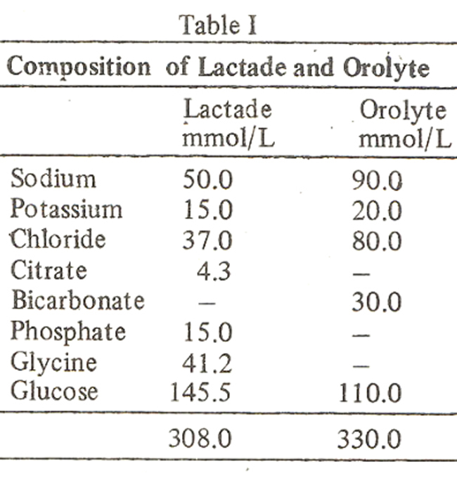
Progress with oral replacement therapy in the two groups was assessed by maintaining the following data on admission, after 6 hours, 12 hours, 24 hours, day 2 and day 3; oral fluid input; weight; urine output (±); vomiting (±); diarrhoea (±); mouth moist or dry; skin elasticity; fontanelle and eyes; laboratory data; hemoglobin, PCV, serum sodium, K, Chloride, and bicarbonate. Comments by the medical and nursing staff on the acceptability of solutions and on going drug treatment, if any, were also recorded. Statistical analysis was done by applying the student test.
Results
The mean age of one hundred and forty four children was 12.5 months. Their nutritional status was poor. As per Harvards’ growth standards27 only 4 children had standard body weight for their ages, 24 were in grade I, 42 in grade II, 30 in grade III and 44 in grade IV. 75 children received Lactade and 69 had orolyte.
Fluid Intake: Total fluid intake during the first 24 hours of treatment in the lactade group was 1378 ± 625 ml. (139.3ml/kg) and 1403 ± 602 ml (172.5m1/kg) in orolyte group and on day 3 it was 757 ± 425 ml. (120 ml/kg) and 823 ± 467 ml (119.6 mi/kg) respectively.
Weight: The mean weight of children on admission in Lactade group was 6.2 kg and 6.6 kg in Orolyte group. Mean weight at the end of first 24 hours of treatment was 6.4 kg and 6.8 kg and on day 3, 6.5kg and 6.9 kg respectively.
Packed Cell Volume: Mean PCV on admission in Lactade group was 37.7 and 32.1 in Orolyte group (Fig. 1).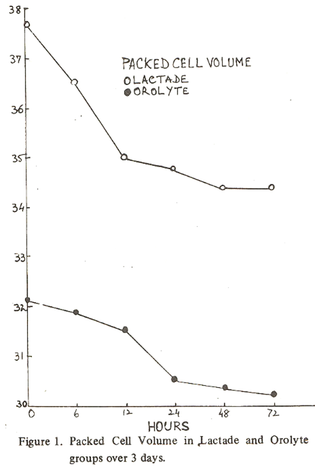
It will be observed that in Lactade group the maximum fall in PCV occured in first 12 hours of rehydration and thereafter it remained steady throughout indicating quick absorption of Lactade whereas with Orolyte fall in PCV was slow and gradual upto 72 hours.
Serum Sodium: The serum sodium status of the two groups on admission in shown in Table II.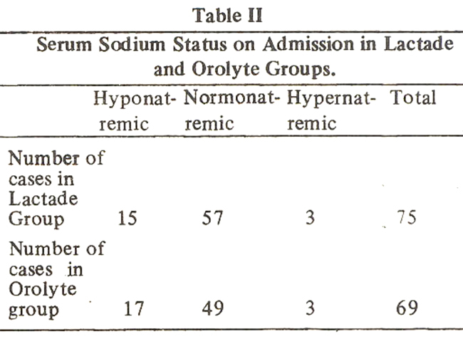
It will be observed that the two groups were more or less similar.
Mean values of serum sodium on admission were 135.8 and 134 mmol/L in Lactade and Orolyte groups respectively. Subsequent progress is shown in Fig. 2.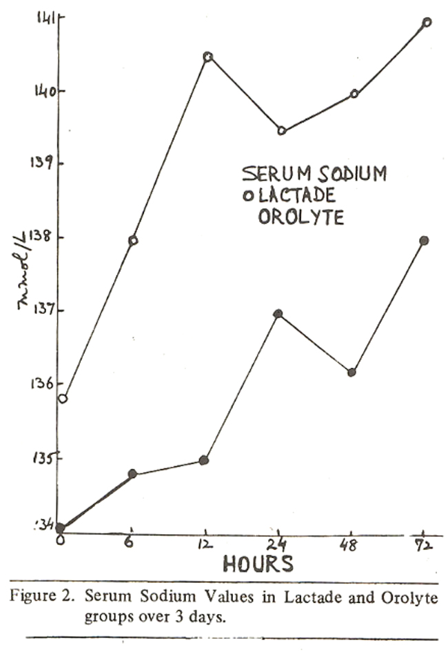
It will be observed that serum sodium levels rose early and were maintained at a higher level throughout with Lactade indicating a better absorption. Rise in serum sodium levels were slower with Orolyte and sustained at a lower level. When the mean serum sodium values in the two groups from 12 hours to day 3 were compared, as children by then were expected to be sufficiently rehydrated, the difference was found to be significantly favourable for Lactade (Lactade group 140.2 ± 5.6 : Orolyte group 136.6 ± 5.6, P 1 .00 1). Three children who were hypernatremic in each group on admission became normonatremic during rehydration period (Fig. 3 and 4).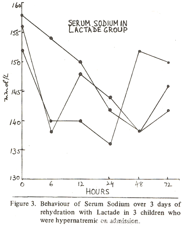
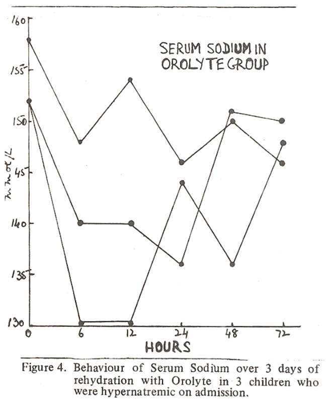
On day 3 it was observed that 10 children in Lactade and 5 in Orolyte group who were normonatremic in the first 48 hours had developed hypernatremia, range 152 162 and 152 158 mmol/L respectively. Oral rehydration was stopped and they were shifted to the general ward for further management. However, none had any untoward effect or convulsions. This. Rspect will be discussed later.
Serum Potassium: Mean values of serum K on admission were 4.5 and 4.8 mmol/L in Lactade and Orolyte groups respectively (Fig.5).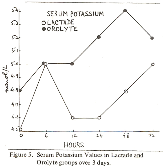
K levels rose steadily with Orolyte and were maintained within normal limits. In lactade group serum K levels rose quickly but between 12 - 24 hours there was a fall and then a steady rise perhaps indicative of quick rehydration with Lactade. Orolyte contains more K (Table I). The difference between the means from 12 hours to day 3 was significantly favourable for Orolyte, P <.001.
Serum Bicarbonate:The initial mean value of serum bicarbonate (Lactade group 18.9 and Orolyte group 18.4 mmol/L) showed a favourable response with Lactade and Orolyte in the first 6 hours (Fig. 6).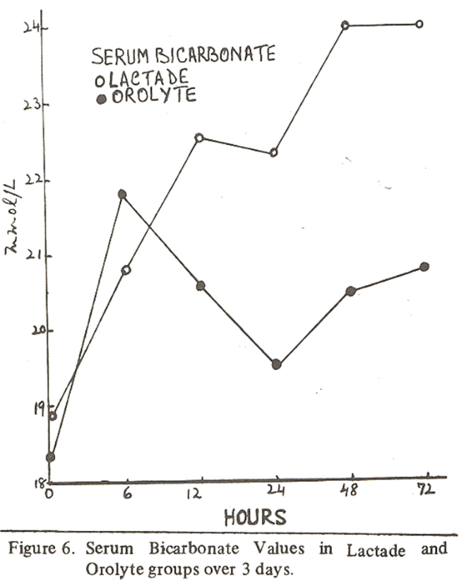
Thereafter serum levels were maintained throughout in the Lactade group only. The difference between the means from 12 hours to day 3 (Lactade group 23.2 ± 3.9, Orolyte group 20.3 ± 3) was significantly favourable for Lactade (PL.0Ol). This despite the fact that Lactade does not contain sodium bicarbonate.
Serum Chloride: Initial mean values in Lactade and Orolyte groups were 108.8 and 101.4 mmol/L respectively (Fig.7).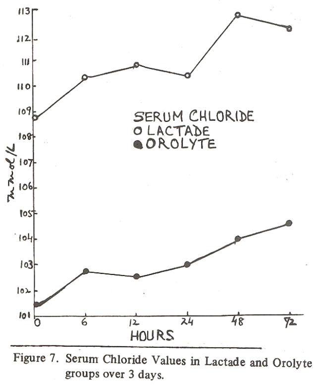
It will be observed that serum chloride levels rose with both the mixtures but the rise was significantly higher in Lactade group. The difference between the means from 12 hours to day 3 (Lactade gr. 111.2 ± 6.4 and Orolyte gr. 101.5 ± 13.4) was significant P-L. X)01. At the end of first 24 hours of rehydration therapy both breast and artificial feeding were started as most of the these children were malnourished and this is the usual routine in our department. Few children were on breast and as the yield was small it was not calculated. Artificial formulae were regraded starting with quarter strength. The latter amounts were included in their daily fluid intake.
All the children survived. Those who were rehydrated and diarrhoea was under control were discharged. Some who required further treatment for parenteral infections were shifted to Geqeral wards. During rehydration period altogether 17 cases were given antibiotics when blood or muccus was present in the stools and pathogenic Ecoli or Shigella infection was suspected.
Discussion
This trial has clearly shown the success of oral rebydration therapy in children with diarrhoea associated with Protein calorie malnutrition under supervision. Figures 1,2,5,6,7, demonstrate the superiority of Lactade in restoring hydration and maintaining electrolyte balance. There was rapid fall in PCV within 12 hours of starting the treatment. The serum levels of Na, Cl and bicarbonate were better maintained over 3 days and the difference with Orolyte group was significant. K levels remained within normal limits in both the groups. On day 2 and 3 stool output was less in Lactade group.
On day 3 ten patients in Lactade group and five in Orolyte group developed hypernatremia. This was expected with Orolyte as it contained 90 minol/L of sodium, but in the case of Lactade it was an indicator of better absorption from the gut Though most of the children were rehydrated by 24 hours after admission in the Lactade group we continued the study for a full assessment for 3 days and no free water was allowed. Following this observation we suggest that after 24 hours of rehydration Lactade should be diluted with double the quantity of water to keep the level of sodium no more than 25 rnniol/L. If the same is done for Orolyte the Na content would still be high at 45 mmol/L for maintenance.
What are the objectives and what should be the design of a formula for oral rehydration in non cholera diarrhoeas?
Regardless of the route of administration the objectives in managing a case of acute gastroenteritis are: (a) Prevention of salt and water depletion, (b) Correction of salt and water deficit and consequent specific failures of homeo. static control of body composition and (c) maintaining fluid and electrolyte balance during rehabilitation phase. Shock when present has to be treated with IV fluids. IV procedures are available only in well equiped centres with trained personnel. A search for an ideal oral hydration solution has been going on in the last decade to treat the vast majority of cases of gastroenteritis at home. The aim has been to prevent complications if the solution is administered early or when less than 10% dehydration is present, it should be able to replace the gastrointestinal losses and not allow the broad limits of homeostatic control to be disturbed. It should be remembered that there are inherent limitations with oral fluid therapy: vomiting limits the intake and diarrhoea reduces the effectiveness of intestinal absorption; there is reduced blood flow through the G.I., tract during dehydration altering the active and passive transport of nutrients; water and electrolyte deficits sometimes may be in excess of what can be replaced by oral route in a reasonable period of time and it is difficult to provide adequate nutritional supplements orally in the acute phase of the illness. The problem in designing a solution is the fact that no one solution can be designed to meet adequately the requirements of treatment and maintenance. In diarrhoea associated with malnutrition no solution can provide caloric require. ments and prevent negative nitrogen balance. It is obvious that development of an oral solution is a compromise.24 It should be safe and suitable for the majority of cases keeping in mind the etiological pattern, social conditions and least health resource commitment.
As stated earlier, early studies on oral rehydration were conducted on patients with cholera. We do not have cholera in Pakistan. Most of the cases are those of non toxigenic diarrhoea. Differences between toxigenic and non toxigenic diarrhoea must be clearly understood for a sound therapeutic approach. In cholera (toxigenic diarrhoea) oral electrolyte therapy has been successful if dehydration was less than 10%. The reason for success is the specific features of toxigenic diarrhoea; intestinal mucosa has the capacity to absorb electrolytes and fluid and the mechanism of glucose enhancing the sodium absorption is intact. In rehydration trials it was also observed that fecal losses of chloride, bicarbonate and K were also decreased under the perfusion. In the absence of convincing evidence that glucose improved the absorption of these ions also, it is assumed they moved in response to electrical and chemical gradients.7 However, when shock is present oral solutions cannot replace IV therapy.
The non enterotoxigenic diarrhoeas are associated with reduced small bowel motility, reduced mucosal integrity, increased anaerobic flora and excess of free bile acids (not observed in cholera); absorption of glucose traps fluid in flaccid ioops of bowel; fecal volume is reduced with fasting and recurrence with premature refeeding (diarrhoea of cholera not affected by fasting); abdomen is distended with succussion splash (in cholera abdomen is scaphoid); X-Ray abdomen reveals air fluid levels in distended ioops. Because of these limitations oral solutions may not be as effective as in toxigenic diarrhoea.24 However, one has to consider what type of oral solution is likely to be successful.
If the primary objective of oral rehydration is the replacement of fecal salt and water losses, a review of the nature of these losses is essential with reference to cholera and non cholera diarrhoeas as shown in Table III.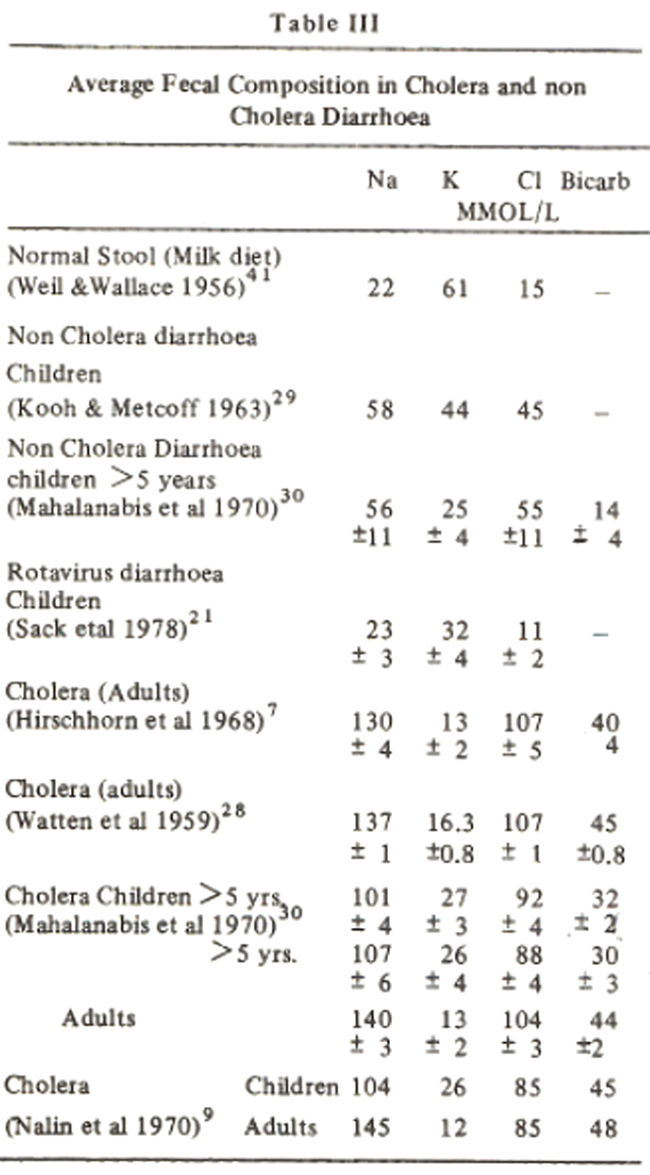
Notable features in these data are the high rates of sodium excretion in cholera stools which are consistently greater in adults. Fecal K concentrations in children are not affected by age or etiology of diarrhoea, but were twice as high with cholera compared to adults with cholera. The sum of Na + K in cholera stools is equal to that of total serum sodium. The concentrations of these ions were fixed when fecal volume exceeded 40 mI/kid. Below this level a reciprocal relationship existed between these two ions. This constancy of sodium and potassium concentrations in cholera stool is not observed in non cholera diarrhoea where the sum is approximately 1 1027 This problem of osmolar gap was studied by Torres-Pinedo.28 He found large quantities of Lactate and organic acids in non cholera diarrhoeas. These fell to low values after 24 hours fasting with low stool volume and they are probably the result of colonic bacterial action on carbohydrate. When colonic transit rates are extremely rapid the fecal electrolyte pattern resembles that of ileal fluid. The conclusion was that ileal pattern of electrolyte composition of stools in cholera was a function of high rates of fecal losses.
Carrazza29 in Brazil studied this aspect in non cholera diarrhoea. He divided his patients in two categories, those with severe diarrhoea, fecal volume greater than 60 ml/k/d and those with mild diarrhoea. 75% of total measurements of fecal volume fell below 60 g/k/d. 60 mmol/L/d. In fact fecal electrolyte pattern could be predicted from fecal volume with reasonable accuracy. Clinical characteristions were also different in these groups. In moderate diarrhoea, dehydration developed in 3-5 days (fecal Na 5-60, Ave. 46 mmol/L) and in severe diarrhoea dehydration was noted within 24 hours (fecal Na 60 120 mmol/L).
It is obvious from these studies and that of others, that there is great variability in stool sodium concentration in diarrhoeal diseases and that sodium concentration is a function of fecal volume. Hence treatment has to be individualised. For the majority of cases with moderate diarrhoea and dehydration amenable to oral treatment an oral solution containing 40-50 mmol/L of sodium should suffice. In those with severe diarrhoea (fecal volume > 60 ml/k/d) a high solute formula might be justified but in such situations obviously IV therapy is required. Since cholera stools have a high rate of sodium excretion with high fecal volume the oral rehydrating fluid had to be designed in a way that it maintained positive electrolyte balance. In various trials on cholera patients6-9 the solution contained 90 - 133 mmol/L of sodium equal or slightly greater than that of stools except in respect of sodium in adult stools because adults could receive volumes of fluid greater than their stool volume. The fecal excretion rates in non cholera diarrhoea as presented in Table III do not justify the use of 90 mmol/L of sodium in mixtures. Even a maximum of 50 is a compromise. Next question is that of maintaining the fluid and electrolyte balance during the rehabilitation phase when diet is being gradually introduced. It is difficult sometimes to demarcate this phase from the previous one. If the solution is used for maintenance hyper-natremia is likely to occur in some cases as happened in our trial and those of other workers30,31 where sodium content in the fluids was only 40 mmol/L. Following such reports it was generally agreed that a solution containing 25-30 mmol/L of sodium could be suitable for both the phases. However, following our trial we felt that if Lactade were diluted in 2 litres of water after 24 hours of rehydration the sodium content would have been at a safe level of 25 mmol/L for maintenance. If the same dilution were to be applied to Orolyte the sodium content would stifi be high, 45 mmol/L not safe for maintenance.
In our child population the effect of preexisting malnutrition and anemia has to be considered while administering rehydration mixtures. It is our practice to restrict salt in severly anemic children till their hemoglobin improved to a reasonable level with hematinics and proper diet. When confronted with diarrhoea and dehydration in such children it is difficult to imagine how one could prescribe a high solute formula with 90 mmol/L of sodium. In a Guatemala Study32 no increased fecal wastage of sodium was noticed in children with kwashiorkor. In anemic malnourished children sodium in excess of 5 mmol/k /d has resulted in heart failure.33 Sudden deaths have been reported in malnourished infants who received 7 mmol/k/d of sodium.34 In those who died altered renal function appeared to be responsible for this intolerance of a large sodium load. 32
This brings us to the consideration of the concentrating ability of the kidneys in diarrhoeal disease. Infants and malnourished children have limited concentrating ability especially when they are recovering from severe dehydration. When single high solute formula is used it has the potential for dehydrating the patient. Further, there is reduced margin of safety in subjects with increased insensible water loss (LWL) and decreased renal concentrating ability (non cholera diarrhoea and malnutrition). This is a problem with fixed intake; however, it can be overcome by increasing the intake to reduce the total water expended for IWL. It is not without risk if a child receives ad libitum a high solute formula to satisfy water requirements especially in unsupervised conditions at home. In the rehabilitation phase when nutritional adequacy is being established introduction of protein further increases the water obligatory requirements. Thus the high solute formula does not lend itself to a gradual transition to full milk formula whereas a low solute formula will not tax renal concentrating ability. 24
As stated earlier electrolyte solutions for oral rehydration can contain either glucose or sucrose. Sucrose supplies twice the number ot calories for the same osmotic load. In an attempt to supply greater energy for the much needed nutritional support at no extra osmotic cost during the acute episode, Sandhu and his colleagues used an electrolyte solution containing 90 mmol/L of sodium, as recommended by the World Health Organisation, alongwith glucose Polymers (G-PES), osmolality 300 masmol/kg, in 7 well nourished children with acute diarrhoea and dehydration: Water was allowed ad libitum tifi symptoms abated.One child developed hypernatremia and convulsions, was treated with IVfluids and completely recovered. However, the trial was abandoned. This report again points to the risk attached to the use of high sodium formula.
In this country we have experience with WHO recommended Orolyte packed in sachet and distributed only through the hospitals. We found lot of discrepancies in preparing the solution by parents at home. Few could do it accurately. A measuring glass in litres was not to be found at home. They used vessels marked in ounces or seers or they thought 4 teacups or drinking glasses equivalent to one litre. Some used 1/4 or 1/2 the sachet in less amount of water. The prescribed quantity of the mixture was not administered. Some children simply refused fluid, others did not like the taste. We have observed oedema after its use in young infants. Hutchins et al23 also noticed marked inaccuracy in preparing solutions at home in a developed country like England. In many samples they found the osmolality unacceptably high. This happened when the sodium content of the formula supplied to the parents was no more than 25-35 mmol/L. Ironside36 has already drawn attcnon to the risk of hyperosmolar solutions being administered specially when glucose was used. The WHO recommended formula has been marketed in Pakistan by several private companies in Polythene bags and thick paper bags to be mixed in a litre of water. The ingredients become discolored and crusty and are unlikely to be effective. The National Health Centre supplies the Hospitals half the quantity of the same formula is sachet to be diluted in 1/2 litre of water. Some chemists are measuring out the ingredients in plastic containers. To avoid confusion should every home have a sachet and a measuring vessel. This ideal is far from reality”.
Acknowledgement
We are grateful to Mr. Mahmood Ali, Managing Director Beecham Pakistan Limited for supplying Lactade and his generous support for this project. We are thankful to Mr. Moulvi, Liason Officer, to the staff of the Pediatric Department and Laboratory of the National Institute of Child Health, Karachi and Mr. M. Safdar of Bureau of Statistics and Mr. Abid Siddiqui of PMRC for statistical help.
References
1. Riklis, E. and Questel, J.H. Effect of cations on sugar absorption by isolated surviving giunea pig intestine. Can. J. Biochem. physiol., 1958; 36: 347.
2. Schultz, S.G., Zalusky, R. Ion transport in isolated rabbit ileum. II. The interaction between active sodium and active sugar transport. J.Gen.Physiol., 1964;47: 1043.
3. Crane, R.K. Natdependent transport in the intestine and other animal tissues. Fed. Proc., 1965;24: 1000.
4. Schedl, H.P. and Clifton, J.A. Solute and water absorption by human small intestine. Nature, 1963; 199: 1264.
5. Levinson, R.A. and Schedi, H.P. Absorption of sodium, Chloride, water and simple sugars in rat small intestine. Am J. Physiol., 1966; 211 939.
6. Phillips, R.A. Water and electrolyte losses in cholera. Fed. Proc., 1964; 23: 705.
7. Hirschhom, N., Kinzie, J.L. Sachar, D.B., Northrup, R.S., Taylor, J.O., Ahmed, S.Z. and Phillips, R.A. Decrease in net stool output in cholera during intestinal perfusion with glucose-containing solutions. N. Engl. J. Med., 1968; 279: 176.
8. Sack. R.B., Cassells, J., Mitra, R., Merritt, C., Butter, T., Thomas, J., Jacobs, B., Chaudrui, A. and Mondal, A. The use of oral replacement solutions in the treatment of cholera and other severe diarrhoea! disorders. Bull. WHO., 1970; 43: 351.
9. Nalin, D.R., Cash, R.A. and Rahman, M. Oral or nasogastric Maintenance therapy for cholera patients in all age-groups. Bull. WHO., 1970; 43: 361.
10. Hirschhorn, N., Mccarthy, B.J., Ranney, B., Hirschhorn, M.A., Woodward, S.T., Lacapa, A., Cash, R.A. and Woodward, W.F. Ad libitum oral glucose-electrolyte therapy for acute diarrhoea in Apache children. J. Pediatr 1973; 83: 562.
11. Nalin, D.R. Mata, L., Vargas, W., Loria, A.R., Levine, M.M., Cespodes, C.D., Lizano, C., Simhon, A., Mohs, E. Comparison of sucrose with glucose in oral therapy of infant diarrhoea. Lancet, 1978; 2: 277.
12. Esposito, G., Faelli, A. and Capraro, V. Influence of the transport of amino acids on glucose and sodium transport across the small intestine of the Albino rat incubated in Vitro. Experentia (Base!), 1964;20: 122.
13. Schultz, S.G. and Zalusky, R. Interactions between active sodium transport and active amino acids transport in isolated rabbit ileum. Nature 1965;205: 292.
14. Adibi, S.A. The inter relationship between leucine absorption rate and net nat and water fluxes in the human jejunum. Clin. Res., 1967; 15:458.
15. Annegers, J.H. Some effects of hexoses on the absorption of amino acids. Am. J. Physiol., 1966;210: 701.
16. Grady, G.F., Madoff, M.A., Duhamel, R.C., Moore, E.W. and Chalmers, T.C. Sodium transport by human ileum in Vitro and its reponse to cholera enterotoxin. Gastroenterology, 1967; 53: 737.
17. Fordtran, J.S., Rector, F.C. J L and Carter, N.W. The mechanisms of sodium absorption in the human small intestine. J. Clin. Invest., 1968,47: 884.
18. Nalin, D.R., Cash, R.A., Rahman, M. and Yunus, M. Effect of glycine and glucose on sodium and water absorption in patients with cholera. Gut, 1970; 11:768.
19. Nalin, D.R. and Cash, R.A. Oral or nasogastric maintenance therapy for diarrhoea of unknown aetiology resembling cholera. Trans. Ro. Sec. Trop. Med. Hyg., 1970;64: 769.
20. Davidson, G.P., Gall, D.G., Petrii, M., Butler D.G., Hamilton, J.R. J. Clm. Invest., 1977; 60: 1402.
21. Sack, D.A. Eusof, A., Merson. M.H., Black, R.E. Chaudury, A.M.A.K., ALIM, MA., Islam, S., Brown, K.W oral hydration in rotavirus diarrhoea. A double blind comparison of sucrose with glucose electrolyte solutions. Lancet, 1978; 2: 280.
22. Taylor, P.R., Merson, M.H., Black, R.E. Mizanur Rahman, A.S., Yanus, M.D., Alim, A.R. and Yolken, R.H. Oral rehydration therapy for treatment of rotavirus diarrhoea in a rural treatment centre in Bangladesh. Arch. Dis. Child., 1980;55: 376.
23. Hutchins, P., Wilson, C., Manly, J.A. and WalkerSmith, J.A. Oral solutions for infantile gastroenteritis - varitions in composition. Arch. Dis. Child., 1980;55: 616.
24. Nickols, B.L. and Soriano, H.A.A. critique of oral therapy of dehydration due to diarrhoea! syndromes. Am. J. Clin. Nutr., 1977; 30: 1457.
25. Wharton, B.A. Gastroenteritis in Britain, management at home. Br. Med. j., 1981; 283: 1277.
26. Bywater, R.J. Evaluation of an oral glueose -glycine - electrolyte fromulation and amoxycillin for treatment of diarrhoea in calves. Am. J. Vet. Res., 1977;38: 1983.
27. Finberg, L., Cheung, C.S. and Fleishinan, E. The significance of the concentrations of electrolytes in stool water during infantile diarrhoea. Am. J. Dis. Child., 1960; 100: 809.
28. Torres - Pinedo, R., Lavastida, M., Rivera, C.L., Rodriguez, H, and Ortiz, A. Studies on infant diarrhoea. l.A Companison of the effect of miJk feeding and intravenous therapy upon the composition and volume of the stool and urine. J. Clin. Invest., 1966;45:469.
29. Carrazza, F.R. Contribuico para 0 estudo da excrecac fecal de electrolitos em lactentes durante a recuperacao da desidratacao por diarreia. Brazil ITniversidade de Sao Paulo, 1975; (Thesis).
30. Colli, E., Ayoub, E and Raib, R. Hypertonic dehydration (hypernatrenia) the role of feedings high in solutes, Pediatrics, 1958; 22: 5.
31. Franz, M.N. and Segar, W.E. The association of Various factors and hypernatremic diarrhoeal dehydration. Am. J. Dis. Child., 1958; 97: 298.
32. Nickoils, B.L. Alvarado, J., Kimzey, S.L., Haziewood, C.F. and Viteri, F. Anomalies of the regulation of salt and water in protein-calorie malnutrition, in endoerine aspects of malnutrition. ed ited by L.I. Gardner and P. Amacher. Santa Ynez. Calif., The KROC Foundation, 1973, p. 363.
33. Wharton, B.A., Howells, G.R. and McCance, R.A. Cardiac failure in kwashiorkor. Lancet, 1967;2: 384.
34. Senecal, J. The treatment and prevention of kwashiorkor in French West Africa. Ann. N.Y. Acad.Sci., 1958;69: 916.
35. Sandhu, B.K., Jones, B.J. Brook, C.G. and Silk, D.B. Oral rehydration in acute infantile diarrhoea with a glucose-polymer electrolyte solution. Arch. Dis. Child., 1982;57: 152.
36. Ironside, A.G. Gastroenteritis of infancy. Br. Med.J., 1973;1: 284.
Journal of the Pakistan Medical Association has agreed to receive and publish manuscripts in accordance with the principles of the following committees:


