

Mirza Naqi Zafar ( Zafar Research and Diagnostic Centre, 7/14 Rimpa Plaza, M.A. Jinnah Road. Karachi. )
August 1985, Volume 35, Issue 8
Original Article

Abstract
FAB classification on ALL describes three morphological variants Li, L2 and L3. Employing scoring criteria outlined by the French American and British co-workers on Romanosky stained bone marrow and peripheral blood films of eighty cases of ALL all aged below fifteen years, the following types were observed Ll 55%, L2 40% and L3 nil. A small minority of cases could not fit the criteria outlined by FAB. This comprised 5% of cases with a heterogenous population of blasts with a high nuclear cytoplasmic ratio and indistinct nucleoli. These cases are either a separate variant of ALL or are undifferentiated blast cell leukaemias. We report a high frequency of L2 in our population and since Li is associated with better explain the poor prognosis observed in our children prognosis within ALL, this high frequency of L2 may with ALL. (JPMA 35 : 233, 1985).
Introduction
The morphologic classication of the acute leukaemias essentially began with the description of the myeloblast by Naegeli1. For many years three distinct varieties of acute leukaemia were generally accepted: lymphoblastic, myeloblastic and monoblastic. However before effective chemotherapy of acute lymphoblastic leukaemia (ALL) was introduced by Farber2, the classification had no prognostic implications but were merely exercises in toxonomy. Although the therapy of ALL has resulted in longer remissions and possible cures in many children, still a large number do not respond to therapy. Efforts have been made over the years to identify this group of ALL who respond poorly to usual chemotherapy regimes. A number of factors such as age, sex, initial leucocyte counts, visceral involvement and cell surface markers have been reported to be useful prognostic indicators3,4,5,6 Morphological sub-classification of ALL has been proposed by a number of workers in an attempt to establish another prognostic indicator. Mathe7 suggested a subdivision of ALL into four morphological sub-types which appeared to have a prognostic significan’e. However others were unable to confirm his classification and the survival in patients with ALL8,9,10 Pantazopoulos and Sinks11 devised a system of semi-quantitative estimates of cell size and nucleolar number and prominance and also found it to be reproducible and related with prognosis. Again others were unable to confirm their findings12,13 In 1976 the French-American and British coworkers (FAB) proposed a classification of ALL14 and described three variants of ALL: Ll, L2 and L3. Ll a homogenous population of blasts with indistinct or small nucleoli regular nuclear membrane outline and high nuclear cytoplasmic ratio. L2 a heterogenous population of blasts with prominant nucleoli , irregular nuclear membrane outline and low nuclear cytoplasmic ratio. L3 a homogenous population, with prominent nucleolf basophilic cytoplasm and cytoplasmic vacuolation. After 1976 several workers found prognostic value of this classification15,16,17,18,19. They reported a better prognosis of LI as compared to L2. In 198120 FAB group devised a scoring system for Li and L2 to get better concordance among workers while criteria for L3 were the same as 1976 due to high concordance among workers. In this study we have applied FAB criteria on eighty children with ALL all below fifteen years of age to determine the frequency distribution of Li, L2 and L3 in Karachi children.
Material and Methods
Pretreatment samples of peripheral blood and bone marrow slides were obtained from various hospitals in Karachi. Romanosky stained films were made and scoring criteria outlined by FAB were applied on each case. The following set of features were used for the classification of ALL sub-types (1) nuclear cytoplasmic ratio N/C ratio (2) nucleolii presence, prominance and frequency (3) nuclear membrane outline and (4) cell size. The scoring system utiised for N/C ratio and nucleoli, criteria for positive (+) and negative (-Ve) scores (Table I).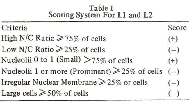
Intermediate and indeterminate criteria were not scored. The maximum positive score in any case is two (+2) and maximum negative score is four (-4). A total score of 0 to +2 establishes a diagnosis of Li whereas a total score of -ito -4 makes L2.
Results
The eighty cases studied were scored by the criteria outlined by FAB group. The frequency determined was Li 55% (figure 1),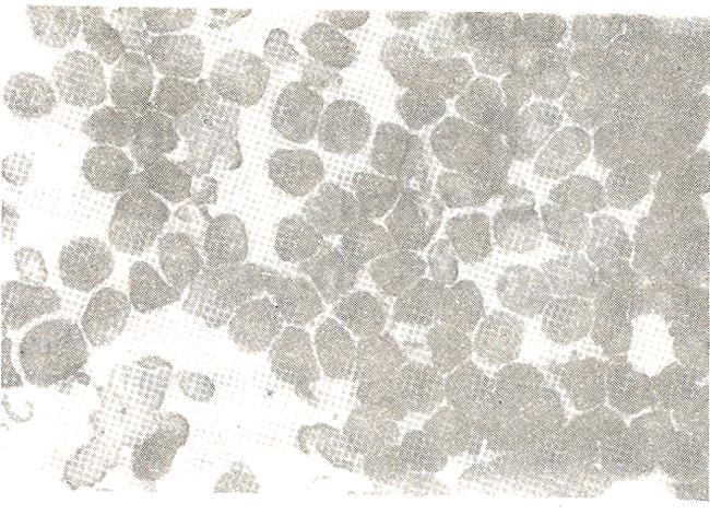
L2 40% (figure 2 and 3)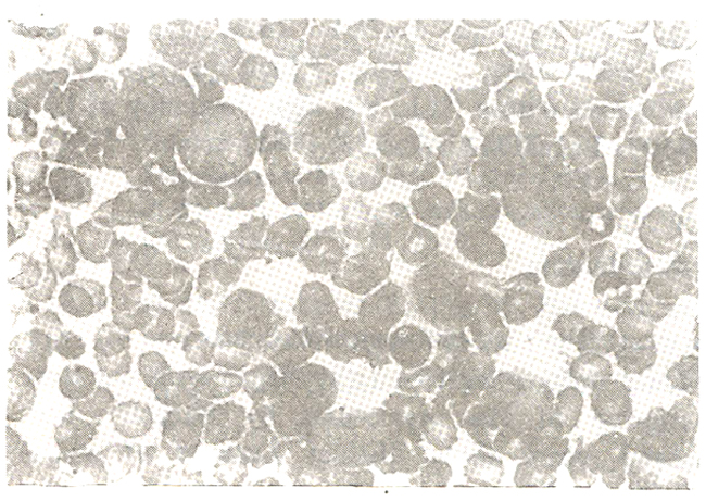
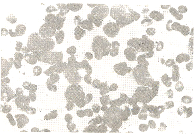
and L3 nil. 5% of the cases could not fit the criteria, this comprised cases with high N/C ratio, heterogenous population and indistinct nucleolii (figure 4).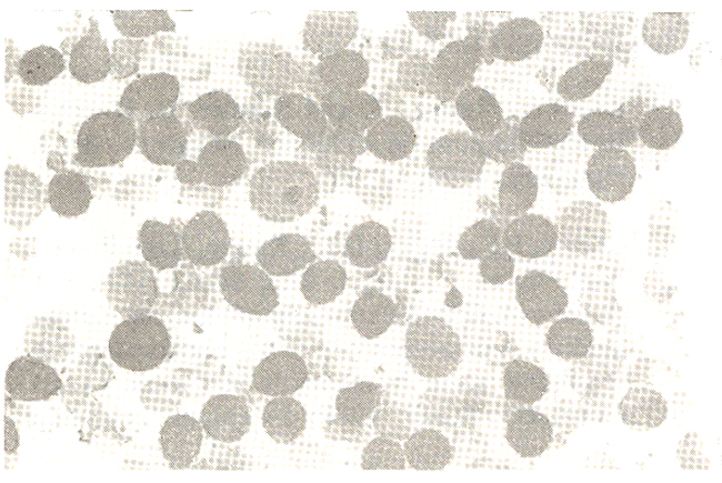
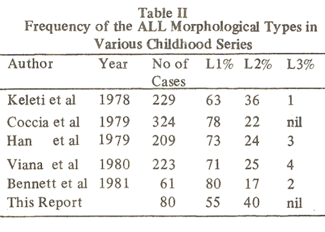
Table II gives a comparision of various childhood series in Europe and America with the present report.
Discussion
All studies in Table II have shown a significantly better prognosis for Li than L2. Keleti16 showed a better survival for Li unrelated to other risk factors, WBC count, mediastinal mass or age. Coccia17 and Viana18 reported higher relapse rates in L2 indpendently of the WBC count and Han15 described a significantly Longer duration of first remission in Li. We have observed a higher frequency of L2 in our population which may explain the poor response and remissions and high relapse rates observed in Karachi children treated with similar regimes. A small group of cases could not be classified, they perhaps are a separate variant of ALL or undifferentiated blast cell leukaemias. Furthermore there could be continental variations in morphological sub-types of ALL, since reduction of 75% limit set by FAB to 60% would classify these cases as Ll .A larger series of these cases and their follow up in the coming years is needed to evaluate further the morphological sub-types in our population.
Acknowledgements
This project was funded by the Pakistan Medical Research Council. I am grateful to Dr. Huma Qureshi, of PMRC Research Centre for doing bone marrows and to Dr. Ejaz Vohra, and Dr. Malik of Dr. Ziauddin Hospital, Karachi, Professor Ghaffar Billo, and Dr. D.S. Akram, of Civil Hospital, Karachi, and Professor Mushtaq, and Dr. M.A. Arif, of National Institute of Child Health, Karachi for sending patients for making this study possible.
References
1. Naegeli, 0. Ueber rothes Knochemmarlc and Myeloblasten. Deutsche Medizinische Wochenschrift, 1900; 26: 287.
2. Farber, S., Diamond, L.K., Mercer, R.D., Sylvester R. F. Jr. and Wolff, J.A. Temporary remissions in acute leukaemia produced by folic acid antagonist, 4-aminopteroyl-glutainic acid. N. Engl. ‘J. Med., 1948; 238: 787.
3. Friereich, E.J., Gehan, E.A., Sulman, D., Boggs, D.R. and Frei E. III. The effect of chemotherapy on acute leukaemia in the human. J. Chronic Dis., 1961; 14: 593.
4. Hardisty, R.M. and Till, M. Acute leukaemia 1959-64; factors affecting prognosis. Arch. Dis. Child., 1968;43: 107.
5. Pierce, M. 1., Borges, W.H., Heyn, R., Wolff, J.A. and Gilbert, E.S. Epidemiological factors and survival experience in 1700 children with acute leukaemia. Cancer, 1969; 23: 1296.
6. Dow, L.W., Boreila, L., Sen, L., Aur, R.J.A., George, S.L., Mauer, A.M. and Simone, J.V. Initial prognostic factors and lymphoblasterythrocyte rosette formation in 109 children with acute lymphoblastic leukaemia. Blood, 1977;50: 671.
7. Mathe, G., Pouillart, P., Sterescu, M., Amiel, J.L., Schwarzenberg, L., Schneuder, M, Hayat, M., De Vassai, F., Jasmain, C. and La Fleur, M.Subdivision of classical varieties of acute leukaemia. Correlation with prognosis and cure expectancy. Eur. J. Clin. BioL Resh., 1971; 16: 554.
8. Bennett, J.M., Kiemperer, M. and Segal, G. Survival prediction based on morphology of lymphoblasts. Recent results. Cancer Resh., 1973;43: 23.
9. Flandrin, G., Daniel, M.T. and Coudere, 0. Classification cytologique des leucemias aigues Iymphoblastiques incidences cliniques et prognostiques. Actual. Hematologiques, 1973; 7: 25.
10. Flandrin, G. and Bernard, J. Cytological classification of acute leukaemias. A survey of 14000 cases. Blood Cells, 1975; 1: 7.
11. Pantazopoulos, N. and Sinks, L.F. Morphological criteria for prognostication of acute lymphoblastic leukaemia. Br. J. Haematol., 1974; 27: 25.
12. Lee, S. L., Kopel, S. and Glidewell, 0. Cytomorphological determinants of prognosis in acute lymphoblastic leukaemia of children. Semin. Oncol, 1976; 3: 209.
13. Murphy, S.B., Borella, L. Sen, L. and Mauer, A. Lack of correlation of lymphoblast cell size with presence of T-cell markers or with outcome in childhood acute lymphoblastic leukaemia. Br. J. Haematol., 1975 ;31: 95.
14. Bennett, J.M., Catovsky, D., Daniel, M.T., Flandrin, G., Galton, D.A.G., Gralnick, H.R. and Sultan C. Proposals for the classification of theacute loukaemias. Br. J. Haematol., 1976; 33: 23.
15. Hann, I.M., Evans, D.I.K., Palmer, M.K., MorrisJones, PJ. and Haworth, C. The prognostic signi ficance of morphological features in childhood acute lymphoblastic leukaemia. Clin.Lab. Haematol., 1979; 1: 215.
16. Keleti, J. Revesz, T. and Schuler, D. Morphological diagnosis in childhood leukaemia. Br. J. Haematol., 1978;40: 501.
17. Coccia, P.F., Miller, D.R., Kersey, J.H., Bleyer, W.A., Gross, S., Siegel, S.E., Sather, H.N. and Hammond, G.D. Relationship of blast cell surface markers and morphology (FAB classification) in childhood acute lymphocytic leukaemia.(ALL) (Abstract). Blood, 1979; 54 (suppl. 1): 182a.
18. Viana, M.B. Maurer, H.S. and Ferenc, C. Subclassicification of acute lymphoblastic leukaemia in children analysis of. the reproducibility of morphological critena and prognostic implications. Br. J. Haematol., 1980;44: 383.
19. McKenna, R.W., Brynes, R.K., Nesbrr, M.E.,Bloomfield, C.D., Kersey, J.H., Spanjer, E. and Brunnings, R.D. Cytochemical profiles in acutelymphoblastic leukaemia. Am. J. Paediatr. Haematol. Oncol., 1979; 1: 263..
20. Bennett, J.M., Catovsky D., Daniel, M.T., Flandrin, G., Galton, D.A.G., Gralnick, H.R. and Sultan, C. The Morphological Classification of acute lymphoblastic leukaemia; concordance among observers and clinical correlations. Br. J.Haematol., 1981;47:553.
Journal of the Pakistan Medical Association has agreed to receive and publish manuscripts in accordance with the principles of the following committees:


