

M. Perwaiz Iqbal ( Department of Biochemistry, Aga Khan University Medical College, Karachi. )
Sheldon P. Rothenberg ( Department of Medicine, Brooklyn Veterans Administration Medical Center, Brooklyn, New York 11209. )
August 1985, Volume 35, Issue 8
Original Article

Abstract
By using ligand-binding radioassay for the enzyme dthydrofolate reductase (DHFR), a new form of this enzyme has been identified in L1210 leukemia cells. This form of DHFR displays very low affinity for the anticancer drug, methotrexate (MTX). Further enzyme kinetic studies using a sensitive radioenzymatic assay for DHFR revealed that this form of DHFR had catalytic activity for the reduction of dihydrofolic acid to tetrahydrofolic acid. Studies using gel filtration chromatography with Sephadex G-75 at pH 5.0 and pH 7.2 on crude L1210 leukemia cell lysate and purified L1210 DHFR, both complexed with radiolabelled MIX, provided additional evidence that the MIX binding results were not due to some experimental artifacts. This low affinity form of enzyme was also found in two human colon tumor tissues. We suggest that, presence of a low affinity form of the enzyme in certain cancer cells may be one of the underlying causes of resistance to MIX therapy in these cells. (JPMA 35 : 237, 1985).
Introduction
Dthydrofolate reductase (E.C.1.5.1.3) is the target enzyme of folate inhibitor methotrexate (MTX) which is clinically useful as an antineo. plastic agent1. Inhibition of cellular DHFR by MTX leads to depletion of thymidylate and the interruption of normal replication. The affmity of the enzyme for this inhibitor is considerably greater than its affinity for substrate, dthydrofolate (FH2).2,3
A number of studies using different experimental neoplastic cell systems have clearly demonstrated that it is necessary to accumulate an intracellular concentration of MIX in excess over that bound to DHFR in order to completely inhibit regeneration of tetrahydrofolate from dthydrofolate4,5,6,7,8 For this reason, it had been suggested that such cells, in addition to a form of DHFR with high affinity Ki 10-9 M) for MIX, may also have a form with low affinity for MTX.
Multiple forms of enzyme have been identified in bacteria9, cultured hamster kidney cells10 and murine leukemia cells11,12,13 In almost all these reports a low affinity form of the enzyme has been identified in cells which were resistant to MIX. In our laboratory, studies were undertaken to identify heterogeneous forms of the enzyme in L1210 leukemia cells which were sensitive to MIX.
Material and Methods
Chemicals and Animals:
[3H ] MIX (specific activity, >5 Ci/mmol) and [3H ] folic acid (specific activity, 24 Ci/mmol) were purchased from Amersham/Searle Corp., Arlington Heights, ifi, NADPH, folic acid and dthydrofolic acid (FH2,) were purchased from Sigma Chemical Co. St. Louis, Mo. MTX was obtained through the courtesy of Lederle laboratories, Inc. and purified by anion exchange chromatography.14 All other chemicals were of reagent grade. BDF1 mice were purchased from Jackson Laboratory (Bar Harbor, Me.). L1210 DHFR was purified by affinity chromatography as described previously.15
Preparation of L1210 leukemia lysates:
MTX sensitive L1210 leukemia cells were obtained from the ascitic fluid of BDF1 mice. The lysates of these cells were prepared in 0.06M citrate buffer, pH 7.4 as described by Rothenberg & IqbaL1,3
Preparation of tissue extracts:
Extracts of various tissues used in this study were prepared by a procedure similar to one described by Rothenberg et al.17 Human tumor tissue and normal tissue from the same organ were rinsed with cold 0.15 M NaC1, dried on filter paper and weighed. These tissues were suspended in 0.06M sodium citrate buffer, pH 7.4 (approximately 3 ml of buffer per gm of tissue) and . homogenized in a Waring blender for 2 minutes. The insoluble debris was then pelleted by centrifugation at 30,000 x g for 30 minutes at 4°C and supernates were analyzed for MIX binding at pH 5.0 and 7.2. The protein concentration of these extracts was measured by the method of Lowry et al.18
MIX binding studies:
Binding of MTX by L1210 leukemia cell lysates and cancerous tissue cell lysates was carried out at pH 5.0 and pH 7.2 as described by Rothenberg et al19. A total reaction volume of 0.5 ml in 0.06M citrate, pH 5.0 or pH 7.2, contained NADPH 48 uM, 2-mercaptoethanol 5.8 mM, [3H] MTX 1.8 nM, unlabelled MIX (0 - 290 nM) and 50ul cell lysate which contained the enzyme. The reaction mixtures were incubated at room temperature for 30 mintues and then the reactions were stopped by the additon of 0.4 ml of 1% Norit A neutral charcoal in 0.5% Dextran (molecular weight 10,000). After centrifugation, the radioactivity in 0.5 ml of supernatant solution which contained bound MIX was counted in a liquid scintillation counter using 15 ml of scintillation solution containing 5 gms of PPO (2,5- Diphenyloxazole) and 75 ml of Bio-solv. BBS-3 solubiizer (Beckman Instruments Inc., Palo Alto, Calif.) per liter of toluene.
Enzyme kinetic studies:
A sensitive radioenzymatic assay for DHFR20 was used to determine the apparent Km arid Vmax of crude L1210 lysate in the presence and absence of free MTX.
The reaction mixture with free MIX in a total volume of 0.5 ml contained an aliquot of crude L1210 lysate, total MIX 23 nM, bound MTX 15 nM, free MTX 8.3 nM, NADPH 120 pM and [3 H ] FH2 ranging rom 5.6 - 90 nM. The reaction velocity was linear during the first 2 minutes, therefore, the reactions were terminated after a 2 minute incubation by the addition of 0.1 ml of 0.1N HC1, 0.2 ml of unlabelled folic acid (0.28 mM) and 0.2 ml of 0.3N ZnSO4. After centrifugation, 0.5 ml of supernatant solution containing [3H ] FH4 was counted as described above. Control reactions, however, were similar to the above mentioned reaction mixtures except that they contained no MTX.
Gel filtration:
Sephadex G-75 was equilibrated with 0.06M citrate, pH 5.0 or pH 7.2 and packed into a column (1.5 x 75 cm) using a constant flow rate of 12 ml/hr. [3H ] MIX complexed with L1210 lysate or affinity purified L1210 DHFR, was filtered through the gel and eluted with 0.06M citrate, pH 5.0 or pH 7.2 containing 48 pM NADPH. 1 ml fractions were collected and the radioactivity in each of these fractions was counted in a liquid scintillation counter.
Results
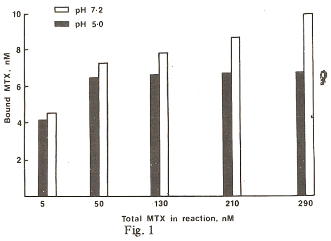
Figure 1 ifiustrates the direct binding of MIX with L1210 cell lysate at pH 5.0 and 7.2. As the total concentration of MIX in the binding reaction is increased, there is more binding of MIX by the cell lysate at pH 7.2 whereas at pH 5.0 the binding of MIX reaches a maximum value stoichiometrically and this value is less than the maximum amount of MIX bound at pH 7.2. This provides indirect evidence that L1210 cell lysate contains atleast 2 forms of enzyme, one having a low affinity for MIX.
To prove that the ‘low affinity form of enzyme also has catalytic activity, a sensitive radioenzymatic assay for DHFR was used to deter. mine the Km and Vmax of L1210 lysate in the presence and absence of MIX. Lineweaver-Burk plots of the enzyme catalysis with and without MIX are shown in Figure 2.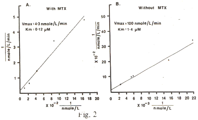
In reactions with L1210 lysate containing MIX in excess of the binding capacity of the lysate, we could measure the enzymatic reduction of FH2 to FH4 within the first 2 minutes. If there would have been a single species of DHFR in L12l0 cell lysate with a Ki of about l09 M, then in the presence of free MTX with a concentration greater than that of FH2 there would not have been any reduction of this substrate to FH4 within 2 minutes. We, obviously, could not compute a Km for the reduction of FH2 by this apparently “low affinity” form of DHFR because the reaction mixture contained free MIX. Nevertheless, the apparent Krn for FH2 reduced in the presence of free MIX (graph A) is 0.12 uM with a Vmax of 4.3 nmole/L/min. This compares with a Km and Vmax of 1.4 uM and 100 nmole/L/min respectively obtained for the cell lysate which contained no MIX (graph B). Thus, it appears that this species of enzyme with low affinity for MIX has greater affinity for FH2 than the DHFR contained in the native lysate.
To obtain further confirmation that the quantitative binding experiments at pH 5.0 and 7.2 using coated charcoal are valid, we compared the binding of [3H] MIX by the L12l0 lysate and the purified DHFR using gel filtration to separate the bound and free inhibitor.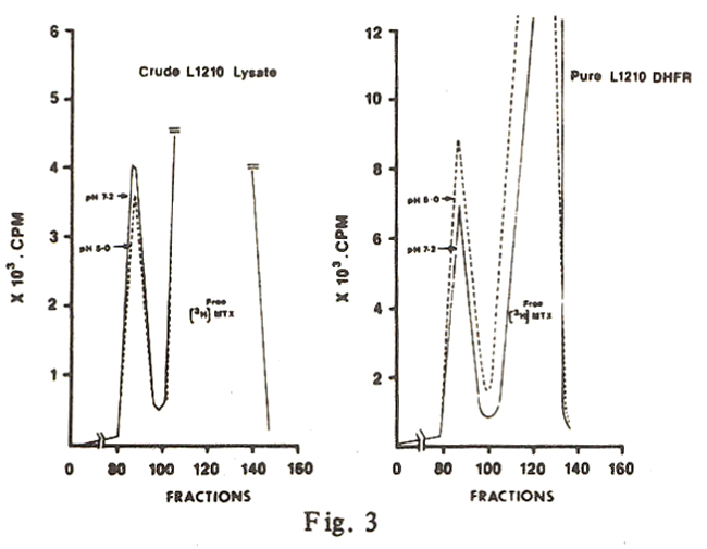
The results shown in Figure 3 confirm that at pH 7.2 the crude L1210 lysate binds more[3H] MIX than at pH 5.0, wher as the purified L1210 enzyme binds more MIX at pH 5.0 where as the purified L12l0 enzyme bind more MIX al-PH 5.0 than at pH 7.2, as would be expected with a single order of enzyme having greater affinity at the low pH.
We also measured MIX binding by extracts of some human normal and tumor tissues from the same organs that were obtained at the time of surgery. The results are shown in figure 4.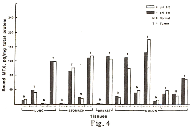
It can be seen that in only two colon tumor samples (2 and 3), the binding of MIX at pH 7.2 is substantially greater (> 25%) than the binding measured at pH 5.0 (the coefficient of variation of our MIX-binding assay is ~5%). In fact, in most of the other samples assayed, the binding of MIX is greater at pH 5.0, as was expected with the kass to be greatest (or Ki lowest) at this pH.
Discussion
While other investigators have reported the existence of a low affinity form of DHFR in M1’X-resistant cells, our results indicate that this form of enzyme is also present in MTX-sensitive cells.
Analysis of crude preparations of human tissues for DHFR activity would be of immense clinical value in selecting tumors for treatment with M1’X. The results in Figure 4 indicate that the binding of MTX by two samples (No. 2 & 3) of human colon cancer is more at pH 7.2 than at pH 5.0. This greater binding of MIX at pH 7.2 (presuming that only DHFR binds this folate analogue) suggests that these tissues contain a species of DHFR with lower affinity for MTX.
The presence of a form of DHFR with decreased affinity for MTX has long been considered to be one of the underlying mechanisms of resistance to MTX in many mammalian cell lines10,11,12,13 If a tumor cell has high concentration of this form of enzyme, it appears likely that this cell is going to be less responsive to the normal therapeutic doses of MIX. In this situation high-dose MTX therapy would be an obvious choice for treatment. Though high-dose MIX therapy has been shown to be effective in various types of leukemia, in osteocarcinoma, in non-Hodgkin’s lymphoma, in adenocarcinoma of the head and neck, breast and ovary and in small-cell lung carcinoma, it has, however, failed to improve the therapeutic results obtained with conventional MIX doses in the treatment of head and neck cancer and lung carcinoma21 It is possible that in these tumor cells the low affinity form of DHFR is absent and, hence, high doses of MIX could not be expected to improve the clinical picture.
References
1. Bertino, J.R. The mechanism of action of folate antagonists in man. Cancer Res., 1963;23:1286.
2. Werkheiser, W.C. Specific binding of 4-amino folic acid anlogues by folic acid reductase. J. BioLChem., 1961;236: 888.
3. Bertino, J.R., Booth, B.A., Bieber, A.L., Cashmore, A. and Santorelli, A.C. Studies on the inhibition of dihydrofolate reductase by folate antagonists. J.Biol. Chem. 1964: 239: 479.
4. Goldman, I.D. The mechanism of action of methotrexate. I. interaction with a low-affinity intracellular site required for maxirnum-inhibition of deoxyribonucleic acid synthesis in L-cell mouse fibroblasts. Mol. Pharmacol., 1974; 10 : 257.
5. Goldman, I.D. and Fyfe, M.J. The machanism of action of methotrexate. II Augmentation by vincristine of inhibition of deoxyribonucleic acid synthesis by methotrexate in Ehrlich ascites tumor cells. Mol. Pharmacol., 1974; 10:275.
6. White, J.C., Loftfield, S. and Goldman, I.D. The mechanism of action of methotrexate. HI. The requirement of free intracellular methotrexate for maximal supression [14C ] -formate incorporation into nucleic acids and proteins. Mol. Pharmacol., 1975;11:287.
7. Sirotnak, F.M. and Donsback, R.C. The intracellular concentration dependence of antifolate inhibition of DNA synthesis in L1210 leukemia. cells. Cancer Res., 1974; 34:3332.
8. Roberts, D, and Wodinsky, I. On the poor correlation between the inhibition by methotrexate of dihydrofolate reductase and of deoxynucleoside incorporation in DNA. Cancer Res., 1968: 28 : 1955.
9. Otting, F. and Huennekens, F.M. TPNH-dependent binding of amethopterin by dihydrofolate reductase from Lactobacillus casei. Arch. Biochem. Biophys., 1972; 15 2:429.
10. Hanggi, UJ. and Littlefield, J.W. Isolation and characterization of the multiple forms of dihy drofolate reductase from methotrexate-resistant hamster cells. J. Biol. Chem., 1974; 249: 1390.
11. Blumenthal, G. and Greenberg, D.M. Evidence for two molecular species of dihydrofolate reductase in amethopterin resistant and sensitive cells of the mouse leukemia L4946. Oncology, 1970; 24: 223.
12. Dedhar, S. and Goldie, J.H. Overproduction of two antigenically distinct forms of dthydrofolate reductase in a highly methotrexate-resistant mouse leukemia cell line. Cancer Res., 1983; 43: 4863.
13. Duffy, T.H., Beckman, S.B. and Huennekens, F.M. Multiple forms of L1210 dthydrofolate reductase differing in affinity for methotrexate. Biochem. Biophys. Res. Commun., 1984; 119: 352.
14. Gallelli, J.F. and Yokoyama, G. Assay of methotrexate in the presence of its decomposition products and other folic acid analogs. J. Pharm. Sci., 1967;56: 387.
15. Iqbal, M.P. and Rothenberg, S.P. A nonfunctional protein in human leukemia cells reacts with antiserum to dihydrofolate reductase from L1210 leukemia cells. Life Sci., 1981; 29: 689. Rothenberg, S.P. and Iqbal, M.P. Identification of a high-molecular-weight nonfunctional protein in L1210 leukemia cells with common antigenic determinants to dihydrofolate reductase. Cancer Res., 1983;43: 529.
17. Rothenberg, S.P., Iqbal, M.P. and daCosta, M. Effect of folate compounds on the accumulation of methotrexate and the activity of dihydrofolate reductase in liver, kidney and small intestine of the mouse. J. Pharmacol. Exp. Ther., 1982; 223: 631.
18. Lowry, O.H., Rosenbrough, NJ.. Farr, A.L. and Randell, R.J. Protein measurement with the folin phenol reagent. J. Biol. Chem., 1951; 193: 265.
19. Rothenberg, S.P., daCosta, M. and Iqbal, M.P. Ligand-bmdini radioassay for the antifolate compounds. Application in patients receiving methotrexate. Cancer Treat. Rep., 1977; 61: 575.
20. Rothenberg, S.P. , Iqbal, MY. and daCosta, M. A simplified radioenzymatic assay for dihydrofolate reductase using [3H] dihydrofolate. Anal. Biochem., 1980; 103: 152.
21. Jolivet, J., Cowan, K.H., Curt, G.A., Clendeninn, NJ. and Chabner, B.A. The pharmacology and clinical use of methotrexate. N. Engi. J. Med., 1983;309: 1094
Journal of the Pakistan Medical Association has agreed to receive and publish manuscripts in accordance with the principles of the following committees:


