

N.P.N. Buchholz ( Departments of Urology , The Aga Khan University, Karachi. )
F. Abbas ( Departments of Urology , The Aga Khan University, Karachi. )
M. Afzal ( Departments of Radiology, The Aga Khan University, Karachi. )
R. Khan ( Departments of Urology , The Aga Khan University, Karachi. )
I. Rizvi ( Departments of Radiology, The Aga Khan University, Karachi. )
J. Talati ( Departments of Urology , The Aga Khan University, Karachi. )
January 2003, Volume 53, Issue 1
Original Article

Abstract
Objective:Silent and not yet discovered stones of the upper urinary tract are potentially dangerous, since in due course they may cause infection, obstruction and renal damage. The aim of this study was to determine the prevalence of such silent kidney stones in a representative Pakistani population of Karachi.
Subjects and Methods: We studied 201 consecutive subjects at our hospital who underwent additional kidney screening whilst undergoing abdominal ultrasound. All these subjects did not have a history or symptoms of urolithiasis.
Results:We found silent kidney stones in 3% of subjects. All stone bearers were males. Most stones were in the left kidney. Notably, multiple stones and stones of a considerable size went unnoticed.
Conclusion:In addition to the usual figures of incidence and prevalence of stone disease drawn from patient data, there is a prevalence of 3% silent stones that may only be discovered incidentally or by screening. This is true for a “stone country” like Pakistan. Figures for other regions have yet to be determined. Due to socioeconomic reasons, we believe that a general kidney screening for urolithiasis is, however, not indicated, at least in our country (JPMA 53:24;2003).
Introduction
Stone disease is a worldwide common problem with an enormous socio-economic impact.1 Amongst other countries, it is most prevalent in Pakistan2,3 where stone patients represent the bulk of all urological patients. In our clinic, more than 50% of all urological patients are stone cases. Up to date, it is not known how exactly stones form in the renal system and how rapidly they may grow to a clinically significant size.4 Stones may be clinically silent for a long time. However, when they grow beyond a size of spontaneous clearance through the urinary tract, they may cause infection, obstruction, permanent kidney damage, and finally loss of the kidney. Therefore, it would be most helpful to find stones in early stages of their growth, where they are not yet clinically symptomatic. An easy to use, harmless and accurate means for such a screening would be a renal ultrasound. However, to assess the cost effectiveness of such a screening, the expected frequency of silent stones to be detected by ultrasound screening has to be established first.
Subjects and Methods
For this study, 201 consecutive adult patients who underwent an abdominal diagnostic ultrasound at the Department of Radiology of the Aga Khan University Hospital in Karachi! Pakistan between August 1997 and March 1998 had an additional screening of the kidneys. Pregnant patients, and patients with known kidney or urinary stone diseases were excluded from the study. All ultrasounds were performed by one experienced radiologist to exclude inter-observer variation. Age, sex, the indication
for ultrasound, any pathological finding, and, in particular, of urolithiasis, were recorded. In patients in whom a stone was discovered, urine analysis and X-ray data were matched with the ultrasonographic finding of urolithiasis. Data were entered in a Microsoft Excell spread sheet.
Results
Of the total 201 subjects, 101 were males and 100 females. The average age was 44 years (15-81) for all subjects, 45.2 years (15-77) for males, and 42 years (16-81) for females.
All subjects underwent renal ultrasound screening through one experienced ultrasonographer. The abdominal ultrasound examination was performed for various reasons. Indications and findings are listed in Table.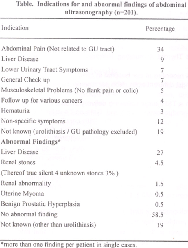
On preultrasound assessment, none of the patients reported previous kidney or urinary stone disease. Yet, stones were discovered in 9 patients (4.4%). On further intensive questioning, 3 subjects recalled having a long forgotten stone or history of stone disease. Thus, the true incidence of clinically silent and yet undiscovered kidney stones was 3% (n6) in our study population. Interestingly, all incidental stones were detected in males accounting for 6% of the male population studied. The mean age of male stone formers was 48.4+17.6 years (23-72 years). There were 1.4 stones per subject on average.1-3 Six subjects had 1, two had 2 and one had three stones. Stones were predominantly located in the left kidney (7) as compared to the right (2). Stones had an average longitudinal diameter (LD) of 7.4 mm (1-25 mm), a transversal diameter (TD) of 4.4 mm (1-10 mm), and a stone surface area (SSA = LD x TD in mm2) of 43.8 mm2 (2-250 mm2). In only two cases, additional X-rays were taken and the stones confirmed. Out of four patients tested, three had microhaematuria as a further indication for the presence of a stone. Consequently, one patient was treated by means of extracorporeal shock waves (ESWL), all others observantly.
Discussion
Today, the lifetime chance for an individual to have a stone is estimated at approximately l2%.1 The prevalence of urinary stone disease is estimated at 2-3%.5 It has an enormous socio-economic impact through treatment and recovery-related costs, time lost from work, and attendant morbidity.’ This is particularly true for countries like Pakistan, where whole families may crucially depend on one earner whose afunctionality may mean an economic catastrophe. Stone disease accounts for more than a third of all urological admissions at our university hospital. There is a known high incidence of stone disease in Pakistan as this country belongs to the so-called stone belt.2,3 In and around Karachi, the climate is hot and dry. The surrounding countryside may best be described as desert area. Both factors may contribute to endemic urolithogenesis.6 Additionally, dietary factors may play an important role3,7 with our population consuming a lot of animal protein and related lithogenic items. Urinary stones have the potential for becoming clinically symptomatic and could lead to infection, obstruction, renal damage, and, in the worst scenario, to the loss of a kidney. This, however, might be effectively prevented if potentially significant stones could be detected prior to the onset of symptoms and be treated appropriately. On the background of a general increased risk of stone formation for our population, we attempted to assess, therefore, the prevalence of clinically silent and yet undiscovered stones in order to judge whether screening for renal stones would be justified. Such a screening would have to be done using a reliable, reproducible, cost-effective, easily available and easy to handle method of examination that does not utilise ionising radiation. That makes ultrasound the method of choice.8
Out of 201 ultrasonographically screened subjects, we identified9 stone bearers. On further questioning, 3 of them remembered a past history of stone disease. However, there remains a true incidence of clinically silent unknown stones of 3%. This is in accord with the, to our knowledge, only other study which looked for silent kidney pathologies. Reisman et al9 examined 171 male prostatitis patients by abdominal ultrasound screening. Five patients (2.7%) had silent kidney stones.
It must be emphasised that these 3% silent stones are in addition to all the actual stone patients on whose figures estimations of incidence and prevalence of urolithiasis are usually based. It is not astonishing that all stone bearers were men. This corresponds with the known difference in incidence that is three times as high in men as in women.3 The question why most of the stones occurred on the left side has to be left unanswered. It is notable that multiple stones and those of a considerable size went unnoticed.
Yet, in a health system where costs have to be born directly by the patient in most instances, and patient affordability at large remains a key issue, screening for silent renal stones with a yield of 3% seems difficult to justify and institute. However, our findings complement the existing epidemiological data on urolithiasis. Comparative data from other parts of the world have to be assessed.
References
1.Resnick Ml, Prsky L. Summary of the National Institutes of Arthritis, Diabetes, Digestive and Kidney Diseases Conference on Urolithiasis: state of the art and future research needs. J Urol 1995;153:4-9.
2.Talati J, Khan F, Drago H, et al. Epidemiology of urolithiasis in Pakistan. in: Talati J, et al (eds.). The management of lithiasis. London: Kiuwer Academic PubI 1997, pp. 21-33.
3.Menon M, Parulkar BG, Drach GW. Urinary lithiasis: etiology, diagnosis and medical management. In: Walsh PC, Retik AB. Vaughan Jr. ED, et al. (eds.). Campbell’s Urology, 7th ed. Philadelphia: Saunders, 1998, pp. 2659-2734.
4.Buchholz NP, Grover PK, Rutishauser G. Modem surgical treatment and basic research in urolithiasis: exclusive or complimentary? An overview. Arch Urol 1996;18:1-13.
5.Uribarri J, Man S, Carroll JFI. The first kidney stone. Ann Intern Med 1989;111:1006-9,
6.Finlayson B. Renal lithiasis in review. Urol Clin North Am 1974;1:181-212.
7.White RW. Minerals in the urines of stone formers and their spouses. In: Hodgkinson A, Nordin BEC (eds.). Proceedings of the renal Stone research symposium. London: Churchill Ltd., 1969.
8.Ablett MJ, Coulthard A, Lee RE, et al. How reliable are ultrasound measurements of renal length in adults? Br J Radiol 1 995;68: 1087-9.
9.Reisman EM, Kennedy TJ, Roehrborn CG, et al. A prospective study of urologist-performed sonographic evaluation of the urinary tract in patients with prostatism. J Urol 1991;145:1186-9.
Related Articles
Journal of the Pakistan Medical Association has agreed to receive and publish manuscripts in accordance with the principles of the following committees:


