

A. Zahid ( Department of Anatomy, Allama Iqbal Medical College,Lahore. )
T. S. Abidi ( Department of Anatomy,Edward Medical College, Lahore. )
January 2003, Volume 53, Issue 1
Original Article

Abstract
Objective:To see the effect of chioroquine on liver weight of developing albino rats.
Methods:Twenty four pregnant female albino rats were used and divided in 4 groups. They were kept in Animal House of Post Graduate Medical Institute, Lahore. Total gestational period in rats ranges from 20-22 days, which in this study was divided into three trimesters, each of seven days. Using oral dose of chloroquine 700mg/kg body weight in first second and third weeks of pregnancy.
Results:Chioroquine caused decrease in liver weight in offsprings specially in those which were exposed to drug during second and third week of pregnancy.
Conclusion:The use of chioroquine should be avoided during pregnancy (JPMA 53:21 ;2003).
Introduction
Despite the introduction of many new antimalarial drugs, chloroquine is still the most widely prescribed drug for prophylaxis and treatment of malaria. It is considered to be the safe antimalarial in pregnant women.1-5 Chioroquine crosses the placenta to the fetus with foetal concentrations approximately the same as in mother . It is excreted in human breast milk,6,7 Chloroquine is extensively bound to body tissues with the liver containing 500 times the blood concentration.8 Chloroquine should therefore be used cautiously in patients with hepatic dysfunction. Higher or more frequent dose schedules may precipitate clinical hepatotoxicity with elevated serum liver transaminase levels.9 Hepatotoxicity caused by chioroquine may be due to direct toxic effect on hepatocytes causing leakage of lactic dehydrogenase from hepatocytes. 10
Chronic exposure to rats of 10 mg/kg of chloroquine phosphate during intra-uterine and postnatal life resulted in a marked decrease in neonatal and postweaning body and organ weight. The observed growth retardation suggested transplacental poisoning and poisoning through milk transfer.11 Liver cell, especially Kupifer cells are known to accumulate lysosomotropic agents like chloroquine, which
can cause overload of liver lysosomes by non-digestible material, increased size and number of liver lysosomes
inhibition of several lysosomal enzymes, increased autophagy and fusion disturbances.12 Administration of chloroquine significantly increases the hepatic and biliary lysosomal enzyme activities.13 Chioroquine causes hepatonecrosis and increased transaminase levels in rats at dose of 970mg/kg body weight.14
Chioroquine administration causes poor development of the yolk sac vasculature resulting in low oxygen levels delivered to the embryo which may contribute to growth retardation. Such low oxygen tension in the tissues has been suggested to cause cell degeneration and necrosis,leading to dysmorphogenesis and growth retardation.15
Materials and Methods
In this experimental study, twenty four adult female rats and eight adult male rats of Albino Wistar strain were used.Weight of female rats was between 250-300 gms and that of male rats between 300-3 50 gms. For conception three female rats and one male rat were kept together in a cage for a week and then the male rat was removed from the cage. Female rats were observed daily for signs of pregnancy which was confirmed by presence of vaginal plug and taken as day one of pregnancy. After conception 24 female rats were divided into four groups A, B, C and D, containing six animals each.Total gestational period in rats ranges from 20-22 days, which in this study was divided into three trimesters , each of seven days.The rats were weighed and marked. Chloroquine phosphate was used in the powdered form.
Group A
This was a control group containing 6 animals which were fed on normal diet throughout pregnancy. They were allowed to complete their gestational periods without drug intake.
Group B, C and D
Each containing 6 animals , were given oral dose of chloroquine 700mg/kg body weight during first, second and third trimester of pregnancy, respectively.
After the control and experimental groups had delivered, their offsprings were selected at random (about 5/adult rat). Before dissecting the offsprings they were weighed and observed for any gross malformations. Newborn rats on day one were anaesthetized by cotton plugs soaked in chloroform. After 3-5 minutes, while the rats were still breathing, a ventral midline abdominal incision was made to expose the abdominal viscera. It was then dissected out and was placed on a blotting paper. Liver was weighed on an electrical balance and weight was recorded in proforma.
Statistical Analysis
Data was collected and appropriately compiled. Mean of all variables were expressed as Mean + Standard Deviation. Difference in mean values of control and experimental groups were analysed using student’s t-test (two tailed) and compared for significance using two tailed probability points of the t distribution.
Results
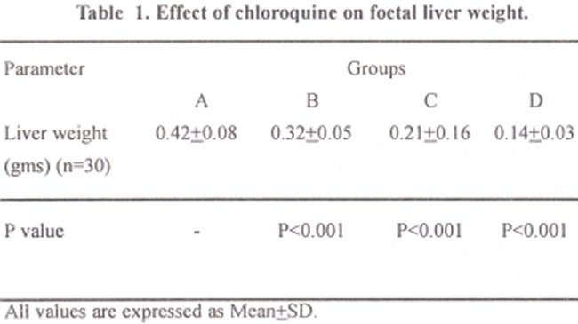
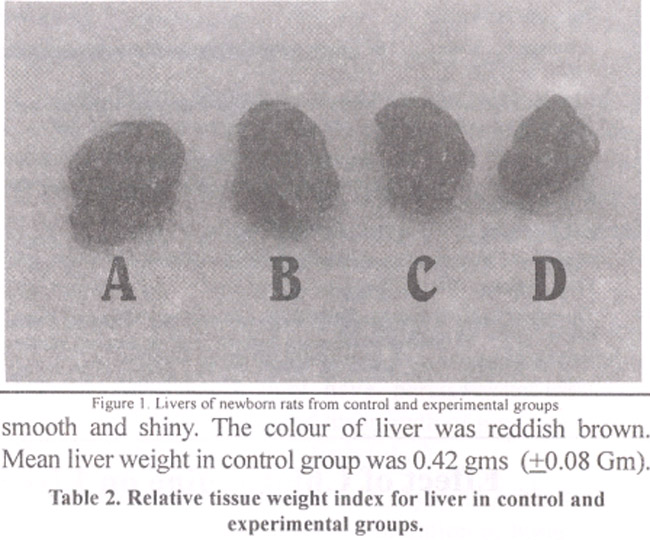
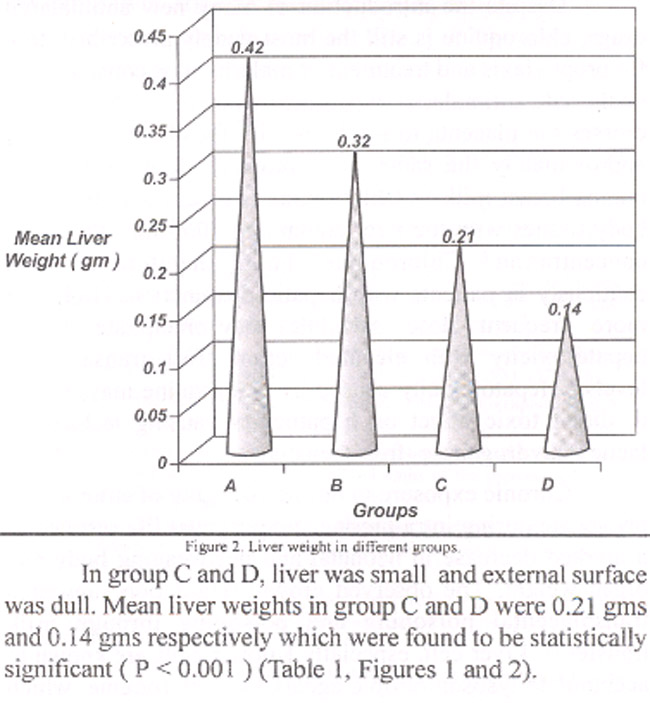
In control group A, the external surface of liver was smooth and shiny. The colour of liver was reddish brown. Mean liver weight in control group was 0.42 gms (±0.08 Gm).
In group B, the external surface of liver was smooth and glistening but size was decreased as compared to control group. Mean liver weight was found to be 0.32± 0.05 (Gm) which was statistically significant (P< 0.00 1).
In group C and D, liver was small and external surface was dull. Mean liver weights in group C and D were 0.21 gms and 0.14 gms respectively which were found to be statistically significant ( P <0.001) (Table 1, Figures 1 and 2).
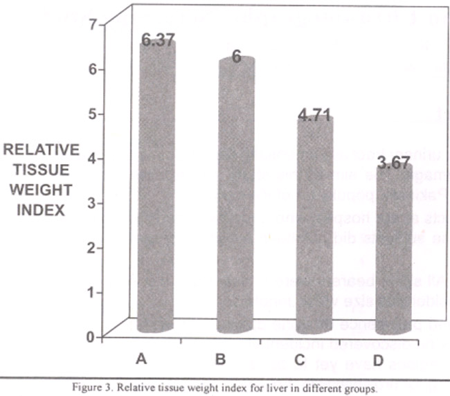
Relative tissue weight index
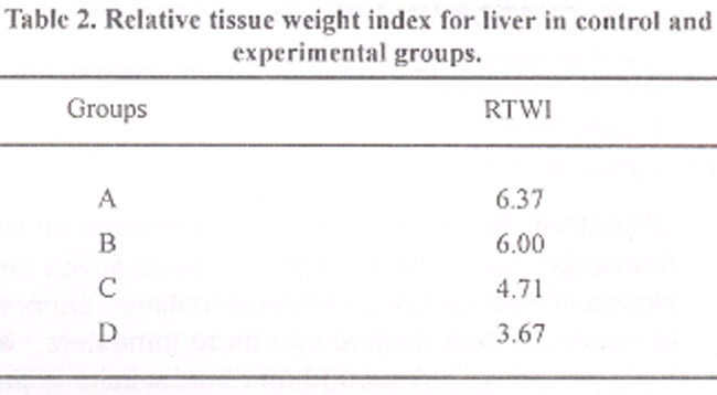
Relative tissue weight index was calculated for each group which showed decrease as compared to control group.
Relative Tissue Weight Index was obtained by the formula:
RTWI = Mean weight of liver x 100
Mean body weight
Relative tissue weight index of all groups is shown in Table 2 and Figure 3.
Discussion
The results of present research have revealed that chloroquine caused decrease in liver weight of offsprings of rats, exposed to the drug during their intrauterine life.
Liver from experimental animals showed significant decrease in weight as compared to their control. Decrease in liver weight was more marked in groups which were exposed to chioroquine during second and third week gestation.The decrease in weight can be explained on embryological basis . In rats , development of liver bud and hepatic cords occur on 10th to 12th day of gestation.16 During the last 3 days before birth in the rat, the liver undergoes a striking acceleration ofgrowth. During this time glycogen deposition in hepatocytes increases dramatically, the volume of individual parenchymal cells triples and the total protein content of the liver increases twofold. Chloroquine administration during this period interfered with the development and growth of hepatocytes and caused destruction of already formed parenchymal tissue.
Chloroquine is a lysosomotropic agent that is selectively taken up into lysosomes causing increase in size and number of liver lysosomes; inhibition of several lysosomal enzymes; secondary increase in the activity of some lysosomal enzymes; increased autophagy and fusion disturbances.12 Autophagy by liver lysosomes may be responsible for reduction in liver weight in the present study.
Chioroquine stimulates the synthesis of nitric oxide in liver cells. Nitric oxide is a free radical that acts as mediator of tissue injury. It is cytotoxic,and bacteriocidal. It contributes to the process of tissue injury by directly damaging the tissue or by initiating additional immunologic reactions that result in damage.17,18 This may be responsible for reduction in weight of livers of offsprings. Mgbodile” also reported that chloroquine administration during intrauterine and postnatal life resulted in marked decrease in neonatal and postweaning body and organ weights. Chloroquine induced hepatotoxity may be due to its action on glutamate metabolism in liver, which is inhibited by this drug.19
In conclusion, chloroquine causes significant decrease in liver weight of animals which were exposed to chloroquine during their intrauterine life. So its use should be avoided during pregnancy.
References
1.Briggs Si , Freeman RK , Yafife SJ .Chloroquine. In: Drugs in pregnancy and lactation. 4th ed. Baltimore: Williams and Wilkins 1994, PP. 158-60.
2.Diro M, Beydon SN. Malaria in pregnancy. South Med J I 982;75:959-62, 68.
3.Rose SR.Pregnancyandtravel. Emerg MedClin NorthAm 1997; 15:93-111.
4.Beers MH, Berkow R. Extraintestinal protozoa. In: Merck manual of diagnosis and therapy. 17th ed, 1999; Sec 13, chapter 161.
5.Phillips HA , Dawn W.The safety of antimalarial drugs in pregnancy. Drug Safety 1996;14:l31-45.
6.Akintonwa A , Gbajumo SA, Mabadeje AF. Placental and milk transfer of chloroquine in humans. Ther Drug Monit 1998;10:147-9.
7.Essien EE, Afamefuna GC. Chloroquine and its metabolites in human cord blood, neonatal blood and urine after maternal medication. Clin Chem 1882;28:1148-52.
8.Craig CR, Stitzel RE. Antimalarial drugs. In: Modem pharmacology with clinical applications. 5th ed. Boston: Little Brown 1997, Pp. 643-44.
9.Goodman L, Gilman AG. Drugs used in the chemotherapy of prozoal infections. In: The pharmacological basis of therapeutics. 9th ed. New York: McGraw Hill, 1996, pp. 965-70.
10.Akesson A, Akesson B . Hepatotoxic effects of antirheumatic drugs in cultured rat hepatocytes. Scand J Rheumatol 1984;13:198-202.
11.Mgbodile MU. Effects of perinatal exposure of albino rats to chioroquine. Biol Neonate1987;51 :273-6.
12.Schneider P, Korolenko TA, Busch U. A review of dnsg induced lysosomal disorders of the liver in man and laboratory animals. Micros Res Tech 1997; 36:253-75.
13.Flideaki F, Kyoshi 0, Nagakawa J1, et al. Effect of rabeprazole, a gastric proton pump inhibitor, on biliary hepatic lysosomal enzymes in rats. Jpn .J Pharmacol 1998;6: 279-88.
14.Dass EE, Shah KK. Paracetamol and conventional antimalarial drugs induced hepatotoxocity and its protection by methionine in rats. Indian J Exp Biol 2000;38: 1138-42.
15.Miki A, Fujimoto E, Ohsaki T,et al. Effects of oxygen concentration on embryonic developing rats: a light and electron microscopic studyusing whole embryo culture techniques. Anat Embryol 1988;t78:337-43.
16.Godlewski G, Gaubert CR, Rouy S, et a!. Liver development in the rat and in man during embryonic period. Micros Res Tech 1997;39: 3 14-27.
17.Laskin JD, Heck DE, Gardner CR. et al. Prooxidant and antioxidant functions of nitric oxide in liver toxicity. Antioxid Redox Signal 2001;3:261-71.
18.Ura.zaev AK, Zefirqv AL.The physiological role of nitric oxide. Usp Fiziol Nauk 1999;30:54-72.
19.Jarzyna R, Lenarcik E. Bryla J. Chloroquine is a potent inhibitor of glutamate dehydrogenase in liver and kidney cortex of rabbit. Pharmacol Res I 997;35:79-84.
Related Articles
Journal of the Pakistan Medical Association has agreed to receive and publish manuscripts in accordance with the principles of the following committees:


