

M. Ahmed ( Department of Anatomy, Baqai Medical College, Karachi )
Z. Janjua ( Department of Anatomy, Baqai Medical College, Karachi )
January 2003, Volume 53, Issue 1
Original Article

Abstract
Objective:To study the effect of induced hypothyroidism and thyroxin replacement on bone growth.
Design:An animal study carried out on experimental Albino rats.
Place and duration of study: The study was carried out in the Department of Anatomy, Basic Medical Sciences Institute (BMSI), Jinnah Postgraduate Medical Centre (JPMC), Karachi, from June 1999 to May 2002.
Method:Pregnant female Albino rats obtained from the animal house of Basic Medical Sciences Institute, JPMC, Karachi were treated with carbimazole and carbimazole plus thyroxin from 10th day of gestation till parturition. Another group of pregnant rats did not receive any treatment and acted as controls. Pups born to the treated as well as control animals were sacrificed on 10th postnatal day and fixed in formal saline. They were then processed through 95% ethanol and acetone, bulk stained with alizarin red and cleared in 4% KOH to reveal their bony and cartilaginous elements. The ulna and tibia of both sides were disarticulated from the treated and control animals and measured for intact bone length and diameter. The measurements of the three groups were then compared statistically.
Results:The retardation in length observed at the end of experimental period in ulna was by 13.67% and in tibia by 27.84% in carbimazole treated group while in carbimazole plus thyroxin treated group the reduction in length of ulna was 5.08% and of tibia 3.91% when compared with their age matched controls.
Conclusion:Prenatal hypothyroidism has an adverse effect on bone growth and results in reduction of long bone length (JPMA 53:18;2003).
Introduction
Thyroid hormones exert an important effect on growth and differentiation of tissues in many mammalian species. They are particularly essential, optimal and critical for normal growth and maturation of skeletal, nervous and reproductive tissues.1 For development and growth of these tissues in fetus the hormone comes from the fetal thyroid gland and not the maternal hormone crossing placenta. Congenital deficiency of thyroid hormones is associated with short stature and mental retardation. Delay and retardation in skeletal growth have been described in human cretins at birth.2 Thyroid deficiency therefore has an adverse effect on skeletal growth.3 Carbimazol is a thionamid compound and a good agent to produce hypothyroidism.4
The purpose of this paper is to evaluate the effect of prenatally induced hypothyroidism on growth of long bones in albino rats and the effect of simultaneous thyroxin replacement.
Materials and Method
Nine adult virgin female albino rats, weighing 175-200 gms, were mated with adult male albino rats of same age and strain in a ratio of three females with one male rat in one cage.5 Next morning each female rat was examined for any sign of mating in form of blood stained vagina or a vaginal plug.6,7 Absence of any apparent sign of mating led to vaginal wash which was obtained on glass slide and observed under microscope for presence of spermatozoa. Their presence was again considered as day zero of pregnancy.
The pregnant albino rats were then separated from male rats and divided into three groups, P1, P2 and P3 each comprising three rats. Animals of group P1 were given carbimazole (neomercazole) at a dose of 6µg/gm body weight subcutaneously from 10th day of pregnancy to parturition.8 Group P2 animals were given carbimazole subcutaneously at a dose of 6µg/gm body weight as well as thyroxin 5µg intraperitoneally9 daily from 10th day of gestation until parturition. Group P3 animals received 0.5 c.c unossified bone reniained unstained.
Appendicular skeleton was then separated from the axial skeleton and long bones of fore and hind limbs were disarticulated. Gross length and diameter of the ulna and tibia of two sides were then measured with the help of an electronic digital caliper with a measuring range of 0-300mm (up to 12 inches) and an error of ±0.03mm of indicating value.
Results
Intact bone length
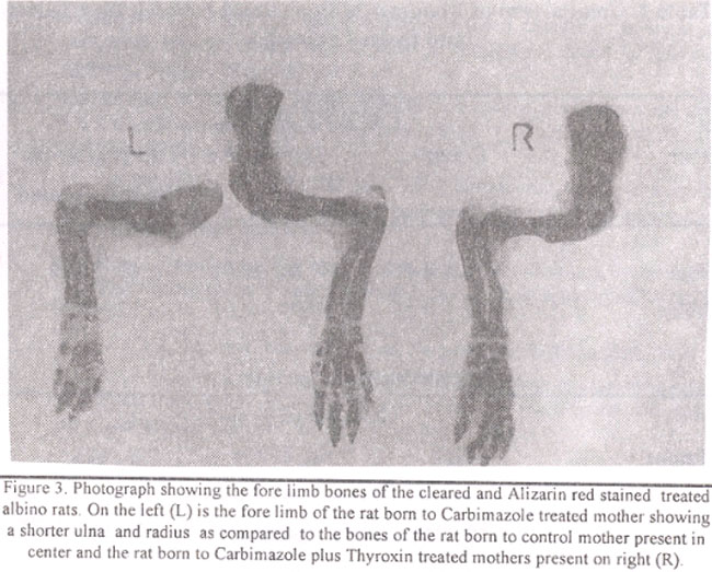
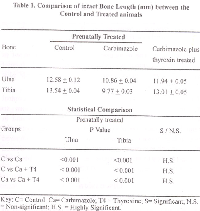
Ulna:The mean bone length of ulna in pups born to control mothers was 12.58± 0.0 1mm while in pups of carbimazole
treated mothers it was 10.86+0.04mm and in pups born to carbimazole plus thyroxin treated mothers it was 11.94±0.05mm. The decrease in the length of ulna was 13.67% in carbimazole treated group as compared to controls (P<.001). The decrease was 5.08% in carbimazole plus thyroxin treated group when compared with age matched controls (P<.001).
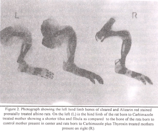
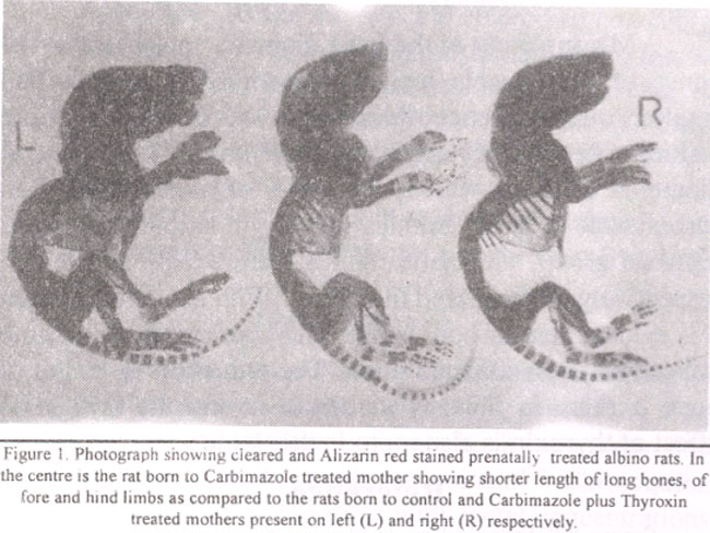 Tibia:The mean bone length of tibia in pups born to control mothers was 13.54±0.04mm while in pups of carbimazole treated mothers it was 9.77+0.03mm and in pups born to carbimazole plus thyroxin treated mothers it was 13.01±0.05mm. The decrease in the length of tibia was found to be 27.84% in carbimazole treated group (P<.001) and 3.91% in the carbimazole plus thyroxin treated group
(P<.001).
Diameter of the bone
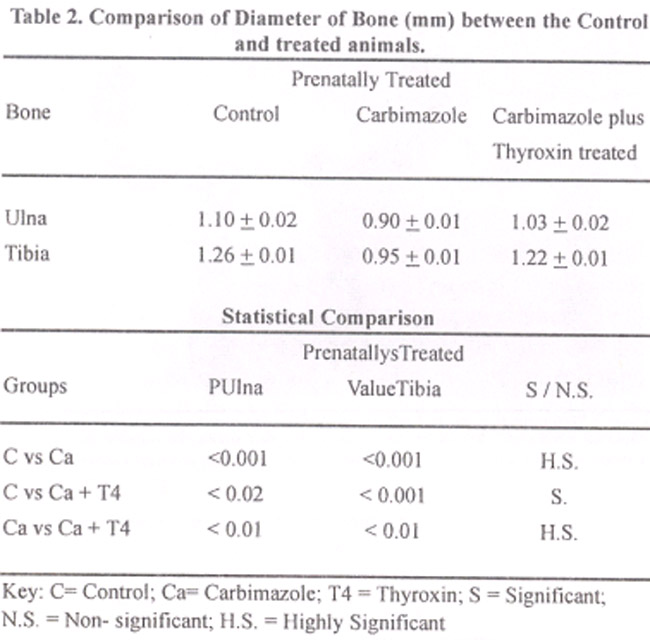
Ulna:The mean diameter of ulna in the pups born to control mothers was 1.10±0.02mm while in pups born to carbimazole treated mothers was 0.90±0.02mm and in pups born to carbimazole plus thyroxin treated mothers was 1.03±0.02mm. The reduction in diameter as compared to age matched control was 18.18% in carbimazole treated group (P<0.001) and approx: 6.36% in carbimazole plus thyroxin treated group (P<0.02).
Tibia:The mean diameter in tibia in pups born to control mothers was 1.26±0.01mm while in pups born to carbimazole treated mothers it was 0.95±0.01mm and in pups born to carbimazole plus thyroxin receiving mothers it was 1.22±0.01mm. The reduction in diameter as compared to age matched control was 24.60% in carbimazole treated group (P<0.00l) and approximately 3.17% in carbimazole plus thyroxin treated group (P<0.02).
Discussion
The carbimazole administered in pregnant rats crosses placenta.12,13 and concentrates in the thyroid of developing pups. It thus blocks the synthesis of fetal thyroid hormones which are ultimately required for growth and differentiation in developing pups since maternal thyroxin crosses the placenta in a very limited amount.14,15 Placenta acts as a very effective barrier, preventing maternal iodothyronins from reaching the fetus. This would be a consequence of both poor permeability and active deiodinating mechanism of fetal members.16 However some maternal T4 and T3 do get transferred to the fetus in both rats and humans. But administered carbimazole in pregnant rats also results in marked reduction in maternal levels of circulating T3 and T417 thus reducing the chances of maternal hormones for crossing the placenta to nil. The effectiveness of carbimazole as a chemical method of inducing hypothyroidism in rats is shown by studies conducted by Redmond et a14 and Berthier et al.18
We have observed in our study that prenatally induced hypothyroidism reduced the growth of long bones in the albino rat. The reduction was however more in tibia (27.84%) than in ulna (13.67%). This may have been due to the fact that there is a postnatal dominance in growth of hind limb bones.19 Patton et al20 have also reported a hind limb dominance in growth postnatally. Although we administered the antithyroid drug during gestation but the data was collected on 10th postnatal day. It is likely that the effect of carbimazole may have persisted up to 10th postnatal day and had affected the growth of hind limb bone (tibia) more than that of fore limb (ulna).
Simultaneous treatment with thyroxin mitigated the effect of carbimazole but the full restoration of bone length was not achieved. There was still a decrease in length of the tibia by 3.9% and that of ulna by 5.08% in pups born to carbimazole plus thyroxin treated mothers as compared to those of control mothers.
Measurement of the bone diameter revealed that the bones of carbimazole treated (hypothyroid) animals had smaller circumference than those ‘of the control and carbimazole plus thyroxin treated animals. Reduction in diameter of tibia was 24.6% and of ulna 18.18% in carbimazole treated animals while in carbimazole plus thyroxin group. The same figures were 3.17% and 6.36% respectively as compared to control. With above discussion it is evident that for both length and diameter the hind limb bone (tibia) was affected more. The reduction in length of bone is more in tibia by carbimazole and the mitigation effect of thyroxin is also more in this bone.
References
1.Ganong WF. Endocrinology. In: Review of medical physiology. 19th ed. Stanford: Appleton and Lange 1999, p. 303.
2.Scow RO, Simpson ME, Asling CW, et al. Response by the rat thyroparathyroidectornized at birth to growth hormone and to thyroxin given separately or in combination. Anat Rec 1949,104:445-63.
3.Lewinson D, Hare Z, Shenzer P, et al. Effect of thyroid hormone and growth hormone on recovery from hypothyroidism of epiphyseal growth plate cartilage and its adjacent bone. Endocrinology 1989;124;937-45.
4.Redmond 0, Tuffery AR. Thyroid proliferation, body weight and thyroid hormones in chronic carbimazole treatment in rats. J Anat1981;137:37-47.
5.Rough R. Reproductive system. In: The mouse. 2nd ed. Minneapolis: Burgess Publishing Company, 1968, pp. 269-99.
6.Patton J, Kaufman H. Timing of ossification of limb bones and growth rates of various long bones of the fore and hind limbs of the prenatal and early postnatal laboratory mouse. J Anat 1995;186:175-85.
7.Smerdely P. Pitsiavas V. Boyages SC. Methimazole inhibits FTRL-5 thyroid cell proliferation by inducing-S-phase arrest of cell cycle. Endocrinology 1993;133 2403-6.
8.Haynes R.C Jr and Murad F. Thyroid and anti-thyroid drugs in Goodman and Gillman’s Pharmacological basis of therapeutics. 6th ed. New York: McGraw Hill, 1986.
9.Inauwa I, Williams MA. Morphometric study on the uterine horn and thyroid gland in hypothyroid and thyroxin treated hypothyroid rats. J Anat 1995; 1 88:383-93.
10.Ronning 0, Kantoma T. The growth pattern of the clavicle in the rat. J Anat l988;159:173-9.
11.Culling CFA. Handbook of histolopathological and histochemical techniques. 3rd ed. London: Butterworth, 1974.
12.Greenspan FS, Dong BJ. Thyroid and anti-thyroid drugs. In: Katzung BG (ed.), Basic and clinical pharmacology. 4th ed. London: Appleton and Lange 1989, pp. 466-78.
13.Calvo R, Obregon MJ, del Rey FE, et at. The rat placenta and the transfer of hormones from the mother to the foetus: effects on maternal thyroid status. Endocrinology 1992;131 357-65.
14.De escobar GM, Obregon MJ, Del Rey FE. Comparison of maternal to foetal transfer of 3,5,3 triiodothyronine versus thyroxin in rats as assessed from the 3,5,3 triiodothyronine levels in foetal tissues. Acta. Endocrinology 1989; 120:489-90.
15.Vulsma T, Gons MN De Vijlder J. Maternal foetal transfer of thyroxin in congenital hypothyroidism due to total organification defect or thyroid agenesis. NEngi J Med 1989;321:3-16.
16.Roti E, Fang SI, Green K. Human placenta is an active site of thyroxine and 3,3,5 triiodothyronine tyrosol ring deiodination. J Clin Endocrinol Metab 1981;53:498-501.
17.De escobar GM, Calvo R, Del Rey, FE. Differential effect of thyroid hormones on growth and thyrotropic hormones in rat foetuses near term. Endocrinology 1993; 132:2056.
18.Berthier C, Lamarchand BT. Importance of thyroid iodine and cyclic AMP and TSH concentration on goitre formation in rats. Acta Endocrinol l978;89 567-80.
19.Arey B. Developmental anatomy. 7th ed. Philadelphia: WB Saunders, 1974.
20.Patton J, Kaufman H. Timing of ossification of limb bones and a growth rates of various long bones of the fore and hind limbs of the prenatal and early postnatal laboratory mouse. J Anat 1995;186:175-85.
Related Articles
Journal of the Pakistan Medical Association has agreed to receive and publish manuscripts in accordance with the principles of the following committees:


