

Muhammad Qamar Masood ( Section of Endocrinology, Department of Medicine, The Aga Khan University Hospital, Karachi, Pakistan. )
Nanik Ram ( Section of Endocrinology, Department of Medicine, The Aga Khan University Hospital, Karachi, Pakistan. )
Syed Ahsan Ali ( Section of Internal Medicine, Department of Medicine, The Aga Khan University Hospital, Karachi, Pakistan. )
February 2015, Volume 65, Issue 2
Case Reports

Abstract
Tumour-induced osteomalacia (TIO) is a rare paraneoplastic syndrome usually presenting with bone pain, fracture of bones and muscle weakness. It is caused by high serum levels of fibroblast growth factor 23 (FGF-23), which is a hormone-regulating phosphate, and vitamin D. FGF-23 is secreted by several tumours, especially benign mesenchymal tumours which are very small and difficult to locate. There is a significant delay from onset of symptoms to the diagnosis of this entity due to occult nature of this disease. We present a case of young male who presented with long history of progressively worsening muscular pain and weakness, rendering the patient confined to bed. Our aim of presenting this patient as a case report is to make physicians realise that any patient with unexplained muscular weakness and pain must undergo workup for TIO, including serum phosphate measurement, as this is a rare but potentially curable disease.
Keywords: Tumour-induced osteomalacia, paraneoplastic syndrome.
Introduction
Tumour-induced osteomalacia (TIO), also known as oncogenic osteomalacia, is a rare paraneoplastic syndrome usually presenting with bone pain, recurrent fractures of long bones and muscle weakness generally in adults.1 It is caused by high blood levels of fibroblast growth factor 23 (FGF-23), which is a hormone-regulating phosphate, and vitamin D. FGF-23 is secreted by several tumours, especially benign mesenchymal tumours which are typically very small and difficult to locate. FGF-23 acts at the renal tubules and impairs phosphate reabsorption and 1a-hydroxylation of 25-dihydroxyvitamin D (25(OH)2 D), leading to hypophosphataemia and low levels of 1, 25-dihydroxyvitamin D (1,25(OH)2 D).2,3
The diagnosis of TIO is challenging. There is a significant delay from onset of symptoms to the diagnosis of this entity due to occult nature of this disease. Even after diagnosis of TIO, location of tumour takes an average period of five years.3 Treatment of choice for this disease is resection of tumour. Such intervention, if possible, leads to rapid normalisation of biochemical abnormalities. If such intervention is not possible then treatment with vitamin D and phosphorus supplements normalises not only the biochemical abnormalities, but also leads to healing of osteomalacia. However, this treatment regimen has potential risk of stimulation of parathyroid hormone with resultant hypercalcaemia. Therefore, close monitoring of serum and urinary calcium, renal function along with parathyroid hormone is essential during treatment.1 Here we present a case of a young male who presented with tumour-induced osteomalacia.
Case Report
This is a case of a 35-year-old young married male, having two children, non-smoker, referred by a neurologist for evaluation of weakness and pain in all four limbs which the patient had for about the preceding eight years. Initially, the patient had pain only in the left calf, but later this pain extended to involve legs, arms and the trunk. This pain and weakness worsened with the passage of time and rendered the patient bound to wheel-chair and bed. Patient was unable to sit by himself without support. His bowel and bladder functions were normal. There was no fever, weight-loss or any mass lesion noticed by the patient. No family member had similar illness. At the time of presentation, in June 2012, he was on calcium supplements, tramadol and esomeprazole. On examination, he was in obvious distress and pain. His blood pressure was 160/110mmHg. No physical finding was there in abdomen, chest or heart. No mass lesion was found, especially in the extremities. All limbs were severely tender. Power was about 3/5 with weakness more pronounced proximally. Sensations were intact. No signs of active synovitis were noted. Laboratory reports revealed serum calcium 9.9mg/dl (8.6-10.2mg/dl), serum phosphate 1.1mg/dl (2.5-4.5mg/dl), repeat serum phosphate 1.2mg/dl (0.38mmol/L), alkaline phosphate 511 (45-129IU/L), serum albumin 4.6g/dl (3.5-5.2g/dl ), parathyroid hormone (PTH) 227pg/ml (16-87pg/ml), creatine phosphokinase (CPK) 56IU/L (46-171IU/L), serum magnesium 2.3mg/dl (1.6-2.6mg/dl), serum sodium 141mEq/L, potassium 3.7mEq/L, chloride 105mEq/L, bicarbonate 24mEq/L, serum creatinine 0.8mg/dl (0.8-1.1mg/dl), uric acid 6.4mg/dl (3.5-7.2mg/dl ). Urine was negative for amino acids, spot urinary creatinine 140mg/dl, spot urinary phosphate 126mg/dl (40.69mmol/L), fractional excretion of phosphate (FePO4) 66% (normal: 5-20%), FGF 23 level 800RU/ml (up to 150RU/ml), thyroid stimulating hormone (TSH) 0.7uIU/ml (0.4-4.2uIU/ml), vitamin B12 536pg/ml (>200pg/ml). Whole body magnetic resonance imaging (MRI) revealed bilateral femoral neck fractures, multiple looser zones seen in the ribs, proximal bilateral femurs medially and an old fracture of right fibula. Octreotide scan showed a well-defined rounded area of increased tracer uptake over centre of left knee joint (Figure-1).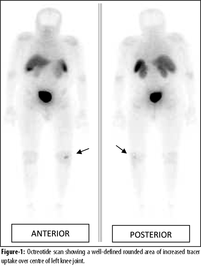
In view of clinical suspicion, this was suspected as a possible tumour site. MRI of the left knee showed irregular abnormal intensity lesion anterior to the lower end of femoral condyle in the intercondylar notch just below the level of patella (Figure-2).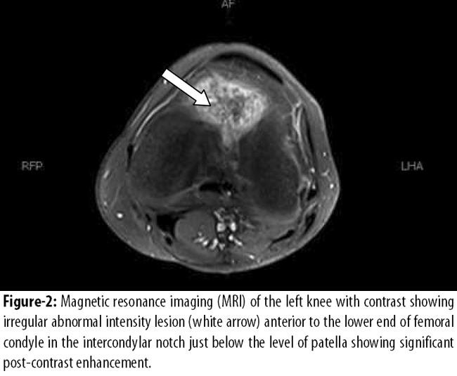
This lesion was showing significant post-contrast enhancement. This area of abnormal signal intensity corresponded with abnormal uptake on octreotide scan over the left knee and most likely represented mesenchymal tumour. The patient was advised for surgery for the possible tumour after biopsy, but he refused.
Discussion
The earliest case of TIO was described in 1947.4 TIO is a rare condition with around 300 cases reported in literature until now.5 It is characterised by longstanding muscle and bone pain and weakness and can lead to recurrent fractures. Average time from the onset of symptoms and diagnosis is usually more than 2.5 years. Reasons for this delay in diagnosis are the occult nature of the disease, non-specific symptoms and the fact that serum phosphate levels is not part of routine chemistry panel.3 In our patient the diagnosis was delayed by eight years until he came to us when the final diagnosis was made. Due to delay in the diagnosis, our patient had suffered so much that he is now totally dependent on others for his daily chores. Even after diagnosis of this syndrome, the definite treatment is usually delayed by an average of five years due to inability to locate the causative tumour.3 In our patient we were able to localise the tumour and were hoping that resection of that tumour will cure the disease, but the patient refused surgery.
TIO is typically caused by mesenchymal tumours which are slow-growing occult tumours located in either soft tissues or bones. These tumours are benign in nature. Even if they are found to have histologically malignant, local recurrence or distant metastasis is extremely rare.6 They are located mostly in lower extremities, but arms, face, skull and neck are other potential sites.7 The tumour of our patient is located in the left femur, which is the most common site of tumours causing TIO reported in literature. To locate these tumours several imaging modalities have been mentioned in literature which are used. These include computed tomography (CT), MRI, Positron emission tomography-computed tomography (PET-CT), octreotide and sestamibi scans, and even bone scintigraphy.8 Our patient initially underwent whole body MRI which did not reveal any tumour. Later, somatostatin receptor imaging was done which revealed a possible tumour near the left knee. MRI of the knee joint then showed a mesenchymal tumour in the left femur.
Laboratory investigations in TIO show low or inappropriately normal serum 1,25(OH)2 D (which should be elevated in hypophosphatemia). Serum calcium and 25(OH)2 D are normal. Serum PTH is occasionally elevated. Serum alkaline phosphatase is typically elevated and is derived primarily from bone.3
Complete tumour resection is the definite treatment of this rare disease. This can lead to normalisation of biochemical abnormalities and remineralisation of bone.3 If the tumour remains obscure then medical treatment is necessary. This includes phosphorus and vitamin D supplementation. However, close monitoring is necessary as this treatment regimen can lead to hypercalcaemia and even tertiary hyperparathyroidism.3 Therefore monitoring of serum calcium, phosphorus, creatinine, alkaline phosphatase and PTH, and urinary calcium is recommended every three months at least.3
Although TIO is a rare disease, but still it is a curable disease. A physician can overlook this disease due to absence of phosphate in routine chemistry panel and rarity of this disease.
Conclusion
Our aim of presenting this case as a case report is to make physicians realise that any patient with unexplained muscular weakness and pain must undergo workup for TIO, including serum phosphate measurement, as this is a rare but potentially reversible disease.
References
1. Drezner MK. Tumour-induced osteomalacia. Rev Endocr Metab Disord 2001; 2: 175-86.
2. Chong WH, Molinolo AA, Chen CC, Collins MT. Tumour-induced osteomalacia. Endocr Relat Cancer 2011; 18: R53-77.
3. Jan de Beur SM. Tumour-induced osteomalacia. JAMA 2005; 294:1260-7.
4. Mc CR. Osteomalacia with Looser\'s nodes (Milkman\'s syndrome) due to a raised resistance to vitamin D acquired about the age of 15 years. Q J Med 1947; 16: 33-46.
5. Jiang Y, Xia WB, Xing XP, Silva BC, Li M, Wang O, et al. Tumour-induced osteomalacia: an important cause of adult-onset hypophosphatemic osteomalacia in China: Report of 39 cases and review of the literature. J Bone Miner Res 2012; 27: 1967-75.
6. Chokyu I, Ishibashi K, Goto T, Ohata K. Oncogenic osteomalacia associated with mesenchymal tumour in the middle cranial fossa: a case report. J Med Case Rep 2012; 6: 181.
7. Munoz J, Michel Ortega R, Celzo F, Donthireddy V. \'Tumour-induced osteomalacia\'. BMJ Case Rep 2012; 2012.
8. Gandhi GY, Shah AA, Wu KJ, Gupta V, Shoraka AR. Tumour-induced osteomalacia caused by primary fibroblast growth factor 23 secreting neoplasm in axial skeleton: a case report. Case Rep Endocrinol 2012; 2012: 185454.
Journal of the Pakistan Medical Association has agreed to receive and publish manuscripts in accordance with the principles of the following committees:


