

Zaibunnisa Memon ( Department of Obstetrics and Gynecology, Aga Khan University Hospital, Karachi, Pakistan. )
Sana Sadiq Sheikh ( Department of Obstetrics and Gynecology, Aga Khan University Hospital, Karachi, Pakistan. )
February 2015, Volume 65, Issue 2
Case Reports

Abstract
Tubo-omental pregnancy is a very rare form of ectopic pregnancy. Here we present a case of tubo-omental pregnancy diagnosed at surgical exploration. A 26-year-old woman presented with abdominal discomfort, nausea and vomiting. There was ectopic pregnancy with viable foetus in the right adnexa and haemoperitonium. Laparotomy was performed and on exploration tubo-omental pregnancy was found attached to fimbria at one side and omentum at the other. Fimriaectomy and partial omentectomy was performed. The patient had a successful post-operative recovery.
Keywords: Ectopic pregnancy, Tubo-omental pregnancy.
Introduction
Extra-uterine abdominal pregnancy is very rare and is associated with high maternal mortality and morbidity, largely due to haemorrhage following placental separation. Extra-uterine pregnancy can be tubal ectopic, abdominal ectopic attached to any of the viscera, peritoneum or omentum and, very rarely, tubo-omental ectopic. Tubo-omental pregnancy is extremely rare and only one case is reported in literature.1
We present an unusual case of tubo-omental pregnancy, with placental attachment to the omentum and fimbrial end of the fallopian tube.
Case Report
A 26-years-old, low-risk primigravida, first antenatal visit at 7 weeks of gestation. She was asymptomatic at booking visit and was advised routine antenatal test and ultrasound for dating and viability. She presented in emergency at 10 weeks of gestation with the complaint of vomiting and abdominal discomfort since morning. She had pulse of 84b/min, blood pressure (BP) 110/70 mmHg; afebrile, abdominal examination was unremarkable. Ultrasound scan was advised again along with routine antenatal laboratory investigations. She presented at 11 weeks for routine check-up. She complained of nausea, vomiting and abdominal discomfort for one week which reduced over the preceding few days. On examination, her pulse was 100b/min, BP 117/66mmHg and haemoglobin was 6.2 g/dl. Abdomen was soft, non-tender, fundal height was 12 weeks and foetal heart sounds were audible with sonicaid. Ultrasound scan showed right adnexal mass measuring 6.40*5.2cm (Figure-1).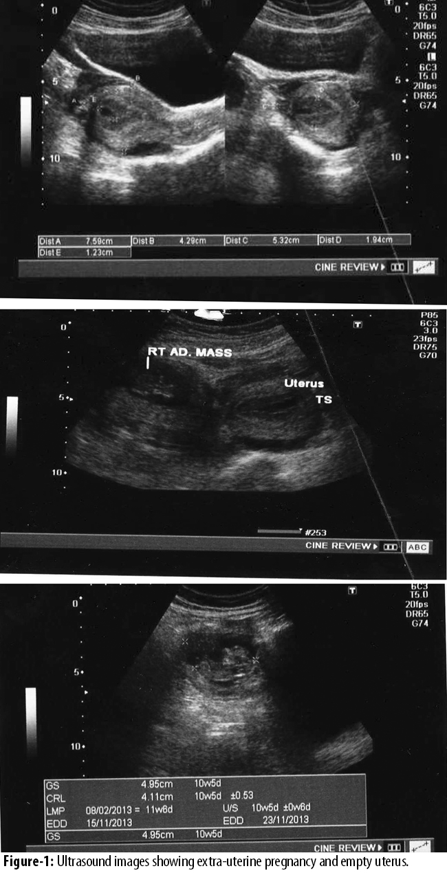
A gestational sac was noted inside measuring 4.9cm, foetal pole measuring 4.1cm, with positive foetal cardiac activity. Small amount of free fluid was present around the uterus and the adnexa. The findings suggested right-sided ectopic pregnancy with a foetus corresponding 10-week gestation.
On laparotomy, haemoperitonium was found on entry into abdomen. A mass of about 5*6cm was found in the right adnexa containing ectopic pregnancy. Placenta was found implanted on the omentum and fimbrial end of the fallopian tube. A complete intact foetus was removed during the handling of the adnexal mass. Blood was trickling from friable adnexal mass. Approximately 400-500ml blood was found in the pelvis. Blood clots were found adherent on surface of gut, omentum and pelvic walls. Omentum, caecum, ascending colon, right fallopian tube and broad ligament were found inflamed and friable. Part of omentum where the placenta was attached was removed and haemostasis was secured. Ovary was found adherent to the fimbrial end of the fallopian tube which was separated gently from the tube so as to apply clamp for removal of that end of ectopic. A part of fimbrial end of the right fallopian tube was removed along with placenta, but a small part of placenta was left attached due to friable tissue and to avoid possible need of removal of ovary in case it bled. Blood clots were removed from the pelvic cavity, and abdominal lavage was performed. Thorough review of gut and omentum was carried out and it was found to be normal. Uterus and both ovaries were normal. Besides, 1500ml of packed cell volume was transfused.
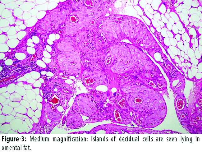
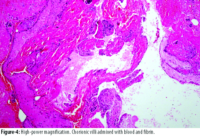
Postoperative recovery was uncomplicated and haemoglobin at discharge was 11.7g/dl. In view of some placental tissue left close to the fimbrial end, the patient was followed up with serial beta human chorionic gonadotropin (hCG) until it reached non-pregnant levels. Histopathology confirmed the diagnosis of tubo-omental pregnancy (Figures-2, 3, and 4).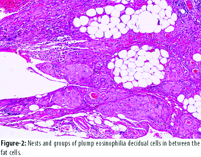
Discussion
Tubo-omental pregnancy is a rare event, but is associated with significant morbidity and mortality. Only few cases are reported in literature. The diagnosis of the reported case was made on emergency caesarean section (CS) for non-progression of labour and the patient ended up in hysterectomy because of the uncontrollable bleeding.
The incidence of abdominal pregnancy varies widely with geographical location, degree of antenatal attendance, level of medical care and socioeconomic status. Moreover, 98% of all extra-uterine pregnancies are intra-tubal, one per cent is ovarian and the rest are primary or secondary peritoneal implantations. A review of the literature showed that only few cases of omental pregnancies have been reported to date, and most were secondary omental pregnancies.
From an analysis of 11 abdominal pregnancy-related deaths and an estimated 5221 abdominal pregnancies in the United States, we estimated that there were 10.9 abdominal pregnancies per 100,000 live births and 9.2 per 1000 ectopic pregnancies; the mortality rate was 5.1 per 1000 cases.2 The diagnosis of omental pregnancy is usually made at laparotomy.
It is extremely important to detect an extra-uterine abdominal pregnancy because of the associated maternal mortality rate, which is approximately seven times higher than the estimated rate for ectopic pregnancy in general, and about 90 times the maternal mortality rate associated with normal delivery in the United States.3
Clinical history, physical examination, laboratory and ultrasonography findings frequently vary in abdominal pregnancy.
Management of such a pregnancy is dependent on gestational age, and the site of placental attachment. Abdominal pregnancy is a life-threatening condition associated with haemoperitonium. Laparotomy is usually required. Complications include haemorrhage, which may be massive, potentiating disseminated intravascular coagulation. Visceral damage can occur, with fistulas due to the presence of foetal bones, faecal peritonitis and venous thromboembolism.4
Conclusion
In view of rarity of the condition, the clinicians should maintain a high index of suspicion for ectopic pregnancy in women of childbearing age as they can be unusual or atypical. Recognising the haemodynamically unstable patient is of critical value.
References
1. Holzer E, Pickel H. Ectopic pregnancy carried to full term with a living infant]. Zentralbl Gynakol 1976; 98: 52.
2. Atrash HK, Friede REW, Hogue CJR. Abdominal pregnancy in the United States: frequency and maternal mortality. Obstet Gynecol 1987; 69: 333-7.
3. Dahab AA, Aburass R, Shawkat W, Babgi R, Essa O, Mujallid RH. Full-term extrauterine abdominal pregnancy. J Med Case Rep 2011; 5: 531.
4. Akhtar MA, Navaratnam K, Davies MJ, Sharma SD. An unusual haemoperitoneum secondary abdominal pregnancy. BMJ Case Rep 2012; 2012.
Journal of the Pakistan Medical Association has agreed to receive and publish manuscripts in accordance with the principles of the following committees:


