

Jinfeng Wang ( Department of Orthopaedics, Tianjin 4th Centre Hospital, Tianjin, China. )
Ping Liu ( Department of General Surgery, Tianjin 4th Centre Hospital, Tianjin, China. )
February 2015, Volume 65, Issue 2
Original Article

Abstract
Objectives: To evaluate and compare the long-term outcome of unstable thoracolumbar burst fractures treated using anterior, posterior or combined anterior and posterior approaches.
Methods: The prospective randomised controlled study was conducted at the Tianjin 4th Centre Hospital, Tianjin, China, and comprised patients of unstable thoracolumbar burst fracture operated between July 2004 and July 2006 and followed up for five years. The patients were divided randomly into three groups of anterior, posterior and combined anterior and posterior approaches. Clinical data was analysed using SPSS 17.
Results: Of the 66 cases in the study, 45(68.18%) were male and 21(31.8%) were female, with overall age ranging from 19 to 69 years. There were 22(33.3%) patients in the anterior group, 23(35%) in the posterior group, and 21(32%) in the combined anterior and posterior group. Comparison was made between two respective groups. The combined group was found to have the longest operation time (p<0.02; p<0.01, respectively), larger blood loss (p<0.006; p<0.005, respectively), longer hospital stay (p<0.01; p<0.003, respectively) and higher hospitalisation costs (p<0.004; p<0.001, respectively). The postoperative kyphotic angle was significantly smaller than preoperative one in all groups (p<0.01; p<0.02; p<0.01).
Conclusion: The anterior approach or combined anterior and posterior approach were better options in managing unstable thoracolumbar burst fracture, while the latter should be used only for the burst fracture with a significant posterior column injury.
Keywords: Thoracolumbar burst fracture, Kyphotic angle, Visual analogue scale (VAS), Frankel grades, Approach. (JPMA 65: 201; 2015).
Introduction
Thoracolumbar burst fractures are common spinal injuries that result in spinal instability and acute or delayed neurological deficits.1 According to the Denis three-column concept, an unstable burst fracture is a 2- or 3-column injury,2 and cases with a loss of anterior vertebral body height exceeding 50%, angulation exceeding 20° or canal compromise exceeding 50% need to be treated surgically.3 The current procedures for thoracolumbar burst fractures are usually performed using anterior, posterior or combined anterior and posterior approach,4 each of which has its advantages and disadvantages. However, there is controversy about the optimal surgical approach for the unstable thoracolumbar burst fracture.5 In addition, most cases reported in literature had a short-term follow-up, with few clinical studies opting for long-term follow-up in recent years.
The current study was planned with a five-year follow-up after the surgery done between July 2004 and July 2006. The objective was to evaluate the long-term clinical outcome of cases with thoracolumbar burst fracture and to help spine surgeons in preoperative decision-making.
Patients and Methods
The prospective randomised controlled study was conducted at the Tianjin 4th Centre Hospital, Tianjin, China, and comprised patients of unstable thoracolumbar burst fracture operated between July 2004 to July 2006 and followed up for five years. Those included were patients with thoracolumbar burst fractures along with mechanical instability.6 Radiographic evidence of instability comprised one or more of the following: vertebral height loss greater than 50% on lateral radiography; kyphosis over 20 degrees; and spinal canal encroachment greater than 50% on axial computed tomography (CT).6 Those presenting with burst fracture at two or more levels were excluded and so were patients with concomitant injuries that required hospital admission and active surgical management, or a history of spinal surgery. After approval from the institutional ethics committee, the randomisation was carried out according to a computer-generated sequence and while obtaining informed consent from all the subjects, the patients were divided randomly into Group 1 anterior, Group 2 posterior and Group 3 combined anterior and posterior approach. The patients were not blinded to the type of surgical approach. Clinical characteristics, including age, gender, hospital stay, operation time, blood loss, fracture level, hospital cost, follow-up periods and complications of the surgery were collected by a surgeon blinded to the grouping.
The neurological status of patients was evaluated using Frankel grades,7 and functional status was evaluated using the Visual Analogue Scale (VAS).8 Lateral X-radiographs were used to measure kyphotic angle, which was measured from the superior endplate of the vertebral body above the fractured level to the inferior endplate of the vertebral body below the affected vertebra on lateral radiograms.1 The neurological status, functional status and kyphotic angle of all patients were evaluated by a surgeon blinded to the grouping before surgery, one month after surgery and at the final follow-up.
In Group 1 patients, a left-sided extra pleural-retroperitoneal approach was used to expose fractured vertebrae. A subtotal corpectomy was performed to decompress the spinal canal, and the upper and lower discs, including cartilaginous endplates, were removed. A cylindrical titanium mesh cage filled with autogenous bone was inserted into the vertebra body defect and screw-plate instrumentation was used to stabilise the cage.
In Group 2, the patients were placed in prone position. Using a standard posterior midline approach, pedicle screws were inserted into a vertebral body one level above and below the injured vertebra. The manual lordotic manoeuvre was performed to correct kyphosis, and posterior decompression and posterolateral fusion with autogenous bone graft was performed.
In Group 3 patients, direct decompression of the spinal canal was performed using subtotal corpectomy, and a cylindrical titanium mesh cage filled with autogenous bone was inserted into the vertebral body defect. It was followed by posterior rods and pedicle screw system, posterior decompression and posterolateral fusion.
Data was statistically analysed using SPSS 17. Comparison of clinical variables of the three groups were carried out using one-way analysis of variance (ANOVA), Chi-square test or Kruskal-Wallis test. The comparison of kyphotic angle and VAS score were performed using repeated measures analysis of variance, and the comparison of complication incidence and neurological recovery rate between the groups was performed using Chi-square test. A p value less than 0.05 was considered statistically significant.
Results
Within the study period, 139 patients with thoracolumbar burst fractures were treated. Of them, 66(47.48%) patients met the inclusion criteria and represented the study population; 45(68.18%) males and 21(31.8%) females, with overall age ranging from 19 to 69. There were 22(33.3%) patients in Group 1, 23(35%) in Group 2, and 21(32%) in Group 3. The fractured level were T12 in 12(18%) cases, L1 in 44(67%), and L2 in 10(15%) cases. Concomitant injuries included calcaneal fracture in 8(12%) cases, skull base fractures in 7(11%), ankle fracture in 2(3%) and pelvic fracture in 5(7.5%).
There was no statistically difference among the three groups in terms of age (p<0.28), gender (p<0.84), injury level (p<0.59) and follow-up duration (p<0.51) (Table-1).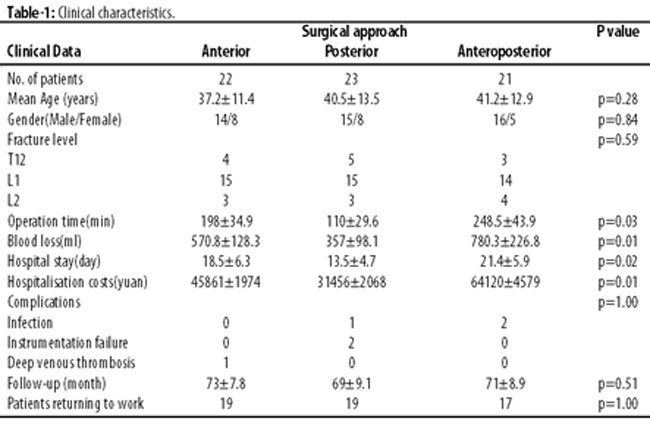
Two groups each were compared. Compared to anterior or posterior approach Groups 1 and 2, the combined anterior and posterior Group 3 reported longer operation time (p<0.02; p<0.01, respectively), larger blood loss (p<0.006; p<0.005, respectively), longer hospital stays (p<0.01; p<0.003, respectively) and higher hospitalisation costs (p<0.004; p<0.001, respectively). In addition, these values were significantly higher (p<0.01; p<0.01; p<0.008; and p<0.01, respectively) in Group 1 compared to Group 2.
Kyphotic angles and VAS score in the three groups were also recorded (Table-2).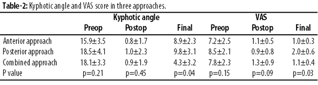
There was no significant difference in the kyphotic angle in the three groups preoperatively (p<0.21) and postoperatively (p<0.45). The postoperative kyphotic angle was significantly lower than the preoperative one in all groups (p<0.01; p<0.02; p<0.01 in Groups 1, 2 and 3, respectively.). At the final follow-up, the angle in Group 3 was significantly lower than those in Groups 1 and 2 (p<0.04; p<0.03, respectively), and the angle in Group 1 was lower than Group 2, but it was not significant (p<0.08).
There was no significant difference in VAS score in the three groups preoperatively (p<0.15) and postoperatively (p<0.09). The postoperative VAS score were significantly lower than the preoperative one (p<0.01; p<0.03; p<0.006 in Groups 1, 2 and 3, respectively). At the final follow-up, the VAS values were the lowest in Group 1 and highest in Group 2. Compared to Group 2, the scores were significantly lower in Group 1 and Group 3 (p<0.03; p<0.04 respectively), but there were no significant difference between the two groups (p<0.35).
Neurological grading was also done (Figure)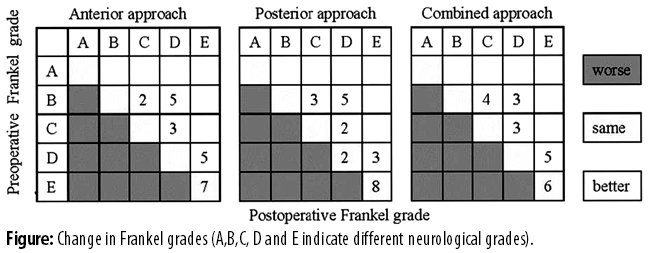
No deterioration was found after surgery in the three groups. In Groups 1, 2 and 3, 15(68.1%), 13(56.5%) and 15(71.4%) patients recovered after surgery respectively. The recovery rate in Group 3 was the highest and Group 2 was the lowest, but there was no significant difference among the three groups (p<0.47).
Wound infection occurred in 3(4.5%) cases: 2(3%) in Group 3, 1(1.5%) in Group 2. Two (3%) patients suffered from instrumentation failure in Group 2, 1(1.5%) patient suffered from deep venous thrombosis (DVT) in Group 1. The incidence of complications was 4.5%, 13.0% and 9.5% in Groups 1, 2 and 3 respectively, but there was no significant difference among the groups (p<1.00). At the final follow-up, 19(86.4%) patients in Group 1, 19(82.6%) in Group 2 and 17(80.9%) in Group 3 returned to work. There was no significant difference among the groups (p<1.00).
Discussion
Thoracolumbar burst fracture is a common spinal lesion. When it is associated with neurological impairment, surgical treatment is indicated for decompression, internal fixation and fusion. The selection of surgical approach remains controversial and the current study was planned to clarify the issue further.
Posterior approach has been a popular and acceptable method to restore spinal stability, which can reduce the fractured vertebral body and improve canal dimensions by indirect methods, but the improvement of the canal dimension is incomplete. In addition, the posterior approach alone can\'t realise fully the reconstruction of anterior or middle column.9 Compared to the posterior approach, direct and complete decompression of spinal canal can be performed completely in the anterior approach.9
Some authors suggested that indirect decompression of the posterior approach is usually effective, and the technique of decompression, direct or indirect, does not affect the rate of neurological improvement.10 But studies.9,11,12 have voiced different viewpoints and suggested that direct decompression would provide better neurological recovery. In the current study, the rate of neurological improvement in Group 1 (anterior) and Group 3 (combined approach) at the final follow-up was higher than that in Group 2 (posterior). Although the difference didn\'t have significance, which may be attributed to the small sample size, but, we support the viewpoint that direct decompression may result in better neurological recovery. From the angle of decompression, the anterior as well as combined approach may have more advantages than the posterior approach.
In addition to the nerve root and cord decompression, other goals of surgical treatment include stabilisation of spine and correction of kyphotic deformity. A biomechanical experiment found that the combined approach provided more rigid fixation than anterior or posterior approaches.13 In the current study, we found that all the three approaches could correct the kyphotic angle effectively, while the loss of kyphotic angle at the final follow-up in Group 3 was significantly lower than those in the two other groups, and the posterior approach presented the largest loss of kyphotic angle. Our study confirmed the viewpoint expressed in literature.13 Moreover, another study4 also confirmed that higher kyphotic correction and improvement of vertebral height were found after combined anterior and posterior approach surgery by a systematic review of literature. We suggest that the combined anterior and posterior approach can provide more stable fixation than anterior or posterior approach.
In terms of VAS score, Groups 1 and 3 presented better score than Group 2 at the final follow-up. In a clinical observation, one study14 found that the anterior approach presented fewer complications, retained sagittal alignment and exhibited a trend towards less pain when compared with posterior surgery. The decompression was more complete in anterior or combined approaches, which may be beneficial to the functional status. As a result, we suggest that unstable thoracolumbar burst fractures should be treated using anterior or combined approach.
In addition, the combined approach provided more stable fixation and reliable decompression, but its disadvantages were also observed in the current study. Although there wasn\'t significant difference in the complication incidence among the three groups, but the significantly longer hospital stay, larger blood loss and longer operation time may increase the risk of the combined approach. In addition, the hospitalisation cost of the combined approach was the highest in the three approaches. Subsequently, we support the viewpoint in literature10,15 that the combined approach should be used only for the burst fracture with significant posterior column injury.
In terms of limitation, the current study had a small sample size. A large sample size would reduce the risk of false negative results, have more statistical significance, and clarify the issues more clearly. Subsequently, more studies need to be performed.
Conclusion
Compared to the posterior approach, the anterior approach or combined anterior and posterior approach are better options in managing unstable thoracolumbar burst fracture, while the combined anterior and posterior approach should be used only for the burst fracture with a significant posterior column injury because of its longer hospital stay, larger blood loss, higher hospitalisation cost and longer operation time.
References
1. Suzuki T, Abe E, Miyakoshi N, Murai H, Kobayashi T, Abe T, et al. Anterior Decompression and Shortening Reconstruction with a Titanium Mesh Cage through a Posterior Approach Alone for the Treatment of Lumbar Burst Fractures. Asian Spine J 2012;6:123-30.
2. Kim HS, Park SK, Joy H, Ryu JK, Kim SW, Ju CI. Bone cement augmentation of short segment fixation for unstable burst fracture in severe osteoporosis. J Korean Neurosurg Soc 2008;44:8-14.
3. Liao JC, Fan KF, Chen WJ, Chen LH. Posterior instrumentation with transpedicular calcium sulphate graft for thoracolumbar burst fracture. Int Orthop 2009;33:1669-75.
4. P Oprel P, Tuinebreijer WE, Patka P, den Hartog D. Combined anterior-posterior surgery versus posterior surgery for thoracolumbar burst fractures: a systematic review of the literature. Open Orthop J 2010;4:93-100.
5. Schmid R, Lindtner RA, Lill M, Blauth M, Krappinger D, Kammerlander C. Combined posteroanterior fusion versus transforaminal lumbar interbody fusion (TLIF) in thoracolumbar burst fractures. Injury 2012;43:475-9.
6. Stancic MF, Gregorovic E, Nozica E, Penezic L. Anterior decompression and fixation versus posterior reposition and semirigid fixation in the treatment of unstable burst thoracolumbar fracture: prospective clinical trial. Croat Med J 2001;42:49-53.
7. Frankel HL, Hancock DO, Hyslop G, Melzak J, Michaelis LS, Ungar GH, et al. The value of postural reduction in the initial management of closed injuries of the spine with paraplegia and tetraplegia. I. Paraplegia 1969;7:179-92.
8. Blondel B, Fuentes S, Metellus P, Adetchessi T, Pech-Gourg G, Dufour H. Severe thoracolumbar osteoporotic burst fractures: treatment combining open kyphoplasty and short-segment fixation. Orthop Traumatol Surg Res 2009;95:359-64.
9. Machino M, Yukawa Y, Ito K, Nakashima H, Kato F. Posterior/anterior combined surgery for thoracolumbar burst fractures--posterior instrumentation with pedicle screws and laminar hooks, anterior decompression and strut grafting. Spinal Cord 2011;49:573-9.
10. Been HD, Bouma GJ. Comparison of two types of surgery for thoraco-lumbar burst fractures: combined anterior and posterior stabilisation vs. posterior instrumentation only. Acta Neurochir (Wien) 1999;141:349-57.
11. D'Aliberti G, Talamonti G, Villa F, Debernardi A, Sansalone CV, LaMaida A, et al. Anterior approach to thoracic and lumbar spine lesions: results in 145 consecutive cases. J Neurosurg Spine 2008;9:466-82.
12. Shi R, Liu H, Zhao X, Liu X, Gong Q, Li T, et al. Anterior single segmental decompression and fixation for Denis B type thoracolumbar burst fracture with neurological deficiency: thirty-four cases with average twenty-six month follow-up. Spine (Phila Pa 1976) 2011;36:E598-605.
13. Kallemeier PM, Beaubien BP, Buttermann GR, Polga DJ, Wood KB. In vitro analysis of anterior and posterior fixation in an experimental unstable burst fracture model. J Spinal Disord Tech 2008;21:216-24.
14. Wood KB, Bohn D, Mehbod A. Anterior versus posterior treatment of stable thoracolumbar burst fractures without neurologic deficit: a prospective, randomized study. J Spinal Disord Tech 2005;18 Suppl:S15-23.
15. Dimar JR 2nd, Wilde PH, Glassman SD, Puno RM, Johnson JR. Thoracolumbar burst fractures treated with combined anterior and posterior surgery. Am J Orthop (Belle Mead NJ) 1996;25:159-65.
Related Articles
Journal of the Pakistan Medical Association has agreed to receive and publish manuscripts in accordance with the principles of the following committees:


