

Ahmet Salvarci ( Konya Hospital, Department of Urology, Konya, Turkey. )
Mehmet Koroglu ( Sakarya University School of Medicine, Medical Microbiology, Sakarya, Turkey. )
Tayfun Gurpinar ( Bayindir Hospital, Department of Urology, Istanbul, Turkey. )
February 2015, Volume 65, Issue 2
Original Article

Abstract
Objective: To evaluate the antimicrobial activity in urinary catheters and silicones in antibiotic-coated prosthetic urinary systems in order to reduce morbidity and mortality caused by catheter-associated infection.
Methods: The study was initiated in 1993 at Houston, USA and continued in Turkey till 1996. A sterile plastic bag was used as kidney in the in vitro urinary system. Physiological renal jet streams (50cc/h) were generated with an intravenous metric pump. The temperature was kept at body temperature. The bladder drainage was achieved at the physiological drainage period of 4-6 hours during the 72-hour experiment. Silicone surfaces coated with pure silicone and impregnated with Minocycline-Rifampin were exposed to the urine contaminated with the targeted bacteria in the in vitro urinary model for 72 hours. Antimicrobial activities occurring in the Eosin methylene blue and blood agar media in the infected silicones were assessed.
Results: Minocycline-Rifampin silicone surfaces exposed to the urine contaminated with Escherichia coli and Pseudomonas aeruginosa reported reproduction. No reproduction was observed in the culture of Minocycline-Rifampin-impregnated silicone surfaces for Proteus mirabilis. The difference with the control group was regarded as statistically significant for Proteus mirabilis (p<0.005). Minocycline-Rifampin-coated silicones were closely monitored only for Proteus mirabilis in the in vitro urinary medium. Although inhibition zones (<10mm) in the cultures were observed for Minocycline-Rifampin-coated silicones for Escherichia coli and Pseudomonas aeruginosa, but the microbial efficacy was not regarded sufficient.
Conclusion: There is still need for evidence-based in vivo and in vitro studies where antimicrobial activity is evaluated on the surface of catheters.
Keywords: Minocycline-rifampin, Silicone material, Urinary catheters, Urinary infection. (JPMA 65: 115; 2015).
Introduction
Forty per cent hospital infections occur in the urinary system.1 The most frequently encountered urinary system infection is 80% associated with urinary catheters.2-4 A urinary catheter is applied in 10-15% of hospitalised patients, with a risk of urinary infection in 25% of these patients.1 Bacteraemia may develop in 1-3% following urinary catheter. More than 30% of patients may die as consequence of bacteraemia.1,5,6 Urinary infections occurring in association with urinary catheter after urinary endoscopic interventions and inadequate antibiotic use are called cross-infections.4 Proteus mirabilis, Escherichia coli and Pseudomonas aeruginosa are cross-infections most frequently encountered in the urinary system.1,6 Urinary tract infections (UTIs) associated with urinary catheterization are a common problem for healthcare institutions. Increase in the urinary system infection increases mortality and morbidity and triggers economic expenditure.7 It is reported in literature that the rate of infection is decreased by up to 45% in urinary catheters coated with antibiotics and antiseptics.8-11 Urinary bacterial agents first stick to the surface of the catheter and form a biofilm layer.7 Microorganisms and adhesion of biofilm layer of organic factors, such as fibrin, fibronectin and microbial polysaccharide that are produced by the body, is observed on the catheter under electron microscopy.1 The urinary infections deriving from the biofilm layer in urinary catheters are 1,000 times more resistant to antibiotics compared to their own planktonic equivalents and their treatment is thus significantly challenging.12 Researchers tried to develop antibiotic-impregnated urinary catheters in order to prevent urinary infection associated with pathological biofilm formation.7 Also in this study it was always claimed that the efficacy of Minocycline-Rifampin (MR), which constitutes the synergistic antimicrobial spectrum that we are using and has again become a current topic in recent years, is better.1
The current study was planned to evaluate the antimicrobial activity in urinary catheters and the silicones in the antibiotic-coated prosthetic urinary systems planned to be used in future at our health facility in order to reduce morbidity and mortality that me be caused by catheter-associated infections in daily practice.
Materials and Methods
The study was initiated in 1993 at Houston, USA and continued in Turkey till 1996. The formation of infection was assessed on the surfaces of MR-coated silicones in the in vitro urinary mechanism lasting 72 hours where drainage was applied with intervals of 4-6 hours without leaving any residue in the in vitro urinary model which we prepared for the first time.
Silicone discs at a thickness of 0.25mm and a diameter of 0.7mm and coated with a dose of 1,000mg of MR were used. The alloplastic silicone surface was covered with tridodecyl methyl ammonium chloride (TDMAC) cationic surfactant and coverage was achieved upon the adhesion with MR, which has an anionic structure antibiotics, with silicone (MD Anderson Tx/USA). Pure silicones (PS) without any antibiotic were used as the control group.
The standard bacteria strains Escherichia coli ATCC 25922, Pseudomonas aeruginosa ATCC 27853 and Proteus mirabilis ATCC 29906 were used for the preparation of contaminated urine and microorganisms. Urine was collected from healthy people and their autoclaved sterility was proven with cultures. Consequently, the urines were used upon being infected with the targeted microorganisms. The biochemical values of urines before and after the autoclave were checked and it was confirmed that there was no deterioration in the physiological structure of the urine. Consequently, Escherichia coli, Pseudomonas aeruginosa and Proteus mirabilis prepared in 10cc suspension separately for each microorganism in 0.5 McFarland 108cfu/ml were mixed with 3,600cc of collected and sterilised urine, thus resulting in a bacterial urine suspension. The bacterial urine suspension was kept in an incubator at 37ºC for 24 hours. Upon confirming that there was reproduction of more than 100,000 colonies per millilitre with the targeted microorganism in the culture prepared, the bacterial urine suspensions were used at the in vitro urinary mechanism. Blood Agar (BA9) and Eosin Methylene Blue (EMB) media were used as culture. Silicone discs exposed to infected urine for 72 hours were consequently spread on the surface of the medium in a similar manner with the Rolling method and placed at the centre of the bacterial cultivation area. For in vitro urinary system model, 5-litre sterile plastic reservoir bags representing the kidneys were used. The intravenous pump set was accepted as the ureter. A 500cc glass bottle was used as the bladder for the storage of the urine and the placement of sterile silicones inside. A hole for the entry of the infected urine inflowing from the metric pump and a drainage hole representing the urethra for draining the urine without any residue were perforated on the cap of the bottle. Freshly collected urine was sterilised and infected with the targeted microorganisms and a new sterile reservoir bag and a new sterile intravenous metric pump set was used each time. The edges of the inflow and outflow sections of the glass bottle were sealed with silicone, thus preventing contamination of the system with the external environment. A heater was placed under the glass bottle and set at 37ºC to ensure body temperature. The glass bottle was sterilised with ethylene oxide prior to each study. Jet streams were achieved via the intravenous metric pump. The pump was adjusted in a manner so as to drain 50ml/h to the bladder, which is the physiological jet stream rate in human kidneys. The exposure of the silicones to the infected urine for 72 hours was ensured. The bottle was fully emptied without leaving any residue at 4-6-hour intervals during this period which is the physiological drainage period.
Four separate experiments were performed for each microorganism. The first two experiments were conducted for PS which was the control group, while the following two experiments were performed for MR-coated silicones. A total of 8 silicone discs, with four silicone discs at the BA medium and four silicone discs at the EMB medium were evaluated. A total of 32 silicone discs, 16 MR and 16 PS, were used for each microorganism. Each experiment lasted 72 hours. This period was accepted as the period required for monitoring the formation of biofilm in silicone surfaces, the formation and prevention of the associated infection.6,7 Cultivation results in the EMB and BA media were evaluated for all three microorganisms. It was taken into consideration that it was necessary for the antimicrobial activity to be >15mm in order to be meaningful for the silicones retrieved from the in vivo and in vitro infected media in the cultures where an inhibition zone was formed.7 The biofilm layer on all surfaces exposed to the infected urine was verified with electron microscopy assessments. The power test was used for determining the number of silicone discs to be utilised in the study. All statistical analyses were performed with the Fisher Exact X2 test. Statistically, p<0.05 was regarded as significant.
Results
An increase of 39 per cent was observed in the disc thicknesses prior to the experiment associated with the biofilm layer in the confocal surface measurements (Figure).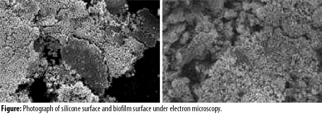
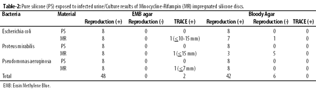
Reproduction was observed in all EMB and BA media for all microorganisms for PS composing the control groups. No statistical difference was observed between the control groups in the culture results in the EMB and BA media in MR-coated silicone materials exposed to urine infected with Escherichia coli (p>0.05). Yet, an inhibition zone (IZ) of <10mm was observed in 1(6.25%) of the 16 MR silicones for Escherichia coli and no reproduction was seen in the BA medium.
In the experiment conducted with Pseudomonas aeruginosa, reproduction was seen in all media in the MR silicones and one <7mm IZ was observed. Compared with the control group for Pseudomonas aeruginosa, the difference was not significant (p>0.05).
A statistical difference was found between the pure silicones and MR-impregnated silicones associated with the reproduction in the media in the Proteus mirabilis experiment (p<0.05). No reproduction occurred in 5(31.25%) BA media in total among the MR-impregnated silicones for Proteus mirabilis. Furthermore, an IZ (+) of >15mm was observed in 1(6.25%) BA medium.
No statistical difference was detected between the culture results of pure silicones and MR-impregnated silicones exposed to urine infected with Escherichia coli and Pseudomonas aeruginosa, while a significant reduction in reproduction was observed on the MR silicones exposed to urine infected with Proteus mirabilis and this difference was statistically significant (Tables-1 and 2).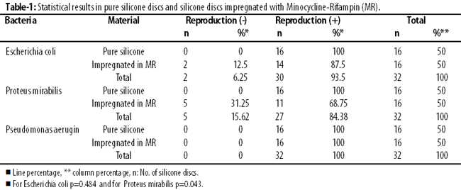
Discussion
The biofilm layer on the surface of the catheters placed in the urinary system was formed on an average between the second and the fifth day.6,7,13 The biofilm layer formed makes it difficult to treat urinary infection associated with catheter. In all studies other than the current one, intermittent drainage was performed and drainage systems were used without leaving any residue.8,9,14 It was reported that the risk of UTIs could be reduced with urinary catheters implanted for a short term and the use of antibiotics constituting a synergistic antimicrobial spectrum in silicone materials.15 It was demonstrated that the efficacy of antibiotic-coated silicones in the urinary system was only on the catheter and that it was not effective against planktonic (suspended) microorganisms.4 While Minocycline is a protein synthesis inhibitor, Rifampin impacts the deoxyribonucleic acid (DNA)-associated ribonucleic acid (RNA) polymerase upon inhibiting it. It was demonstrated both in in vitro and in vivo models that the MR combination was more effective than the other antibiotics such as Rifampicin-Vancomycin, Silver Nitrate and Nitrofurantoin used for the same purpose.16 The fact that MR-coated catheters maintain their efficacy for 12 weeks at room temperature is another superiority over the other combined antibiotic-coated catheters.15
There are studies demonstrating that the gram positive and negative bacterial infection risk is reduced beyond 2 weeks in MR-coated silicones.11 It was also expressed that the migration on the catheter in the in vivo medium was prevented on the 2nd and 5th day and even on the 9th and 34th day in bacteria such as Escherichia coli and Pseudomonas aeruginosa and that the risk for UTI was reduced in catheterisation of no longer than 2-3 weeks at in vivo Nitrofurantoin or MR-impregnated systems.17 We observed that the infection risk on MR-impregnated silicone surfaces obtained upon conducting continuous in vitro drainage did not have an impact on Escherichia coli and Pseudomonas aeruginosa in association with catheter. As was the case in our study, it was recorded that the risk of UTI was not reduced in Silver Nitrate-coated, gold and platinum-coated catheters for Escherichia coli.3,10,18,19 Although a synergistic effect was observed in Escherichia coli and Pseudomonas aeruginosa for in vitro chlorhexidine-protamin sulphate, but we did not find this synergistic effect in MR-impregnated silicones.20,21
New alloplastic biological materials are developed for which it is claimed that an antimicrobial effect will be better with the use of enzyme-loaded polymers in urinary catheters in addition to antibiotics.18 It is still claimed that hydrogel catheters containing MR bonded to silicone or ciprofloxacin liposome will enable the reduction of UTIs.22 It is believed that experimental biocompatibility studies conducted upon the addition of autologous cells on an in vitro biologically degradable matrix will promise great hopes for reducing the risk of infection in the construction of a urinary system (prosthetic bladder, artificial sphincter, penile prosthesis, etc.) and in urinary catheters.22
The common concern with regard to catheters in all studies is the increasing resistance against antibiotics.1,6 The increase in the resistance against antibiotics and multiplication of the strains with increased resistance restrict the chemotherapeutic use in catheter-associated infections and thus the importance of coating catheters with antibiotics and of the designs against adhesive proteins of bacterial fimbriae is increasing day by day.11
Another common finding in literature is that the catheter should be kept for less than one week in the urinary tract if it is aimed at preventing the increase in the risk of infection associated with urinary catheter.1,16 There is still no standard evidence demonstrating that one catheter reduces UTI and is superior to another.9,16 It was demonstrated in randomised and semi-randomised clinical studies that antimicrobial coated silicones reduced UTI but it was expressed that it was not clear whether they provided any clinical benefit.15 The need for new in vitro and in vivo studies as well as the need for adding new drainage systems without leaving any residue and for developing systems releasing antibiotics into the medium indicates that our results are in line with earlier publications.14,16
The efficacy against frequently encountered pathogens in the biofilm layer of MR-impregnated silicone surfaces was not significant except for Proteus mirabilis in our study. Furthermore, antimicrobial efficacy and the prevention of biofilm formation on silicone layers at in vivo and in vitro studies conducted with other antibiotics for Escherichia coli and Pseudomonas aeruginosa were slightly more significant compared to our study.8,19,23
Conclusion
It is necessary to establish a standardization and evidence on the reduction of infection risk in catheters containing antibacterial agents and used in the urinary system. Also, this standardization will have a benefit on the efficacy and cost calculations associated with clinical use. This will provide benefit both at in vivo and in vitro studies. We believe that the in vitro urinary model we have defined for the first time will be beneficial for such type of studies. There is still need for evidence-based in vivo and in vitro studies where antimicrobial activity is evaluated on the surface of catheters, such as more effective drainage, new anti-biofilm mechanisms, different antibiotics and combinations and even systems releasing antibiotics into the planktonic medium and the prevention of the adhesion of the fimbriae of bacteria.
References
1. Isikgoz TM, Durusoy R, Pullukcu H, Sipahi OR, Ulusoy S. 2011 Turkish Nosocomial Urinary Tract Infection Study Group .Hospital acquired urinary tract infection point prevalence in Turkey: Differences in risk factor among patient groups. Ann Clin Microbiol Antimicrob 2013;12:31.
2. Darouiche RO, Mansouri MD, Gawande PV, Madhyastha S. Efficacy of combination of chlorhexidine and protamine sulphate against device-associated pathogens. Antimicrob Chemother 2008; 61: 651-7.
3. Hachem R, Reitze R, Borne A, Jiang Y, Thinkey P, Uthamanthil R et al. Novel antiseptic urinary catheters for prevention of urinary tract infections: correlation of in vivo an in vitro test results. Antimicrob Agents Chemother 2009;53: 5145-9.
4. Ha US, Cho YH. Catheter-associated urinary tract infection: new aspects of novel urinary catheters. Int J Antimicrob Agents 2006;28: 485-90.
5. Parker D, Callan L, Harwood J, Thompson DL,Wilde M, Gray M. Nursing interventions to reduce the risk of catheter-associated urinary tract infection. Part 1: Catheter selection. J Wound Ostomy Continence Nurs 2009;36:23-34.
6. Ozgur BC, Ekic M, Yuceturk CN, Bayrak O. Bacterial colonization of double J stents and bacteriuria frequency. Kaohsiung J Med Sci. 2013; 29:658-61.
7. Cormio L, La Forgia P, La Forgia D, Siitonen A, Ruutu M. Bacterial adhesion to urethral catheters: role of coating materials and immersion in antibiotic solution. Eur Urol 2001; 40:354-8.
8. Chaiban G, Hana H, Dvorak T, Raad I. A Rapid method of impregnating endotracheal tubes and urinary catheters with gendine: a novel antiseptic agent. J Antimikrob Chemother 2005; 55: 51-6.
9. Ha US, Cho YH. Catheter-associated urinary tract infection: new aspects of novel urinary catheters. Int J Antimicrob Agents 2006:28:485-90.
10. Serefko A, Chudzik B, Malm A. In vitro activity of caspofungin against planktonic and sassile Candida sp. cells. POl. J microbiol 2006;55:133-7.
11. Burton E, Gawande PV, Yakandawala N, LoVetri K ,Zhanel GG, Romeo T et al. Antibioflim activity of GImU enzyme inhibitors against catheter-associated uropathogens. Antimicrob Agents Chemother 2006;50:1835-40.
12. Reisner A, Marier M, Jörger M, Kra?se R, Berger D, Haid A, et al. Type 1 fimbriae contribute to catheter-associated urinary tract infections caused by Escherichia coli. J Bacteriol 2014;196:931-9.
13. Maki DG, Tambyah PA. Engineering out the risk for infection with urinary catheters. Emerg Infect Dis 2001;7:342-7.
14. Tenke P, Köves B, Johansen TE. An update on prevention and treatment of catheter associated urinary tract infection. Curr Opin Infect Dis 2014;27:102-7.
15. Costerton JW, Irvin RT, Cheng KJ. The bacterial glycocalyx in nature and disease. Ann Re Microbial 1981;35:299-324.
16. Nicolle LE. Catheter-Related Urinary Tract Infection: Practical Management in the Elderly. Drugs Aging 2014;31:1-10.
17. Esposito S, Noyiello S, Leone S. Catheter-associated urinary tract infections: epidemiology and prevention. Infez Med 2008; 16:130-43.
18. Dayenport K, Keeley FX. Evidence for the use of silver-alloy-coated urethral catheters. J Hosp Infect 2005;60:298-303.
19. Desgrandchamps F. Biomaterials in functional reconstruction. Curr Opin Urol 2000;10:20-6.
20. Bosnahan J, Jull A, Tracy C. Types of urethral catheters for management of short-term voiding problems in hospitalized adults. Cochrane Database Syst Rev 2004; CD004013.
21. Schumm K, Lam TB. Types of urethral catheters for management of short-term voiding problems in hospitalized adults: a short version Cochrane review. Neurourol Urodyn 2008;27:738-46.
22. Eydelnant IA, Tufenkji N. Cranberry derived proanthocynidis reduce bacterial adhesion to selected biomaterials. Langmuir 2008; 24:1273-81.
23. Daye RN, Joshi HM, Venugopalan VP. Novel biocatalytic polymer-based antimicrobial coating as potential urethral biomaterial preparation. Antimicrob Agents Chemother 2011;55:85-53.
Related Articles
Journal of the Pakistan Medical Association has agreed to receive and publish manuscripts in accordance with the principles of the following committees:


