

Abbas Ali Tasneem ( Department of Hepatogastroenterology, Sindh Institute of Urology and Transplantation (SIUT), Karachi, Pakistan. )
Ghous Bux Soomro ( Department of Hepatogastroenterology, Sindh Institute of Urology and Transplantation (SIUT), Karachi, Pakistan. )
Zaigham Abbas ( Department of Hepatogastroenterology, Sindh Institute of Urology and Transplantation (SIUT), Karachi, Pakistan. )
Nasir Hassan Luck ( Department of Hepatogastroenterology, Sindh Institute of Urology and Transplantation (SIUT), Karachi, Pakistan. )
Syed Mujahid Hassan ( Department of Hepatogastroenterology, Sindh Institute of Urology and Transplantation (SIUT), Karachi, Pakistan. )
February 2015, Volume 65, Issue 2
Original Article

Abstract
Objective: To determine aetiology, clinical presentation and predictors of survival in Budd Chiari Syndrome patients.
Methods: The prospective observational study based on non-probability convenient sampling was conducted at the Sindh Institute of Urology and Transplantation (SIUT), Karachi, and comprised Budd Chiari Syndrome patients between January 2004 and December 2013. The patients were evaluated for onset of symptoms, causes, mode of presentation and predictors of survival. SPSS 20 was used for statistical analysis.
Results: Of the 25 patients, 16(64%) were males, and 16(64%) belonged to the paediatric age group. Overall age range was 2-50 years with a mean of 14.7±12.41 years. Presentation was chronic in 14(56%) patients, acute in 10(40%) and acute on chronic in 1(4%). Commonest morphological abnormality involved was hepatic veins alone in 14(56%). Probable aetiologies were hypercoagulable states in 21(84%) patients, infections in 2(8%) and malignancy in 1(4%). Among hypercoagulable states, protein C deficiency was the commonest, affecting 9(36%) patients. Seven (28%) patients died; acute 4(16%) and chronic 3(12%). Causes of death included sepsis 4(16%), fulminant hepatic failure 1(4%), gastrointestinal bleeding 1(4%), and bleeding from liver biopsy site 1(4%). Poor survival was associated with bilirubin >5mg/dl (p<0.031), serum alanine transaminase >40U/L (p<0.005), serum albumin <2.8 g/dl (p<0.008), Child-Turcotte-Pugh score >10 (p<0.001) and absence of varices (p<0.025). Cox regression analysis failed to show any significant independent predictors of survival.
Conclusion: Budd Chiari Syndrome affected young patients more frequently and was associated with high mortality. The commonest aetiology was hypercoagulable state. Survival was poor in patients with decompensated liver disease and those with an acute clinical presentation.
Keywords: Budd Chiari Syndrome, Hepatic venous outflow obstruction. (JPMA 65: 120; 2015).
Introduction
Budd-Chiari Syndrome (BCS) refers to hepatic venous outflow tract obstruction (HVOTO) starting from the level of small hepatic veins (HV) through large HV and inferior vena cava (IVC) to the junction of the IVC and right atrium.1-3 The syndrome was first described by Budd (1845) followed by Chiari (1899) after which a number of case reports and case studies were published around the world concentrating on the underlying aetiology, clinical spectrum and treatment strategies.
BCS may arise secondary to hypercoagulable states, infections, malignancies or due to other conditions, including membranous obstruction of the vena cava (MOVC), polycystic liver disease, sarcoidosis, trauma to hepatic veins or inflammatory bowel disease (IBD). BCS has various anatomical types depending on the level of venous occlusion. The disease may manifest in different kinds of clinical presentation, including chronic, sub-acute, acute or fulminant forms. Most patients present with sub-acute or chronic presentation evolving over three to six months. These patients already have cirrhosis and exhibit complications of chronic hepatic decompensation. Hepatosplenomegaly and ascites are usually present and some patients may experience variceal bleeding. Acute BCS is not uncommon and may present in the first one to two months with abdominal pain, tender hepatomegaly, and ascites. Fulminant BCS is uncommon and has rapid deterioration of hepatic function, encephalopathy and renal failure. Only few of these patients survive without prompt intervention.
BCS is an important cause of mortality related to acute and chronic liver disease. Also, changing spectrum of the disease profile has been reported in a number of studies performed in Asia compared to the West,4 indicating the need to explore the disease patterns in our country. Although extensive research has been conducted around the world, but there is insufficient data regarding the aetiology, patterns of clinical presentation and outcome of BCS in Pakistan. The aim of this study was to identify the causes, types, forms of clinical presentation, course of disease and outcome of such patients in our part of the world.
Patients and Methods
The prospective observational study based on non-probability convenient sampling was conducted at the Sindh Institute of Urology and Transplantation, Karachi, and comprised BCS patients between January 2004 and December 2013. After approval by the institutional ethics review committee, all patients diagnosed as having BCS during the study period were evaluated prospectively. The patients were offered usual standard of care and were followed up. A detailed patient history, including onset of symptoms, history of medications including oral contraceptive pills, previous diseases and surgery, and trauma were noted. Findings of physical examination were also noted. Ultrasonography and computed tomography (CT) scan was performed and analysed by senior radiologists. The sites of lesions were classified as hepatic vein, inferior vena cava (IVC) or combined. The extent of obstruction was noted as either partial or complete. The outcome of the disease and the duration from the day of first presentation to date or death was recorded for each patient.
Haematology and coagulation profile were studied at the time of diagnosis. Protein C, S, anti-thrombin III, factor V Leiden mutation, anti-cardiolipin antibodies and other tests contributing to hypercoagulability were performed. The presence of ascites and oesophageal varices were determined at admission and on subsequent visits. Complications that occurred during the follow-up were recorded.
Mode of presentation was recorded as acute or chronic. The onset of BCS was considered acute if it developed over 1-2 months and presented as abdominal pain, tender hepatomegaly and ascites. It was considered acute fulminant if it presented with abrupt and severe abdominal pain and vomiting, marked hepatomegaly, jaundice, ascites, high serum aminotransferase levels, and rapid deterioration of hepatic function with resulting encephalopathy and renal failure. The onset of BCS was regarded as chronic if it evolved over 3-6 months with signs and symptoms of hepatic decompensation, including ascites or variceal bleeding.
SPSS 20 was used for statistical analysis. Frequency and percentage were computed for different continuous and categorical variables like age, gender and cause of BCS. Kaplan-Meier method was employed to calculate survival, and comparisons were made by log rank test. Significant factors identified by the log rank test were analysed by the Cox regression model to identify the independent predictors of survival. P<0.05 was considered statistically significant.
Results
Of the 25 patients, 16(64%) were males, and 16(64%) belonged to the paediatric age group. Overall age range was 2-50 years with a mean of 14.±12.41 years. Presentation was chronic in 14(56%) patients, acute in 10(40%) and acute on chronic in 1(4%) (Table-1).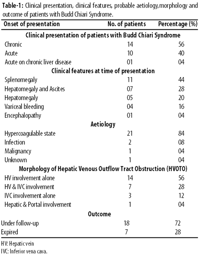
Among patients with acute presentation, 6(24%) had hepatomegaly with ascites, while 4(16%) presented with tender hepatomegaly alone. Among those with chronic presentation, 4(16%) presented with oesophageal variceal bleeding, 7 (28%) with compensated cirrhosis and 3(12%) with advanced cirrhosis.
Commonest morphological abnormality involved was hepatic veins alone in 14(56%). Probable aetiologies were hypercoagulable states in 21(84%) patients, infections in 2(8%) and malignancy in 1(4%). Among hypercoagulable states, protein C deficiency was the commonest, affecting 9(36%) patients, followed by protein S deficiency in 2(8%),anti-thrombin III deficiency in 1(4%), lupus anti-coagulant deficiency in 1(4%) and combined anti-coagulant deficiencies in 8(32%). Two (8%) patients developed BCS as a complication of infections; 1(4%) had abdominal tuberculosis with caudate lobe enlargement causing extrinsic compression of IVC; 1(4%) was an alcoholic who developed a large liver abscess that was complicated by hepatic and portal vein thrombosis; and 1(4%) developed BCS secondary to renal cell carcinoma with IVC invasion. Besides, 14(56%) patients had hepatic vein (HV) involvement alone, followed by 7(28%) with HV and IVC involvement, 3(12%) with IVC involvement alone, and 1(4%) with hepatic and portal vein involvement.
Overall, 7(28%) patients died; acute 4(16%) and chronic 3(12%), and 18(72%) are under close follow-up. Causes of death included sepsis 4(16%), fulminant hepatic failure 1(4%), gastrointestinal bleeding 1(4%), and bleeding from liver biopsy site 1(4%). Of the patients who died of sepsis, 2(8%) developed gram-negative bacterial septicaemia, 1(4%) developed spontaneous bacterial peritonitis with acute renal failure, and 1(4%) developed acute renal failure alone. The patient who died of acute on chronic liver failure had an already existing chronic liver disease secondary to hepatitis B and had developed an acute hepatic insult secondary to HV and IVC thrombosis. One (4%) patient died of intractable oesophageal variceal bleeding. One (4%) patient with protein S deficiency died secondary to intractable bleeding after liver biopsy that was done for the confirmation of the cause of liver failure.
Poor survival was associated with bilirubin >5mg/dl (p=0.031), serum alanine transaminase >40U/L (p<0.005), serum albumin <2.8 g/dl (p=0.008), Child-Turcotte-Pugh (CTP) score >10 (p=0.001) and absence of varices (p=0.025). Cox regression analysis failed to show any significant independent predictors of survival (Table-2).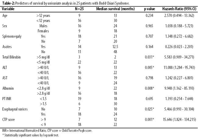
Discussion
The prevalence of BCS appears to vary significantly according to the geographical area under consideration. For instance, it is the leading cause for liver disease related hospital admissions in Nepal,5 whereas it is quite rare in Japan and France.6 The gender and age distribution is also variable depending upon the geographical location. There has been slight predominance of males and a median age of 45 years in Asia, while marked female preponderance and a younger median age (35 years) are seen in the West.7 In our study, male patients were predominant, accounting for 64% of the cases which was in consistence with the previously reported data regarding Asian population. Also, according to previous studies, this condition is uncommon in children compared to adults8 and more than half of the cases of classic BCS occur between ages 20 and 39 years. In one study, only 5% patients were found to be below 12 years of age out of the total 177 cases of BCS.8 In our study, 64% patients were children or adolescents. Further studies are needed to explore the cause of greater prevalence of this disease in the younger age group in our part of the world. Furthermore, previous data suggests that BCS is diagnosed more commonly with a chronic presentation rather than an acute one.9 Patients presenting with sub-acute or chronic symptoms are those with signs evolving over 3-6 months.10 In our study, majority of the patients presented with chronic stage of the disease which was in conformity with reported data.
There is growing evidence that BCS occurs in conjunction with various predisposing factors such as one or more underlying thrombophilic conditions.4 A study of aetiological factors in BCS from neighbouring country India suggested that inherited prothrombotic factors were more important than acquired predisposing factors. An overall underlying risk factor for thrombosis has been found in upto 87% of BCS patients.11 In our study, too, hypercoagulable state was the commonest probable aetiological factor accounting for 84% of all cases. Since the levels of both the coagulation as well as anti-coagulant factors become diminished in chronic liver disease, it is difficult to state that deficiency of natural anti-coagulants is the actual aetiological factor responsible for the development of thrombosis in BCS as majority of our cases were having chronic disease. As the international normalised ratio (INR) was normal or not much elevated, we assumed that hypercoagulable states secondary to deficiency of one or a combination of natural anti-coagulant factors was the probable aetiology of BCS. Furthermore, we did not encounter any case of membranous obstruction of the vena cava (MOVC). This is in sharp contrast to China where more than 70% of BCS cases are due to MOVC.10 The findings of our study are in conformity with its decreasing trend of incidence which has been noticed in India in recent years.10 This can partly be explained by the improvement in hygiene and sanitation in the Indian subcontinent over the last few decades because poverty, malnutrition, recurrent bacterial infections and filariasis have been previously suggested as predisposing factors for IVC obstruction.4
In BCS, reductions in hepatic venous outflow can occur anywhere from the small hepatic venules to the right atrium. The level of obstruction may also differ according to the geographicalarea under evaluation. According to previously reported data, pure HV block predominates in the Western countries in contrast to the Asian countries which predominantly demonstrate combined IVC/HV block.6 Nevertheless, a recent European survey showed an equal distribution of pure HV block and combined IVC/HV block.12 In our study, 56% patients had HV involvement alone, while 28% patients had combined HV/IVC involvement. This finding is not in conformity with the previously reported data and further studies are needed to identify the cause of this pattern of venous involvement in our part of the world.
Main causes of death in BCS are gastrointestinal bleeding, liver failure and intractable ascites with emaciation.13 With a stepwise approach beginning with anti-coagulation therapy followed by transjugularintrahepatic portosystemic shunt (TIPS) and then liver transplantation, an overall five-year survival rate of nearly 90% can be achieved.10 Patients with fulminant BCS awaiting liver transplantation may have mortality rates as high as 50%. In a British cohort dating back to the 1960s when no specific therapy was yet available, 90% patients had died by 3 years.13 There has been continued improvement in outcome and prognosis of BCS over the last 4 decades. In the most recently reported cohorts, overall 5-year survival rates over 80% have been achieved. In our study, 28% patients died. The most common cause of death was sepsis (57.1%) followed by liver failure and variceal bleeding. Sepsis was the most common cause of death probably because most of the patients belonged to poor socioeconomic class with prevailing malnutrition and depressed immune responses. The reason for the high mortality rate in our study can also be explained by the fact that facilities for angioplasty, TIPS and liver transplantation were not available at our centre.
Several studies have been done to study the factors that may affect the survival of BCS patient. A study of 47 consecutive BCS patients found the following factors to adversely affect survival: florid clinical presentation, male gender, no TIPS performed and CTP score.14 An international multi-institutional study included BCS patients treated with a variety of modalities. The following factors were found by multivariate analysis to be independent predictors of 5-year transplant-free survival: presence of ascites, presence of encephalopathy, INR and bilirubin.15 Our study showed that poor prognosis was associated with an acute course of illness as reflected by a high bilirubin, high alanine transaminase (ALT), and absence of varices; and with decompensated liver disease as evidenced by low serum albumin levels and an advanced CTP score. However, in our study male gender, presence of ascites and deranged INR were not found to adversely affect the outcome. This difference may partly be explained on the basis of the relatively smaller number of patients in our study.
Conclusion
BCS is not an uncommon cause of liver disease in Pakistan. The commonest probable aetiology was hypercoagulable state. BCS is associated with high mortality in the paediatric age group. Survival is poor in patients with decompensated liver disease and those with an acute clinical presentation.
References
1. Valla DC. The diagnosis and management of the Budd-Chiari syndrome: consensus and controversies. Hepatology 2003;38:793-803.
2. Menon KV, Shah V, Kamath PS. The Budd-Chiari syndrome. N Engl J Med 2004;350:578-85.
3. Janssen HL, Garcia-Pagan JC, Elias E, Mentha G, Hadengue A, Valla DC. Budd-Chiari syndrome: a review by an expert panel. J Hepatol 2003;38:364-71.
4. Eapen CE, Mammen T, Moses V, Shyamkumar NK. Changing profile of Budd Chiari Syndrome in India. Indian J Gastroenterol 2007;26:22-5.
5. Shrestha SM, Okuda K, Uchida T, Maharjan KG, Shrestha S, Joshi BL, et al. Endemicity and clinical picture of liver disease due to obstruction of the hepatic portion of the inferior vena cava in Nepal. J Gastroenterol Hepatol 1996;11:170-9.
6. Valla D. Hepatic venous outflow tract obstruction etiopathogenesis: Asia versus the West. J. Gastroenterol. Hepatol 2004;19: S204- S211.
7. Pleisser A, Valla DC. Budd Chiari Syndrome. Semin Liver Dis. 2008; 28:259-69.
8. Misra V, Verma K. The Budd Chiari Syndrome in a child: A case report and review of literature. J Clin Diagn Res 2012; 6: 1783-5.
9. Dilawari JB, Bambrey P, Chawla Y, Upjeet K, Bhusnurmatch SR, Malhotra et al. The hepatic outflow obstruction (the Budd Chiari Syndrome). Medicine 1994; 73: 21-36.
10. Stevens WE, Patil A. Vascular diseases of the liver. In: Feldman M, Friedman LS, Brandt LJ. Gastrointestinal and Liver Disease: Pathophysiology, Diagnosis, Management. 9th ed. Philadelphia: Saunders, 2010;pp 1372.
11. Denninger MH, Chait Y, Casadavell N, Hillaire S, Guillin MC, Bezeaud A, et al. Cause of portal or hepatic venous thrombosis in adults: the role of multiple concurrent factors. Hepatology 2000; 31:587-91.
12. Darwish MS, Pleiser A, Hernandez-Guerra H, Fabris F, Eapen CE, Bahr MJ et al. EN-vie (European network for vascular liver disorders of liver). Etiology, management and outcome of Budd Chiari Syndrome. Ann Intern Med 2009;151:167-75.
13. Tavill AS, Wood EJ, Kreel L, Jones EA. The Budd Chiari Syndrome : correlation between hepatic scintigraphy and the clinical, radiological and pathological finding in nineteen cases of hepatic venous outflow obstruction. Gastroenterology 1995 ; 68:509-18.
14. Khuroo M S, Al Suhaibani H, Al Sebayel M, Al Ashgar H, Dahab S, Khan MQ et al. Budd Chiari Syndrome: long term effect on outcome with transjugular intrahepatic portosystemic shunt. J. Gastroenterol. Hepatol 2005;20:1494-502.
15. Horton JD, San Miguel FL, Ortiz JA. Budd-Chiari Syndrome: Illustrated Review of Current Management. Liver Int 2008; 28:455-66.
Related Articles
Journal of the Pakistan Medical Association has agreed to receive and publish manuscripts in accordance with the principles of the following committees:


