

Erhan Yengil ( Department of Family Medicine, Mustafa Kemal University, School of Medicine, Antakya, Turkey. )
Cengiz Cevik ( Department of Otolaryngology, Mustafa Kemal University, School of Medicine, Antakya, Turkey. )
Ibrahim Silfeler ( Department of Paediatric, Mustafa Kemal University, School of Medicine, Antakya, Turkey. )
Erkan Yula ( Department of Microbiology, Mustafa Kemal University, School of Medicine, Antakya, Turkey. )
Ibrahim Ahmet Kurtoglu ( Department of Paediatric, Mustafa Kemal University, School of Medicine, Antakya, Turkey. )
Vefik Arica ( Department of Paediatric, Mustafa Kemal University, School of Medicine, Antakya, Turkey. )
Cahit Ozer ( Department of Family Medicine, Mustafa Kemal University, School of Medicine, Antakya, Turkey. )
February 2015, Volume 65, Issue 2
Original Article

Abstract
Objective: To investigate resistant microorganisms in nasal mucosa of children with Familial Mediterranean Fever.
Methods: The study was conducted from March to May 2013 at Mustafa Kemal University, Turkey, and comprised children with Familial Mediterranean Fever and healthy controls. All subjects had no history of antibiotic or local and/or systemic steroid use within the preceding 2 weeks. Nasal swab samples were obtained from all the subjects. Strain identification was done by using standard methods. SPSS 13 was used for statistical analysis.
Results: Of the 151 subjects in the study, 73(48.34%) were cases and 78(51.65%) were controls. Among the cases, there were 26(35.6%) girls, while among the controls, there were 40(51.3%) girls (p=0.052). The mean age of the cases was 7.78±3.34 years (range: 3-15 years), while it was 8.15±2.71 years (range: 3-16) among the controls (p=0.208). Methicillin-resistant coagulase-negative staphylococcus and methicillin-resistant staphylococcus aureus were isolated in both the groups. The growth rate of resistant bacteria was 63% (n=46) in the cases, in the controls (p=0.003; odds ratio [OR]: 2.7; 95% confidence interval [CI]: 1.4-5.2). Among the controls, history of hospitalisation increased the risk for the presence of resistant bacteria by 7.7 fold (OR: 7.7; 95%CI: 1.4 - 40.4).
Conclusion: Higher rates of resistant bacteria showed that they were at risk of comorbidities related to antibiotic resistance.
Keywords: Familial Mediterranean Fever, Methicillin-resistant staphylococcus aureus, Nasal flora, Colonisation. (JPMA 65: 196; 2015)
Introduction
Familial Mediterranean Fever (FMF) is an autosomal recessive, recurrent, auto-inflammatory disease that is characterised by fever with abdominal pain, pleurisy, arthritis and skin lesion.1-4 It is frequently seen in populations living in the Mediterranean region, particularly among Turkish, Jewish, Arabian and Armenian.4,5 It has been reported that the estimated prevalence of the disease is 0.1% in Turkey.6
Nasal cavity and paranasal sinuses are sterile at birth. After birth, the nasal flora is acquired from mother as well as nursing staff and healthcare team who provide care to the infant in the delivery room and newborn units.6 This flora acquired by respiration and indirect contact colonises into the nasal cavity. These microorganisms are killed by the host or their growth is suppressed after the introduction of mucosal defence mechanisms at nasal mucosa. Nasal bacterial flora of the individual develops by changes in immune resistance over time.8 Gram-positive microorganisms (Staphylococcus species [spp.]) are the major elements of the flora at nasal region. In addition, aerobic and facultative isolates, corynebacterium species, neisseria species, haemophilus influenzae, staphylococcus spp., moraxella spp., micrococcus spp., staphylococcus (S.) epidermidis, Viridans group streptococci and anaerobic isolates can also be present in nasal flora.8
It has been reported that methicillin-resistant Staphylococcus aureus (MRSA) can be present in the nasal flora of healthy individuals without causing any symptom with increasing MRSA rates in recent years.9,10 This microorganism is the most commonly isolated antibiotic-resistant pathogen in hospital-acquired infections (both surgical and non-surgical infections).9 Moreover, S. aureus colonisation at anterior nares is a risky condition for endogenous infection that would occur.11
To our knowledge nasal carriage of MRSA in FMF has not been studied. The aim of the present study was to investigate resistant microorganisms in the nasal mucosa of children with FMF, and to compare it with healthy controls.
Subjects and Methods
The case-control study was conducted from March to May 2013 at Mustafa Kemal University, Turkey, and comprised children with FMF and healthy controls. In children with FMF, the condition was diagnosed according to Tell-Hashomer criteria.12
After obtaining approval from the institutional ethics committee, children without any systemic disease other than FMF who had no nasal pathology and history of antibiotic, nasal spray and/or antihistaminic use within the preceding 2 weeks who hadn\'t been admitted to hospital within the preceding 3 months were included. Healthy children matching the criteria but who had no systemic disease, including FMF, were enrolled in the control group.
Demographic characteristics, age at the onset of the disease, duration of the disease, history of hospital admission, mean duration of drug use (colchicine), number of attacks and data regarding FMF gene mutations were obtained. Nasal swab samples were obtained from nostrils in both the groups.
Nasal swab samples were transferred to laboratory where they were inoculated on to blood agar (BA) by reduction technique. In the suspected colonies, strain identification was done through conventional methods such as gram staining, catalase and tube coagulase tests. A suspension in sterile saline was prepared from fresh cultures of strains according to 0.5 MacFarland standard.13 The suspension was then homogenously inoculated on to Mueller Hinton Agar (MHA) by using a sterile swab. A 30µg cefoxitine disk (Oxoid, UK) was placed on to the surface of growth media after a wait of 5-10 minutes at room temperature. It t was then incubated at 35°C for 18-24 hours. After incubations, inhibition zones were assessed according to Clinical and Laboratory Standards Institute (CLSI) guidelines13 as follows: <21mm methicillin-resistant, and >21mm as methicillin sensitivity.
Data was analysed using SPSS 13. Normal distribution of variables were tested with One Sample Kolmogorov-Smirnov test. For intra- and inter-group comparisons, Chi-square and Fischer\'s exact tests were used for categorical variables, whereas Mann Whitney U test was used for continuous variables. P<0.05 was considered statistically significant.
Results
Of the 151 subjects in the study, 73(48.34%) were cases and 78(51.65%) were controls. Among the cases, there were 26(35.6%) girls, while among the controls, there were 40(51.3%) girls (p=0.052). The mean age of the cases was 7.78±3.34 years (range: 3-15 years), while it was 8.15±2.71 years (range: 3-16) among the controls (p=0.208). Methicillin-resistant coagulase-negative staphylococcus (MRCNS) and MRSA were isolated in both the groups. There were mutations in 64 (98.5%) patients, whereas no mutation was found in 1 patient. The most common mutations were: A165A (40Het, 18Hom), G138G (38Het, 18Hom), R202Q (34Het, 13Hom), M694V (14Het, 2Hom), E148Q (16Het), V726A (6Het), M680I (2Het), P706P (2Het) and R761H (2Het), respectively.
Microorganisms identified in the nasal flora of the controls were: diphteroid spp. n=53 (34.4%), MRCNS n=29 (18.8%), viridans group streptococci n=21 (13.6%), methicillin-susceptible coagulase-negative staphylococcus (MSCNS) n=20 (13%), Bacillus spp. N=15 (9.7%), S. pneumonia n=12 (7.7%), MRSA n=1 (0.7%), candida spp. n=1 (0.7%), gram-negative extended spectrum beta lactamase (ESBL) negative bacillus n=1 (0.7%), gram-negative ESBL positive bacillus n=1 (0.7%). In the nasal flora of the cases, the identified microorganisms were: diphteroid spp. n=22 (20.5%), MRCNS n=43 (40.1%), viridans group streptococci n=11 (10.2%), MSCNS n=6 (5.7%), S. pneumonia n=6 (5.7%), MRSA n=6 (5.7%), methicillin-sensitive staphylococcus aureus (MSSA) n=7 (6.5%), micrococcus spp. n=4 (3.7%), and neisseria spp. n=2 (1.9%).
Bacterial growth was detected in all patients with 3 different types in 3 cases and 2 different types in 31 cases in the nasal flora cultures of the cases, while it was detected in all subjects with 4 different types in 1 case, 3 different types in 19 cases and 2 different types in 56 cases in the controls. The most commonly detected microorganism was MRCNS (n=43), followed by diphteorid spp. (n=22) and viridans group streptococci (n=11) in the cases, while diphteorid spp. (n=53) was followed by viridans group streptococci (n=21) in the controls (Figure).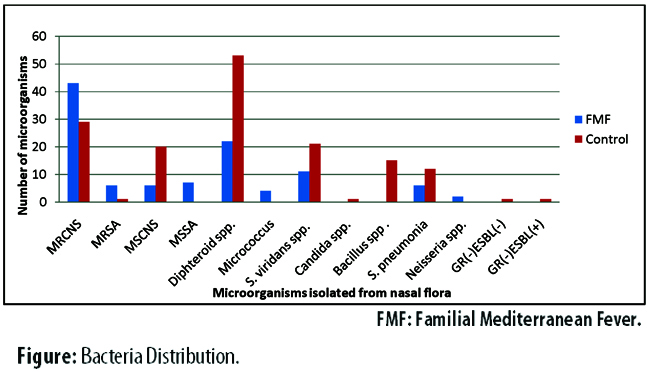
The growth rate of resistant bacteria was 63% (n=46) in the cases, while it was 38.5% (n=30) in the controls (p=0.003; odds ratio [OR]: 2.7; 95% confidence interval [CI]: 1.4-5.2). Among the controls, history of hospitalisation increased the risk for the presence of resistant bacteria by 7.7 fold (OR: 7.7; 95%CI: 1.4-40.4) (Table-1).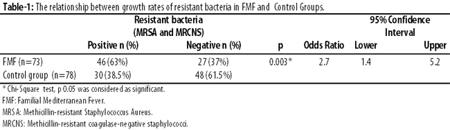
Among the cases, no relationship was found between the presence of resistant bacteria and gender, age, duration of disease, age at the onset of disease, presence of a first-degree relative with FMF, number of attacks within the preceding year and duration of colchine therapy (Table-2).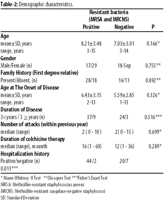
Among the cases, 64 had an attack, while 9 had no attack within the preceding year. The growth rate of resistant bacteria was similar in patients with (65.6%) or without (44.4%) attack within the preceding year (p=0.276).
There was a history of hospitalisation due to any reason in 87.7% of the cases. Resistant bacteria MRSA plus MRCNS was 68.8%) in patients with a history of hospitalisation, while it was 22.2% in those without (p=0.011). The hospital admission increased the risk for the presence of resistant bacteria 7.7 fold (OR: 7.7; 95CI: 1.4 - 40.4.
Discussion
To our knowledge, this is the first study aimed at determining rates of resistant microorganisms in the nasal flora of children with FMF. According to the study, the rate of resistant microorganisms was significantly higher in children with FMF compared to healthy individuals.
FMF caused a trend towards increase in the rate of resistant microorganisms in the nasal flora. Nasal cavity is one of the areas in human body which normally have a flora.8 S. pneumoniae, H. influenza and S. aureus are the most common microorganisms localised at healthy nose and paranasal sinuses. Conditions (nasal surgery, allergic rhinitis, acute and/or chronic sinusitis) leading to alterations in humidity and amount of oxygen in nose and those affecting systemic immunity and/or nasal mucosal defence can cause changes in nasal flora.8
Although S. aureus is colonised at perineum and throat, S. aureus carriage is localised at anterior nasal region in 20% of the general population.14 A study reported that nasal carriage plays an important role in the epidemiology and pathogenesis of S. aureus-related infections.15 In addition, it is one the major risk factors in the development of S. aureus infections in several patient populations (e.g. haemodialysis patients, those with acquired immunodeficiency syndrome [AIDS], intravascular device or those who underwent surgery).16 MRSA is an endemic pathogen that causes approximately 70% of the invasive S. auerus infections that occur in healthcare facilities across the world.16 It is a matter of concern that risk for development of resistance against vancomycine, which is still effective agent in MRSA, because of increasing MRSA rates in the last decade.16 Thus, there is an increasing interest in MRSA because of increased risk for vancomycine resistance and MRSA rates. Moreover, it is important to eradicate S. aureus carriage for both prevention and spread of infection. One study reported that short-term (4-7 days) intranasal mupirocin administration resulted in successful eradication of MRSA with a rate of 90%. In addition, it was shown that the eradication persisted beyond one week and at long-term in 60% patients.14 It has also been reported that mupirocin eradicated MRSA in 73.5% of the carriers.17
History of hospitalisation increased the risk for the presence of resistant bacteria in FMF. FMF patients present to hospital more frequently than normal population. One study assessed MRSA colonisation stratifying risk groups in patients presenting to emergency department.18 It found MRSA positivity in 31.4% patients. It further reported that nasal MRSA positivity rate was 60% in patients with a history of positive MRSA whereas it was 43.9% in patients who were hospitalised for 30 days and/or longer within the preceding 3 months; 41.7% in those who were hospitalised 10 days and/or longer within the preceding 3 months; 26.3% in dialysis patients with renal failure; 34.2% in those with chronic skin disease; and 40% in patients who were admitted to hospital because of an acute disease.18 In our study the rate of resistant bacteria was 68.8% in patients who had a history of hospitalisation.
It has been reported that there is a strong correlation between nasal carriage of S. aureus and relapse in patients with Stegeman Wegener\'s granulomatosis and that sulfamethoxazole-trimethoprim used in therapy reduces relapse frequency by decreasing the nasal carriage.19 Vasculitis is a clinical condition that can be observed in association with FMF.3 However, there was no patient with vasculitis in our study. Although a correlation was found between relapse and nasal carriage of S. aureus in Stegemen Wegener\'s granulomatosis, but no significant relationship was found between number of attacks and presence of resistance microorganism in our study.
S. aureus is the most commonly seen pathogen in wound infections after clean, elective surgery.20 S. aureus is the causative agent in approximately 30-50% of the wound infections after clean surgery.20 In particular, MRSA emerges as the infectious agent in patients with wound infections occurring after surgery.20 It has been reported that the likelihood of MRSA-related wound infection is 44%-65% in patients colonised by MRSA, whereas it is 0.4-2% in those without MRSA colonisation.14,21 One study reported that the detection of carriage by nasal swab before surgery could reduce the risk for wound infection after surgery in patients undergoing gastrointestinal surgery.22 The MRSA-related wound infection causes prolonged hospitalisation, increased risk for morbidity and mortality and hospital costs.20
Conclusion
Resistant bacteria rates were found to be significantly higher in children with FMF compared to healthy controls, suggesting that these children are at higher risk regarding complications and comorbidities resulting from these resistant bacteria. Physicians need to be more alert about resistance, and consider this issue when prescribing antibiotics.
Acknowledgement
We have no financial disclosures for this article.
We have no conflicts of interest to declare.
Disclosure
This study was presented in 7th School in Spring Congress of Family Medicine in 2013 at Antalya, Turkey.
References
1. Ben-Chetrit E, Levy M. Familial Mediterranean fever. Lancet 1998;351:659-64.
2. Livneh A, Langevitz P, Zemer D, Zaks N, Kees S, Lidar T,et al. Criteria for the diagnosis of Familial Mediterranean fever. Arthrit Rheum 1997;40:1879-85.
3. Samuels J, Aksentijevich I, Torosyan Y, Centola M, Deng Z, Sood R, et al. Familial Mediterranean fever at the Millennium Clinical spectrum ancient mutations and a survey of 100 American referrals to the National institutes of Health. Medicine 1998;77:268-97.
4. Sohar E, Gafni J, Pras M, Heller H. Familial Mediterranean fever :A survey of 470 cases and review of the literatüre. Am J Med 1967;43:227-53.
5. Özdemir AL, Sökmen C. Familial Mediterranean fever among the Turkish people. Am J Gastroenterol 1969;51:311-6.
6. Tunca M, Akar S, Onen F, Ozdogan H, Kasapcopur O, Yalcinkaya F, et al Turkish FMF Study Group. Familial Mediterranean fever (FMF) in Turkey: results of a nationwide multicenter study. Medicine (Baltimore) 2005;84:1-11.
7. Bamberger DM. Antimicrobial treatment of sinusitis. Semin Respir Infect. 1991;6:77-84.
8. Haug RH. Microorganisms of the nose and paranasal sinuses. Oral Maxillofac Surg Clin North Am 2012;24:191-6.
9. Manunga J Jr, Olak J, Rivera C, Martin M. Prevalence of methicillin-resistant Staphylococcus aureus in elective surgical patients at a public teaching hospital: an analysis of 1039 patients. Am Surg 2012;78:1096-9.
10. Brook I, Foote PA. Isolation of methicillin resistant Staphylococcus aureus from the surface and core of tonsils in children. Int J Pediatr Otorhinolaryngol 2006;70:2099-102.
11. Del Rio A, Cervera C, Moreno A, Moreillon P, Miró JM. Patients at risk of complications of Staphylococcus aureus bloodstream infection. Clin Infect Dis 2009;48:246-53.
12. Daniels M, Shohat T, Brenner-Ullman A, Shohat M. Familial mediterranean fever: high gene frequency among the non-Ashkenazic and Ashkenazic Jewish populations in Israel. Am J Med Genet 1995;55;311-4.
13. Clinical and Laboratory Standards Institute. Performance Standards for Antimicrobial Susceptibility Testing; Twenty-Second Informational Supplement M100-S22, Vol. 32 No. 3. Wayne, PA: Clinical and Laboratory Standards Institute; 2012.
14. Ammerlaan HS, Kluytmans JA, Wertheim HF, Nouwen JL, Bonten MJ. Eradication of methicillin-resistant Staphylococcus aureus carriage: a systematic review. Clin Infect Dis 2009;48:922-30.
15. Kluytmans J, van Belkum A, Verbrugh H. Nasal carriage of Staphylococcus aureus: epidemiology, underlying mechanisms, and associated risks. Clin Microbiol Rev 1997;10:505-20.
16. European Antimicrobial Resistance Surveillance System. EARSS annual report 2005: ongoing surveillance of S. pneumoniae, S. aureus, E. coli, E. faecium, E. faecalis. Bilthoven, The Netherlands: Rijksinstituut voor Volksgezondheid en Milieu; 2006.
17. Schmid H, Romanos A, Schiffl H, Lederer SR. Persistent nasal methicillin-resistant staphylococcus aureus carriage in hemodialysis outpatients: a predictor of worse outcome. BMC Nephrol. 2013;14:93.
18. Wakatake H, Fujitani S, Kodama T, Kawamoto E, Yamada H, Yanai M, et al. Positive clinical risk factors predict a high rate of methicillin-resistant Staphylococcus aureus colonization in emergency department patients. Am J Infect Control 2012;40:988-91.
19. Stegeman CA, Tervaert JW, Sluiter WJ, Manson WL, de Jong PE, Kallenberg CG. Association of chronic nasal carriage of Staphylococcus aureus and higher relapse rates in Wegener granulomatosis. Ann Intern Med. 1994;120:12-7.
20. Cowie SE, Ma I, Lee SK, Smith RM, Hsiang YN. Nosocomial MRSA infection in vascular surgery patients: impact on patient outcome. Vasc Endovascular Surg 2005;39:327-34.
21. Davis KA, Stewart JJ, Crouch HK, Florez CE, Hospenthal DR. Methicillin-resistant Staphylococcus aureus (MRSA) nares colonization at hospital admission and its effect on subsequent MRSA infection. Clin Infect Dis 2004;39:776-82.
22. Ramirez MC, Marchessault M, Govednik-Horny C, Jupiter D, Papaconstantinou HT. The impact of MRSA colonization on surgical site infection following major gastrointestinal surgery. J Gastrointest Surg 2013;17:144-52.
Related Articles
Journal of the Pakistan Medical Association has agreed to receive and publish manuscripts in accordance with the principles of the following committees:


