

Momna Khan ( Department of Obstetrics and Gynaecology, The Aga Khan University Hospital, Karachi, Pakistan. )
Syeda Seema Sultana ( Department of Obstetrics and Gynaecology, Civil hospital Karachi, Pakistan. )
Nigar Jabeen ( Department of Obstetrics and Gynaecology, The Aga Khan University Hospital, Karachi, Pakistan. )
Uzma Arain ( Department of Obstetrics and Gynaecology, The Aga Khan University Hospital, Karachi, Pakistan. )
Salma Khan ( Department of Surgery, The Aga Khan University Hospital, Karachi, Pakistan. )
February 2015, Volume 65, Issue 2
Original Article

Abstract
Objective: To determine the diagnostic accuracy of visual inspection of cervix using 3% acetic acid as a screening test for early detection of cervical cancer taking histopathology as the gold standard.
Methods: The cross-sectional study was conducted at Civil Hospital Karachi from July 1 to December 31, 2012 and comprised all sexually active women aged 19-60 years. During speculum examination 3% acetic acid was applied over the cervix with the help of cotton swab. The observations were noted as positive or negative on visual inspection of the cervix after acetic acid application according to acetowhite changes. Colposcopy-guided cervical biopsy was done in patients with positive or abnormal looking cervix. Colposcopic-directed biopsy was taken as the gold standard to assess visual inspection readings. SPSS 17 was used for statistical analysis.
Results: There were 500 subjects with a mean age of 35.74±9.64 years. Sensitivity, specifically, positive predicted value, negative predicted value of visual inspection of the cervix after acetic acid application was 93.5%, 95.8%, 76.3%, 99%, and the diagnostic accuracy was 95.6%.
Conclusion: Visual inspection of the cervix after acetic acid application is an effective method of detecting pre-invasive phase of cervical cancer and a good alternative to cytological screening for cervical cancer in resource-poor setting like Pakistan and can reduce maternal morbidity and mortality.
Keywords: Visual inspection, Acetic acid, Cervical cancer. (JPMA 65: 192; 2015).
Introduction
Cervical cancer is one of the most prevalent malignancies among women in developing countries.1,2 Each year over 490,000 new cases of cervical cancer are diagnosed worldwide with 80% in under-developed countries.3 Screening should be provided to all women to prevent or diagnose cervical cancer at an early, treatable stage.3 Screening for cervical lesions has proven successful in the industrialised world, with incidences of cervical cancer reduced by 80% in countries with organised screening programmes.4 The success of these screening programmes can largely be attributed to the use of papanicolaou (PAP) smear.4 In contrast to Western countries, this approach may not be feasible for many decades in low-resource settings like Pakistan due to financial and technical constraints.5 This has led to the development of multiple low-cost technologies like visual inspection of cervix after application of Acetic acid (VIA) and visual inspection after lugol's iodine (VILI), which would overcome the barriers posed by cytological screening.6
VIA is simple, accurate, cost-effective method of rapidly differentiating between diseased and healthy cervix.7 World Health Organisation (WHO) has recommended VIA as an alternative to cytology to identify patients at risk of cervical cancer.8
Due to non-invasive nature, easy applicability with immediate results make VIA a useful screening test in developing countries like Pakistan.5 Initial studies have shown VIA sensitivity to be similar to the PAP smear, but more studies are required to confirm the utility of VIA as a primary screening method.9
The current study was planned to identify an easily applicable, low-cost and sensitive cervical cancer screening test. We used VIA as the primary screening tool taking histopathology as the gold standard.
Subjects and Methods
The cross-sectional study was conducted at Civil Hospital Karachi from July 1 to December 31, 2012 and comprised all sexually active women aged 19-60 years who furnished informed consent. Women with previous cervical procedure, biopsy and overt cervical lesion were excluded. Detailed history was obtained and physical examination was conducted.
The procedure was performed in the outpatient clinics by gynaecology residents of III year and above who were well trained to perform VIA. Patients were placed in lithotomy position, and lubricated Cusco's speculum was introduced into the vagina under good light source to assess cervix for any gross abnormality. Then 3% acetic acid was applied to cervix with the help of cotton swab and observed for acetowhite changes for one minute. The detection of any distinct acetowhite area was considered positive, while no light, faint or doubtful acetowhite areas were considered negative.
Colposcopy-directed biopsy was taken from all VIA-positive women, plus those who were VIA-negative but had abnormal looking cervix. Tissues were sent for histopathology assessment. This served as the reference standard for VIA results. On histology, cervical interstitial neoplasia (CIN) 1, 2, 3 or invasive carcinoma was true positive. A structured proforma was filled by year III or IV residents of gynaecology for baseline variables, VIA and colposcopic findings.
All statistical data was analysed using SPSS 17. Frequencies and percentages were calculated for all categorical variables like parity, marital status, religion, ethnic group. Mean and Standard Deviation (SD) was calculated for quantitative variables like age, duration of marriage. The diagnostic accuracy, sensitivity specificity and positive predictive value (PPV) and negative predictive value (NPV) was calculated using standard statistical formula taking histopathology as the gold standard.
Results
There were 500 subjects with a mean age of 35.74±9.64 years and mean duration of sexual activity of 17.33±9.82 years. Nulliparous women were 41(8%), 241(48%) were single or multiparous (parity 1-5), while 218(44%) were grand multipara (parity 6 to 10) (Table-1).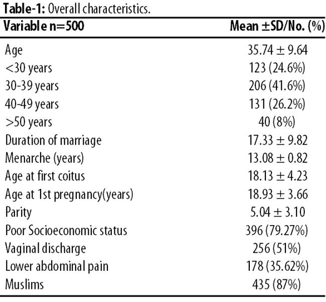
Findings related to the 76(15.2%) VIA-positive women and 28(5.6%) women who were VIA-negative but had abnormal cervix were referred to histopathology. As such, 104(21%) specimen were sent for biopsy, while the remaining 396(79%) women either had negative VIA or normal cervix which did not require biopsy and were considered true negative cases. Biopsy confirmed 62(59.6%) of the 104 cases of CIN or invasive cervical cancer of which 58(93.5%) patients were VIA-positive and 4(6.5%) were VIA-negative. VIA detected 100% (3 of 3) invasive carcinoma, 100% (4 of 4) CIN III, 78.6% (11 of 14) CIN II and 78.1% (25 of 32) CIN I (Figure).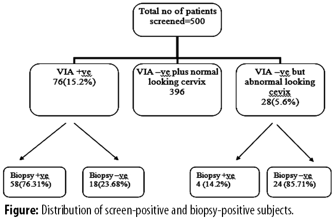
The sensitivity, specificity, PPV, NPV and accuracy of VIA and abnormal looking cervix were 81.13% (95% Confidence Interval [CI]: 68.64-89.41), 35.29% (95%CI: 26.63-19.04), 56.58% (95%CI: 45.39-67.1), 64.29% (95%CI: 45.83-67.1) and 58.65% (95%CI: 51.05-67.65) respectively. Overall validity was worked out while leaving aside 396(79%) VIA-negative cases (Table-2).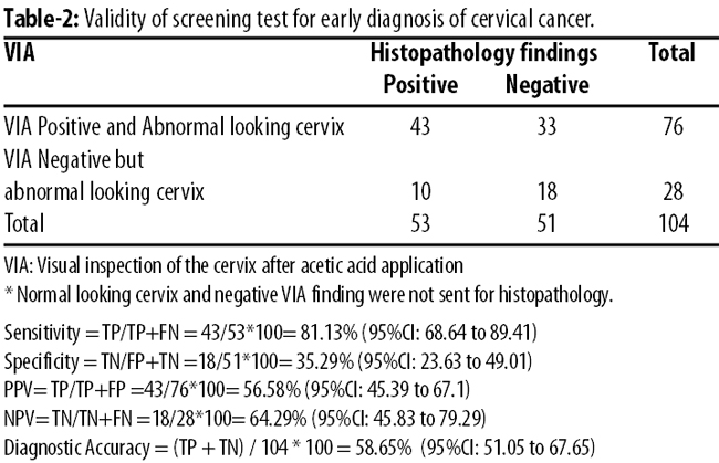
After the inclusion of all negative findings in the analysis (Table-3),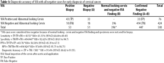
the approximate sensitivity, specificity, PPV, NPV and accuracy of VIA were 93.5%, 95.8%, 76.3%, 99%, and 95.6% respectively.
Discussion
Cervical cancer is one of the common malignancies among women worldwide.10 It is an important women's reproductive health problem, especially in developing countries.10 Each year 500,000 new cases are diagnosed worldwide with 80% of them occurring in resource-poor countries.10 A study reported 3.6% incidence of cervical cancer each year in Pakistan, affecting mainly women in fifth and sixth decades of life.10 Most of our patients presented to the clinic in advance stages.
Although repeated PAP smears have been an established screening test for this disease in the West, but the logistic requirements cannot be met in developing countries in the foreseeable future.12 Therefore VIA has emerged as good alternative to PAP smear in the developing countries.13,14
Our results suggest we can effectively screen most of the cases with cervical pre-cancer and cancer through VIA. In our study, 76(15.2%) women had positive results and 424(84.8%) had a negative results on VIA. Various studies have shown VIA to be positive in 1.7%-29% cases.5,9,15-17 This wide variation in rate is due to different criteria used in different studies because of lack of standardised criteria for positive results. In one study only 3.1% women screened were positive on VIA largely due to only distinct acetowhite areas were considered positive.18 On the other hand, another study reported 28% VIA positives as it took all grade of acetowhite as positive.19 In our study we screened in-hospital symptomatic population, and our VIA positivity rate of 15.2% was slightly higher than that found in other studies.
Sensitivity of VIA in previous studies ranged from 60%-100% and specificity from 36.4%-99.1%.9,12,15,16,20,21 While in our study, VIA was found to be highly sensitive as well as specific compared to most previous studies. Sensitivity of VIA in our study was 81% and specificity 92%. One of the reasons for such variable results is the experience and level of healthcare providers. High sensitivity of VIA reported in our study is attributed to well-trained providers as VIA is provider-dependent. This finding highlights the importance of training and experience for the performance of visual evaluation. The issue should be kept in mind before implementing VIA as a national screening programme. A uniform reproducible system for categorising and reporting VIA findings plus standard training should be provided to all healthcare providers before performing the test independently.
In our study, PPV and NPV of VIA was 76.3% and 99% respectively and accuracy was 95.6%. It is also comparable with other studies.2,9,6,15,16 Due to high NPV, women can be reassured that they are not likely to have a neoplastic cervical lesion.
The non-invasive nature and immediate results make VIA a useful screening test in developing countries like Pakistan. It would reduce the burden of work on the already burdened cytopathology unit by screening out-patients who are VIA-negative and disease-free. Thus, only patients who are VIA-positive would need to undergo further diagnostic tests.
The 'see and treat' protocol could especially be used in rural areas that are far from the cities. Though there is potential hazard of over-treatment, it has to be weighed against the cost-benefit ratio of patients lost to follow-up who eventually present with late stages of the diseases. VIA would reduce patients' anxiety since it would reduce waiting time for the results experienced with PAP smear screening test.
Conclusion
In developing countries like Pakistan, adequate coverage of the entire population by cytology-based screening programme is not at present feasible. Also, women generally are lost to follow-up. In such a situation VIA is a suitable primary screening alternative for a large population. Its high sensitivity, low costs and immediate results overcome the problem of loss-to-follow-up that occurs in cytology-based programme. As in Pakistan, no systemic screening schedule has ever been planned and this screening method will be very helpful in early detection and treatment of cervical pre-cancerous lesion and reduced maternal mortality and morbidity.
Reference
1. Blumenthal PD, Gaffikin L, Deganus S, Lewis R, Emerson M, Adadevoh S. Cervical cancer prevention: safety, acceptability, and feasibility of a single-visit approach in Accra, Ghana. Am J Obstet Gynecol 2007;196:407.e1-e9.
2. Jeronimo J, Morales O, Horna J, Pariona J, Manrique J, Rubiños J, et al. Visual inspection with acetic acid for cervical cancer screening outside of low-resource settings. Revista panamericana de salud pública 2005;17:1-5.
3. Basil JB, Horowitz IR. Cervical carcinoma: contemporary management. Obstet Gynecol Clin North Am 2001;28:727-42.
4. De Vuyst H, Claeys P, Njiru S, Muchiri L, Steyaert S, De Sutter P, et al. Comparison of pap smear, visual inspection with acetic acid, human papillomavirus DNA-PCR testing and cervicography. Int J Gynecol Obstet 2005;89:120-6.
5. Tayyeb R, Khawaja N, Malik N. Comparison of visual inspection of cervix and Pap smear for cervical cancer screening. Journal of the College of Physicians and Surgeons--Pakistan: J Coll Physicians Surg Pak. 2003;13:201-3.
6. Bhatla N, Mukhopadhyay A, Joshi S, Kumar A, Kriplani A, Pandey R, et al. Visual inspection for cervical cancer screening; evaluation by doctor versus paramedical worker. Indian J Cancer 2004;41:32-6.
7. Soutter W, Dina R. Premalignant disease of the genital tract. Gynaecology London: Churchill Livingstone, 2003; pp 569-71.
8. Doh A, Nkele N, Achu P, Essimbi F, Essame O, Nkegoum B. Visual inspection with acetic acid and cytology as screening methods for cervical lesions in Cameroon. Int J Gynecol Obstet. 2005;89:167-73.
9. Goel A, Gandhi G, Batra S, Bhambhani S, Zutshi V, Sachdeva P. Visual inspection of the cervix with acetic acid for cervical intraepithelial lesions. Int J Gynecol Obstet 2005;88:25-30.
10. Munoz N, Bosch FX, de Sanjose S, Herrero R, Castellsagué X, Shah KV, et al. Epidemiologic classification of human papillomavirus types associated with cervical cancer. N Engl J Med 2003;348:518-27.
11. Abbasi AN, Zahid S, Bhurgri Y, Ali N, Karsan F. Nasopharyngeal Carcinoma-An Update of Treatment and Acute Radiation Induced Reactions from a Tertiary-Care Hospital in Pakistan. Asian Pac J Cancer Prev 2011;12:735-8.
12. Bhatla N, Mukhopadhyay A, Kriplani A, Pandey R, Gravitt PE, Shah K, et al. Evaluation of adjunctive tests for cervical cancer screening in low resource settings. Indian J Cancer 2007;44:51.
13. Cronje H. Screening for cervical cancer in the developing world. Best Pract Res Clin Obstet Gynaecol 2005;19:517-29.
14. Szarewski A. Cervical screening by visual inspection with acetic acid. Lancet 2007;370:365-6.
15. Albert S, Oguntayo O, Samaila M. Comparative study of visual inspection of the cervix using acetic acid (VIA) and Papanicolaou (Pap) smears for cervical cancer screening. Ecancermedicalscience 2012;6:262.
16. Bomfim S, Santana-Franco E, Bahamondes L. Visual inspection with acetic acid for cervical cancer detection. Int J Gynecol Obstet 2005;88:65-6.
17. Sankaranarayanan R, Wesley R, Somanathan T, Dhakad N, Shyamalakumary B, Amma NS, et al. Visual inspection of the uterine cervix after the application of acetic acid in the detection of cervical carcinoma and its precursors. Cancer 1998;83:2150-6.
18. Megevand E, Denny L, Dehaeck K, Soeters R, Bloch B. Acetic acid visualization of the cervix: an alternative to cytologic screening. Obstet Gynecol 1996;88:383-6.
19. Belinson J, Pretorius R, Zhang W, Wu L, Qiao Y, Elson P. Cervical cancer screening by simple visual inspection after acetic acid. Obstet Gynecol 2001;98:441-4.
20. Rana T, Zia A, Sher S, Tariq S, Asghar F. Comparative Evaluation of PAP Smear and Visual Inspection of Acetic Acid (VIA) in Cervical Cancer Screening Program in Lady Willingdon Hospital, Lahore. Ann King Edward Med Univ 2010;16:S104-7.
21. Mahmud G, Tasnim N, Iqbal S. Comparison of Visual Inspection with acetic acid and Pap smear in cervical cancer screening at a tertiary care hospital. J Pak Med Assoc 2013; 63: 1013-6.
Related Articles
Journal of the Pakistan Medical Association has agreed to receive and publish manuscripts in accordance with the principles of the following committees:


