

Lubna Akhtar ( Department of Anatomy, FUMC, Islamabad, Pakistan. )
Muhammad Yunus Khan ( Department of Anatomy, CPSP Regional Center, Islamabad, Pakistan. )
Liaqat Ali Minhas ( Department of Anatomy, Rawal Medical College, Rawalpindi, Pakistan. )
February 2015, Volume 65, Issue 2
Original Article

Abstract
Objective: To assess the effects of prenatal administration of valproic acid on the survivability and day of hatching of chick embryo in comparison with age-matched controls.
Methods: The experimental study was conducted at the Department of Anatomy, Regional Centre of College of Physicians and Surgeons Pakistan, Islamabad, from February 2010 to February 2011. Fertilised chicken eggs were divided into two groups, labelled as experimental group-A and control group-B. Group-A eggs were injected with valproic acid, incubated and hatched. Group-B eggs underwent sham treatment using normal saline. The fully hatched chicks were then evaluated for the day of hatching and survivability, on hatching or on day 22 of incubation whichever was earlier. Outcome was statistically compared with the controls using SPSS 10.
Results: The two groups had 30 eggs each. In Group-A 23(76.66%) chicks hatched out, while there were 7(23.33%) dead chicks. In Group-B, 28(93.33%) chicks hatched out and 2(6.66%) were dead. Chicken embryos exposed to valproic acid in ovo showed increased mortality (p<0.001) and delayed hatching (p<0.001).
Conclusion: Prenatal exposure of chick embryos to valproic acid decreased embryo survival and also delayed hatching compared to age-matched controls.
Keywords: Valproic acid, Chick embryo, Survivability and hatching. (JPMA 65: 175; 2015).
Introduction
Epilepsy is a commonly encountered serious neurological problem faced by obstetricians, gynaecologists and primary healthcare physicians. Its importance lies in the fact that most of the epileptic women need to continue taking medication during pregnancy, since uncontrolled seizures may be harmful to the women as well as to the foetuses.
Valproic acid (VPA; 2-n-propylpentanoic acid; chemical formula: C8H16O2) was approved as an antiepileptic drug in 1967 in France.1 It is one of the most frequently-prescribed anti-epileptic drugs worldwide due to its broad spectrum and good tolerability.2,3 It is considered the drug of first choice for the treatment of generalised and focal epilepsies in adults and children. Both the usage and the therapeutic indications of VPA are increasing. Now in addition to epilepsy, it is also employed in the treatment of different pathologies, including schizophrenia,4 bipolar disorders,5 and different forms of headache.6 It is also currently under experimental and clinical investigation as an anti-cancer drug,7 in treating the patients of retinitis pigmentosa (RP),8 autoimmune neuropathies,9 and human immunodeficiency virus (HIV) infection.10
Valproic acid is used in increasing numbers of pregnant patients not just for epilepsy, but also for psychiatric diseases and migraine headaches. Previous studies have shown that the VPA readily crosses the placental barrier to the foetus.11 It has been shown to be teratogenic in animals and humans. Its human teratogenic effects have been reported since 1980.12 Teratogenic effects include neural tube defects (NTDs), heart, craniofacial features, urogenital structures and limbs leading to identification of a distinctive Foetal Valproate Syndrome.13 It has been assigned to pregnancy category D by the US Food and Drug Administration (FDA).14
Many animal studies have been carried out in order to mimic the effects of valproic acid on human embryo. Teratogenic effects of VPA have already been established and the current study was planned to investigate the effects of VPA on the survivability of chick embryos on hatching, and the day on which chicks hatched, by comparing it with the number of alive chicks on hatching in the control group and their day of hatching, because these effects are still being investigated. The chick embryo was selected as the experimental model because it offered obvious advantages such as small size, rapid embryonic development, and can be used in experiments that require synchronised embryo subjects in order to assess the extent to which all embryos are or are not equally affected by the same drug exposure.
Materials and Methods
The experimental study was carried out at the Department of Anatomy, Regional Centre of College of Physicians and Surgeons, Pakistan (CPSP), Islamabad, from February 2010 to February 2011.
It was carried out on two groups labelled as experimental Group-A and the control Group-B. The freshly laid fertilised chicken eggs, belonging to 'Rhode Island Red' breed of Gallus domesticus, were collected from Poultry Research Institute (PRI), Punjab. Cracked eggs and eggs stored for more than 03 days were excluded. The specimen was randomly selected by using the random selection table.
The egg shells were sterilised with 70% alcohol swabs. They were then placed in the racks with the blunt end facing upwards and the pointed end downwards. The eggs were left in this position for 5 to 15 minutes to let them dry. This also allowed the blastoderm to rotate and come to lie above the blunt end, which protected it from getting damaged while the drug was being injected at the lower end. The drug was injected by drilling two holes. The first hole was just one finger above the lower pointed end for injecting the drug and the second one was at the top of the blunt end to allow escape of air (which is normally present at this end) during injection of the drug, otherwise the drug would not stay inside, and leak back. By using insulin syringe, the eggs were injected with 0.4mg valproic acid in 20µl normal saline, in the yolk.14 Similarly, the same volume of normal saline was injected in the eggs of the control group. Soon after the administration of drug and normal saline, the holes in the shell were sealed with melted wax and the eggs were placed in the incubator (manufactured by Memmert Electric Company, Germany). The temperature was maintained at 38±0.5°C, relative humidity was kept between 60-70% and adequate ventilation was maintained. The day when eggs were placed in the incubator was taken as day 0. Daily, the eggs were manually rotated ½ turn twice. The chicks were allowed to develop till hatching or day 22 of incubation, whichever was earlier. The day of hatching was recorded. Afterwards, on day 22 of incubation the remaining chicks were manually taken out by breaking the shell. The number of alive and dead chicks was recorded. Statistical analysis of the data was done with SPSS 10. Chi-square test was applied to detect any significant difference in the survivability of embryos in the control and experimental groups. To detect any significant difference in the day of hatching, Fisher exact test was applied and p<0.05 was considered statistically significant.
Results
The two groups had 30 eggs each. In Group-A 23(76.66%) chicks hatched out, while there were 7(23.33%) dead chicks out of which 6(2-%) were completely formed embryo with limb deformities and failure of retraction of yolk sac into the abdominal cavity (Figure-1).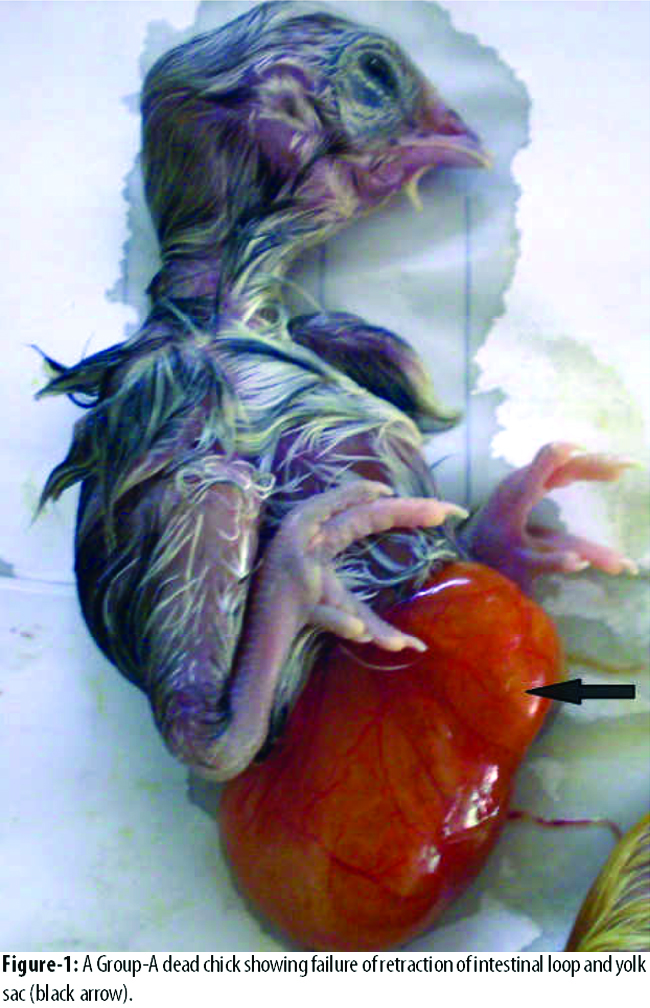
One had arrested growth at early vascularised blastoderm stage. In Group-B, 28(93.33%) chicks hatched out and 2(6.66%) were dead and had arrested growth at early vascularised blastoderm stage (Figure-2).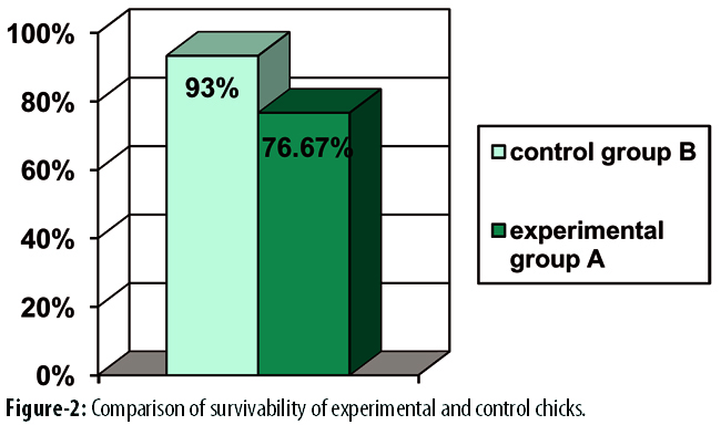
The survival rate of Group-B was significantly higher than that of Group-A (p<0.001) (Table-1).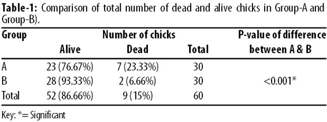
As against the normal gestational period of 21 days, 14(46.66%) Group-A chicks hatched on day 21 while 9(30%) hatched on day 22 of the incubation by themselves. The remaining 7(23.33%) were dead chicks when broken manually on day 22. In Group-B 28(93.33%) chicks hatched on day 21 by themselves, while 2(6.66%) did not hatch, and when broken on day 22, they were found to be macerated early vascularised blastoderms. Chicken embryos exposed to valproic acid in ovo showed increased mortality (p<0.001) and delayed hatching (p<0.001) (Table-2).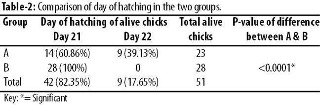
Discussion
Valproic acid is known to have teratogenic effects on the development of neural tube, heart, limbs, genitourinary system, eye, respiratory tract, abdominal wall and skin.
In the present study, the exposure of developing chick embryos of experimental Group-A to valproic acid decreased their survivability compared to control Group-B. This is in accordance with a previous study in which chicken embryos of stage 14-16 were exposed to valproic acid in ovo topically on rostral region after the removal of vitelline membrane. The chick embryos showed increased mortality when examined 24-72 hours later while also increasing the dose of VPA worsened the viability of embryos.15
Despite the fact that it is a known teratogen and is in use since 1967,4 the exact mechanism of valproic acid-induced teratogenicity and mortality is still not clear. However, different clinical and experimental studies proposed different mechanisms. In one of the previous studies, it has been shown that valproic acid interfered with embryonic folate metabolism.16 Folic acid (folate, B9) is a vitamin required for the normal cellular functions. Folate metabolism is important for the transfer of a methyl, methylene, or formyl group, and in the transformation of certain amino acids as well as in the synthesis of purines and pyrimidine needed for the synthesis of dioxyribonucleic acid (DNA). These processes are required by all the rapidly growing cells and are important for differentiation and proliferation during embryogenesis.17 Experimental studies in a number of animal species demonstrated that folate deficiency causes increase the incidence of foetal loss, intrauterine growth retardation and various congenital malformations.18 Therefore, one cause of decreased embryonic survivability could be through the interference of folate metabolism.
Vitamin A (retinol) and its oxidative metabolite, all-trans-retinoic acid affects growth, differentiation and morphogenesis of organs. Both excess and deficiency of vitamin A can cause human embryo toxicity and congenital anomalies.19 In infants, children and adults treated with either valproic acid monotherapy or with valproic acid in combination therapy significant alteration in the retinoid metabolism was noted.20 Another mechanism of embryo toxicity and teratogenicity might be high serum concentration of all-trans and 13-cis retinoic acid.
In the past, different experimental observations have shown that valproic acid affects gene environment by the inhibition of histone deacytylase (HDAC) and alteration of Wnt signalling in human and animal cells.21 HDACs are proteins with a fundamental impact on gene expression. A study found that inhibition of HDACs may result in interruption of cell proliferation, differentiation and apoptosis.22 Therefore, HDAC activity is crucial for embryonic development as is shown by the HDAC1 knockout mice which die early in development due to growth retardation and proliferation defects.23
All of these previous studies strongly suggest that valproic acid interferes with different factors that are vital for the normal development of embryo and thus resulted in the increased rate of mortality of chicks at hatching.
The normal gestational period in chick is 21 days. During normal development, on day 20 of incubation, pipping begins, embryo breaks into air cell and breathing begins; allantois ceases to function and starts to dry up, and embryo hatches on day 21 of incubation. In the present study, the cause of delayed hatching in the experimental group (A), was either the death of the chicks or diminished mobility of the chicks who survived.
Conclusion
Exposure of developing chick to valproic acid decreased the survivability of the embryos and delayed the hatching compared to age-matched controls.
References
1. Peterson GM, Naunton M. Valproate: a simple chemical with so much to offer. J Clin Pharm Ther 2005; 30: 417-21.
2. Loscher w. Basic pharmacology of valproate: a review after 35 years of clinical use for the treatment of epilepsy. CNS Drugs. 2002; 16: 669-94.
3. Gerstner T, Bell N, König S. Oral valproic acid for epilepsy-long-term experience in therapy and side effects. Expert Opin Pharmacother. 2008; 9: 285-92.
4. Horowitz E, Bergman LC, Ashkenazy C, Moscona-Hurvitz I, Grinvald-Fogel H, Magnezi R. Off-label use of sodium valproate for schizophrenia. PLoS One. 2014; 9: e92573.
5. Cipriani A1, Reid K, Young AH, Macritchie K, Geddes J. Valproic acid, valproate and divalproex in the maintenance treatment of bipolar disorder. Cochrane Database Syst Rev. 2013; 10: CD003196.
6. Kalita J, Bhoi SK, Misra UK. Amitriptyline vs divalproate in migraine prophylaxis: a randomized controlled trial. Acta Neurol Scand. 2013; 128:65-72.
7. Gotfryd K, Skladchikova G, Lepekhin EA, Berezin V, Bock E, Walmod PS. Cell type-specific anti-cancer properties of valproic acid: independent effects on HDAC activity and Erk1/2 phosphorylation. BMC Cancer 2010;10:383.
8. Clemson CM, Tzekov R, Krebs M, Checchi JM, Bigelow C, Kaushal S. Therapeutic potential of valproic acid for retinitis pigmentosa. Br J Ophthalmol 2011; 95: 89-93.
9. Zhang Z, Zhang ZY, Fauser U, Schluesener HJ. Valproic acid attenuates inflammation in experimental autoimmune neuritis. Cell Mol Life Sci 2008; 65: 4055-65.
10. Lehrman G, Hogue IB, Palmer S, Jennings C, Spina CA, Wiegand A et al. Depletion of latent HIV-1 infection in vivo: a proof-of-concept study. Lancet 2005; 366:549-55.
11. Brigs GG, Freeman RK, Yaffe SJ. Drugs in Pregnancy and Lactation: A Reference Guide to Fetal and Neonatal Risk. 8th Edition. Philadelphia: Lippincott Williams & Wilkins, 2008.
12. Brown NA, Kao J, Fabro S. Teratogenic potential of valproic acid. Lancet 1980; 1: 660-1.
13. DiLiberti JH, Farndon PA, Dennis NR, Curry CJ. The fetal valproate syndrome. Am J Med Genet 1984; 19: 473-81.
14. Peckman H. The U.S. Food and Drug Adminstration (FDA) has assigned pregnancy category D to valproate. N C Med J 2010 ; 71:396-8.
15. Whitsel AI, Johnson CB, Forehand CJ. An in Ovo Chicken Model to Study the Teratogenic Effects of Valproic acid. Teratology. 2002; 66: 153-63.
16. Wegner C, Nau H. Alteration of embryonic folate metabolism by valproic acid during organogenesis: implications for mechanism of teratogenesis. Neurology 1992; 42:17-24.
17. Czeczot H. Folic acid in physiology and pathology. Postepy Hig Med Dosw (Online). 2008; 62: 405-19.
18. Pickell L, Li D, Brown K, Mikael LG, Wang XL, Wu Q et al. Methylenetetrahydrofolate reductase deficiency and low dietary folate increase embryonic delay and placental abnormalities in mice. Birth Defects Res A Clin Mol Teratol. 2009; 85: 531-41.
19. Lee LM, Leung CY, Tang WW, Choi HL, Leung YC, McCaffery PJ et al. A paradoxical teratogenic mechanism for retinoic acid. Proc Natl Acad Sci U S A. 2012; 109: 13668-73.
20. Chuang CM, Chang CH, Wang HE, Chen KC, Peng CC, Hsieh CL et al. Valproic acid downregulates RBP4 and elicits hypervitaminosis A-teratogenesis--a kinetic analysis on retinol/retinoic acid homeostatic system. PLoS One. 2012; 7: e 43692.
21. Wiltse J. Mode of action: inhibition of histone deacetylase, altering WNT-dependent gene expression, and regulation of beta-catenin: developmental effects of valproic acid. Crit Rev Toxicol. 2005; 35:727-38.
22. Marks PA, Richon VM, Rifkind RA. Histone deacetylase inhibitors: inducers of differentiation or apoptosis of transformed cells. J Natl Cancer Inst 2000; 92:1210-6.
23. Lagger G, O\'Carroll D, Rembold M, Khier H, Tischler J, Weitzer G et al. Essential function of histone deacetylase 1 in proliferation control and CDK inhibitor repression. EMBO J. 2002; 21:2672-81.
Related Articles
Journal of the Pakistan Medical Association has agreed to receive and publish manuscripts in accordance with the principles of the following committees:


