

Huseyin Kurku ( Department of Biochemistry, Konya Training and Research Hospital, Konya, Turkey. )
Murat Kacmaz ( Department of Medical Biochemistry, Faculty of Medicine, Kirikkale University, Kirikkale, Turkey. )
Ucler Kisa ( Department of Medical Biochemistry, Faculty of Medicine, Kirikkale University, Kirikkale, Turkey. )
Ozlem Dogan ( Mardin State Hospital, Unit of Biochemistry, Mardin, Turkey. )
Osman Caglayan ( Department of Medical Biochemistry, Faculty of Medicine, Kirikkale University, Kirikkale, Turkey. )
February 2015, Volume 65, Issue 2
Original Article

Abstract
Objective: To investigate increased oxidative stress in saliva of smokers along with their serum.
Methods: The case-control study was conducted from September to December 2008 in the Department of Biochemistry of the Medical School, Kirikkale University, Kirikkale, Turkey. A blood sample and saliva samples before and after smoking were collected from the smokers, while blood and saliva samples were taken from the controls. All samples were taken concurrently. The samples were measured for total antioxidant capacity, total oxidant stress, oxidative stress index, malondialdehyde, nitric oxide and total sulfhydryl groups. Levels of superoxide dismutase and glutathione peroxidase were also measured on saliva samples. SPSS 13 was used for statistical analysis.
Results: Of the 54 subjects in the study, 27(50%) were smokers with a mean age of 28.4±5.42 years, and 27(50%) were controls with a mean age of 29.7±8.03 years. Total oxidant stress, oxidative stress index, malondialdehyde and nitric oxide levels were found higher in the serum samples of smokers (p<0.05), and the levels of total sulfhydryl groups in smokers were lower compared with the controls (p<0,05). Among the smokers, salivary malondialdehyde levels were higher before and after smoking (p<0.05), glutathione peroxidase levels were lower than the controls, and salivary nitric oxide levels after smoking were higher than both those of the control group and the levels before smoking (p<0.05).
Conclusion: Both acute and chronic increased oxidative conditions may be a significant sign of the destructive effects of smoking. The investigation of disorders in smokers concerning oxidative stress will be beneficial in terms of novel approaches and treatment modalities.
Keywords: Smoking, Saliva, Oxidative stress, Total antioxidant capacity. (JPMA 65: 164; 2015).
Introduction
Cigarette smoke is the most known burnt chemical with organic qualities inhaled into the lungs. It is commonly accepted that the lungs of smokers are exposed to increased levels of oxidants. Free oxygen radicals and oxidants arise after smoking and play a significant role in the pathogenesis of quite a few diseases such as severe tissue damage, cancer, cardiovascular diseases, diabetes mellitus, lung diseases, kidney diseases, rheumatoid arthritis, cataract and nerve tissue disorders.1
Cigarettes include a number of chemicals such as aldehydes, phenols, hydrocarbons, nitric oxide (NO), and quinone and semiquinone radicals. These chemicals directly or indirectly lead to the formation of free radicals caused by oxygen.2-4
Lipid peroxidation caused by the attack of free radicals to unsaturated fatty acids in the cell membrane results in the formation of conjugated dienes and some toxic aldehyde products. Malondialdehyde (MDA) is the most commonly recognised one among these products.2,3
It is expected that oxidative stress (OS) occurring due to smoking leads to some changes in plasma antioxidant defence system. In both serum and erythrocyte in smokers, MDA levels were found to be higher, while erythrocyte superoxide dismutase (SOD) levels were lower. Total sulfhydryl groups (t-SH) constitute a significant part of antioxidant defence against free radicals. Therefore, another goal of free radical damage is the groups of sulfhydryl based on protein and soluble.3,5
A number of studies point out the role of oxidant/antioxidant balance in physiological state and disease pathogenesis. It is claimed that in the pathogenesis of the diseases where smoking is influential, it will be useful to focus on smoking in terms of oxidant/antioxidant balance as regards new approaches and treatment varieties.5,6
The basic functions of saliva can be summarised as lubricating the mucosa in the mouth, facilitating chewing, washing the food residue and bacteria through mechanical means, solving food, facilitating the process of swallowing and digestion, regulating the pH of the oral cavity and antimicrobial effect. In recent studies, it has been reported that saliva contributes to oral mucosa repair with the help of the epidermal growing factors it involves and to haemostasis in the oral cavity through proteins.6
The present study aimed at investigating OS in smokers since smoking is a common activity, and the complications based on this activity are frequently seen. If the oxidative mechanisms of some effects of smoking can be accounted for, we considered that the antioxidant defence system of the body could be strengthened in order to avoid the harmful effects of smoking.
Subjects and Methods
The case-control study was conducted from September to December 2008 performed in the Department of Biochemistry of the Medical School, Kirikkale University, Kirikkale, Turkey. While picking up the subjects and matching controls, the World Health Organisation (WHO) criteria was used.7 The individuals in the smoking group were regular daily smokers, and the controls were life-long non-smokers or those with no smokers among their close environments (family, social and work). Those with a previously known chronic disease like diabetes, hypertension or kidney disease, drug users, any malignancies, pregnant women and those with anaemia or any complaint or disease concerning the oral cavity and passive exposure to non-smoker\'s were excluded.
In smokers, both morning fasting blood and saliva samples before smoking were concurrently collected, and a saliva sample was collected 1h after smoking only one cigarette. Until all saliva samples in the study and control groups, including post-smoking, were collected, no subject ate or drank anything. Fasting blood and saliva (control saliva sample) samples were also collected simultaneously from the participants in the control group. Venous blood samples drawn were centrifuged for 5 minutes after coagulation at 3000g to separate the sera. To collect saliva samples, the participants were asked to chew a piece of previously sterilised natural cotton for 2 min, and the wet cotton was squeezed with a sterilised injector. Having been taken into a glass tube, the saliva samples were centrifuged at 3000g for 5 minutes, and the supernatant liquid was stored at -80°C until the analysis.
When collecting samples from the study sample, data concerning age, gender, duration of smoking if they smoked, how many cigarettes they smoked a day were recorded. The number of cigarettes smoked (packet/day) was multiplied by the number of years to calculate smoking in terms of packet/year and the results were recorded. An approval was obtained from the ethical board of the Medical School of Kirikkale University, Turkey.
The levels of MDA, NO, t-SH, total oxidant stress (TOS), total antioxidant capacity (TAC) in serum and saliva samples, and SOD (EC 1.15.1.1) and glutathione peroxidase (EC 1.11.1.9) (GSH-Px) in saliva samples were examined. Oxidative stress index (OSI) was calculated using TAC and TOS results. MDA was manually studied by modifying Yagi\'s method;8 NO was manually studied by modifying the method inliterature,9 and t-SH levels were also manually studied by modifying the method outlines in literature.10 TOS, TAC, SOD and GSH-Px levels were measured by adjusting related commercial kits to chemical autoanalyser (Roche/Hitachi Modular Analytics System, Modular P800 Module, Roche Diagnostics).
Erel\'s method was used to measure TOS.11 Standard series from H2O2 was prepared using total antioxidant stress Rel Assay Diagnostics commercial kits (Mega Tip San, Gaziantep, Turkey), and the results were calculated in terms of mmol H2O2Equiv/L.
Erel\'s method was used for the measurements of TAC.12 Standard series from glutathione (GSH) was prepared using TAC Rel Assay Diagnostics commercial kits (Mega Tip San, Gaziantep, Turkey), and the results were calculated in terms of mmol GSH Equiv/L.
SOD level was measured using RANSOD commercial kits (RANDOX, Antrim, England). GSH-Px level was measured using RANSEL commercial kits (RANDOX, Antrim, England). Saliva protein levels were measured, and SOD and GSH-Px results were calculated in terms of U/mg protein.
The TOS indices of the samples were compared with TAC values, and OS indices were obtained. When calculating OSI, TAC concentrations were converted into mol GSH Equiv./L; the indices were calculated using the formula OSI = (TOS = mol H2O2 Equiv/L) / (TAC = mMol GSH Equiv/L x 1000) x 100.
Statistical analysis was carried out using SPSS 13. Student t test was used to compare the data between the smokers and the controls; paired samples t test was used to compare pre- and post-smoking saliva samples, and Pearson\'s correlation was used to examine the relationship between the parameters of both groups. P was considered significant at <0.05.
Results
Of the 54 subjects in the study, 27(50%) were smokers with a mean age of 28.4 5.42 years, and 27(50%) were controls with a mean age of 29.7 8.03 years. Of the cases, 15(55.5%) were men, while among the controls, there were 14(51.8%) men. No difference was found between smokers and non-smokers in terms of age and gender (p>0.05 each).
The mean duration of smoking was 10.6±6.9 years; mean amount of cigarette smoking was 16.1±8.0) cigarettes/day and 9.4±8.0 pack/year. A positive correlation was found between the total duration of smoking and three parameters; that is, smokers\' serum OSI levels, pre-smoking salivary TAC levels and post-smoking salivary t-SH levels (r2=0.316, p=0.108; r2=0.435, p<0.05; and, r2=0.341 p=0.056, respectively). TAC, TOS, OSI, MDA, NO and t-SH values of smokers and the controls were kept separately (Table-1).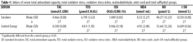
TOS, OSI, MDA and NO levels were found higher in the serum samples of smokers (p<0.05), and the levels of t-SH in smokers were lower compared with the controls (p<0.05). Among the smokers, salivary MDA levels were higher before and after smoking (p<0.05) (Table-2).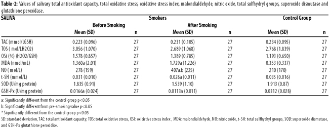
Salivary MDA levels in smokers after smoking was slightly higher than the pre-smoking saliva values, but the difference was statistically insignificant.
Salivary NO levels before smoking in the smokers was slightly higher than those of the controls, but the difference was statistically insignificant (p=0.139). Smokers\' post-smoking salivary t-SH levels were significantly lower than the control group\'s levels (p<0.05).
Smokers\' pre-smoking and post-smoking salivary GSH-Px levels were significantly lower than the control group\'s salivary GSH-Px levels (p<0.05).
No significant difference was detected between the smokers and the controls in terms of their SOD levels. Smokers\' post-smoking salivary OSI levels were slightly higher than those of the control group, but the difference was statistically insignificant (p=0.072).
There was a positive correlation between smokers\' serum TAC levels and pre-smoking and post-smoking salivary TAC levels (r2=0.319; p=0.10). Furthermore, there was a positive correlation between serum NO, and post-smoking salivary NO levels, serum OSI, pre-smoking and post-smoking salivary MDA levels (r2=0.391, p<0.05; r2=0.453, p<0.05; and, r2=0.278 p=0.16, respectively).
Discussion
Smoking is claimed to be a significant factor in the development of OS due to elevated levels of free radicals and to play a role in the pathogenesis of quite a few diseases.5 The antioxidant materials found in cigarette smoke are the sources of oxygen radicals, and they are highly influential in the development of cigarette-related diseases. Cigarette smoke degrades oxidant-antioxidant balance of the organism in favour of the oxidance.13,14
In quite a few previous studies, it was reported that smoking significantly increases serum and plasma MDA levels. In several studies, erythrocyte MDA levels also increased along with serum/plasma MDA levels. In one study it was found that erythrocyte GSH-Px was significantly lower in smokers; erythrocyte SOD was the same in both groups; but MDA was significantly higher in both serum and erythrocyte in smokers.15
Another study found that salivary MDA levels were higher in smokers compared with controls, and it noted that the more this level increases, the more the amount of smoking increases.16 In the present study, smokers\' pre- and post-smoking salivary MDA levels were significantly higher than those of the controls. Salivary MDA levels in smokers after smoking were slightly higher than the values before smoking, but the difference was insignificant. In the light of this finding, we consider that lipid peroxidation develops chronically.
Though a few studies report contradictory findings, our study, consistent with many in the literature, suggests that depending on the fact that cigarette smoke increases polymorphonuclear leucocytes (PMNL) activation, membrane lipid peroxidation occurs. Hence, it may be suggested that MDA level increases as an indication of the mechanism.
Different studies are present in literature investigating TAC, TOS and OSI levels in smokers, and reporting variable findings. While some studies indicate that TAC levels are decreased17 or same,6 others reveal that TOS and OSI levels are generally increased.17
In the present study, no significant difference was found between TAC and TOS levels in the the saliva of smokers and controls. However, one study found that salivary TAC levels were lower in both smokers and in the cases of periodondit and oral lichen planus, compared with controls.18 In our study, OSI values as a sign of OS were found to be generally higher in smokers. This indicates that oxidant load of smokers increase. The measurement of TAC level, however, gives an idea about the whole antioxidant activity in any sample. We assumed that the reason why TAC level remained unchanged is that the organism tries to balance the OS even if the balancing capacity of OS is limited. This is because the participants in our study were relatively young and moderate smokers. However, a study found that TAC was lower in cancer patients who were smokers than in those without cancer.19 These results suggest that smoking can be compensated more easily at initial stages. That smoking can be compensated more easily at initial stages is also consistent with our findings. A study performed in babies reported that low levels of TAC may indicate weaker antioxidant potentials, and babies are unable to compensate for OS.
NO is known to control vascular tonus in salivary glands during rest and following autonomous nerve stimulation. There is increasing evidence for that NO is released into saliva, and saliva might play a physiological part in antibacterial impact of saliva and detoxification of oral carcinogens.
Previous studies reported an increase in NO levels of smokers in serum and respiratory air. A study on respiratory air found that NO levels of smokers in respiratory air was significantly lower than those of controls, but their nitrite + nitrate level was significantly higher. According to pre-smoking values, nitrite + nitrate levels at minute 30 after smoking two cigarettes were significantly higher, but the levels at minute 90 after smoking were similar to pre-smoking values.20 The nitrite + nitrate levels reported are parallel to those in our study.
One study found that serum NO concentration was significantly higher in smokers.21 Another study found no difference in serum NO concentrations of pregnant smokers, but found a difference in cord bloods of pregnant smokers.22
Findings suggest that salivary NO is released against oxidant matter for a short time in a similar way to respiration air NO and becomes effective. These results also indicate that NO is effectively and quickly released as a compensatory element against OS, but it disappears from the medium rather quickly due to its short half-life.
In previous studies, controversial findings related to serum, salivary and erythrocyte t-SH levels were reported. These levels were found to be significantly lower in line with our findings,17 higher6 or unvariable in smokers.23
While finding levels of serum MDA to be higher in smokers, one study found t-SH as unvariable, and also stated that the lack of change in t-SH level might be due to compensatory mechanisms, and that t-SH level should be suspected as an indication of OS.3 However, consistent with our findings, one study found that erythrocyte glutathione levels were significantly low in smokers, and deoxyribonucleic acid (DNA) double-strand breaks were significantly high.24
Glutathione is highly significant in the inactivation of free radicals, and according to our results, t-SH level decreases in contrast to increased oxidant load in smokers. This decrease might be associated with mutations in smokers due to DNA double-strand break, as in an earlier study.24
In previous studies, it was found that compared serum, salivary and erythrocyte SOD and GSH-Px levels in smokers with those of controls, SOD was decreased5,25 or remained the same,15 and GSH-Px decreased15,25 or remained invariable.5
In the present study, in line with low levels of t-SH, salivary GSH-Px levels were low as well. This similarity clearly indicates the importance of glutathione in OS caused by smoking and how the metabolism of glutathione is affected. GSH-Px particularly affects hydrogen peroxites and fatty-acid hydroperoxides. In such antioxidative reactions, glutathione is used as a reductive molecule. Depending on the increase in the number of oxidative molecules in smokers this reaction accelerates, and we consider that glutathione decreases due to the excessive use of glutathione, and that a decrease in glutathione level also leads to a decrease in the activity of GSH-Px, as well as the increase in free radicals in the medium.
SOD, however, turns superoxide into hydrogen peroxide (H2O2) and molecular oxygen and is also significant for phagocytic function of granulocytes. SOD activity is abundant in tissues consuming oxygen much and in PMNL. Therefore, by being compensated, SOD activity is conserved against OS in smokers and may be considered to fulfil its function without a change in its level. It is assumed that SOD levels might only be affected in long-term and heavy smokers.
Saliva is the first line confronting cigarette smoke. The antioxidant system of saliva plays a significant role in the anti-carcinogenic and anti-bacterial effect of saliva.26 Our results indicate that oxidant-antioxidant balance of saliva is degraded in favour of oxidants in smokers. The degradation of the balance may contribute to deterioration of mouth hygiene and oral cancer development in smokers.
The oxidant-antioxidant balance is affected by a number of personal factors such as environment, diet, physical activity, lifestyle and metabolism. Future studies should be based on more standardisation in these factors, and the oxidant-antioxidant balance should be evaluated as a whole rather than assessing only on a molecular basis.
Conclusion
Saliva samples can also be useful in showing oxidant-antioxidant balance in smokers. Given the high performance of saliva in demonstrating the negative impact of smoking both locally and systematically, saliva that is used routinely to measure few parameters may also be a good sample type to be preferred in the diagnosis and monitoring of other pathologies in the organism.
References
1. Rahman I, MacNee W. Oxidant/antioxidant imbalance in smokers and chronic obstructive pulmonary disease. Thorax 1996;51:348-50.
2. Church DF, Pryor WA. Free - radical chemistry of cigarette smoke and its toxicological implications. Environ Health Perspect 1985;64:111-26.
3. Demir S, Özkurt S, Köseo?lu M, Enli Y, Aslan D, Gümü?su N. Sigara içenlerde plazma lipid peroksidasyonu. Solunum 2001;3:57-9.
4. Van der Vaart H, Postma DS, Timens W, ten Hacken NH. Acute effects of cigarette smoke on inflammation and oxidative stress: a review. Thorax 2004;59:713-21.
5. Sekeroglu MR, Aslan R, Tarakçio?lu M. Sigara kullananlarda lipid peroksidasyonu ve antioksidan aktivite. Tüberküloz ve toraks 1997;45:105-9.
6. Zappacosta B, Persichilli S, De Sole P, Mordente A, Giardina B. Effect of smoking one cigarette on antioxidant metabolites in the saliva of healthy smokers. Arch Oral Biol 1999 ;44:485-8.
7. World Health Organization. Guidelines for controlling and monitoring the tobacco epidemic. World Health Organization; 1998.
8. Yagi K. Lipid peroxides in hepatic, gastrointestinal, and pancreatic diseases. Adv Exp Med Biol 1994;366:165-9.
9. Miranda KM, Espey MG, Wink DA. A rapid, simple spectrophotometric method for simultaneous detection of nitrate and nitrite. Nitric Oxide 2001;5:62-71.
10. Sedlak J, Lindslay RH. Estimation of total, protein-bound, and nonprotein sulfhydryl groups in tissue with Ellman\'s reagent. Anal Biochem 1968;25:192-205.
11. Erel O. A new automated colorimetric method for measuring total oxidant status. Clin Biochem 2005;38:1103-11.
12. Erel O. A novel automated direct measurement method for total antioxidant capacity using a new generation, more stable ABTS radical cation. Clin Biochem 2004;37:277-85.
13. Ergün A. Sigara ve sistemik etkileri T. Klin J Med Sci 1998;18:159-63.
14. Yamaguchi Y, Nasu F, Harada A, Kunitomo M. Oxidants in the gas phase of cigarette smoke pass through the lung alveolar wall and raise systemic oxidative stress. J Pharmacol Sci 2007;103:275-82.
15. Gültekin F, Gökduman A, Özgüner F, Akdo?an M, Sütçü R, Deliba? N. Changes in the antioxidant defense system of the smokers. Tuberk Toraks 2001;49:259-64.
16. Kurtul N, Gökpinar E. Salivary lipid peroxidation and total sialic acid levels in smokers and smokeless tobacco users as Mara? powder. Mediators Inflamm. 2012;2012:619293.
17. Aycicek A, Ipek A. Maternal active or passive smoking causes oxidative stress in cord blood. Eur J Pediatr 2008;167:81-5.
18. Miricescu D, Greabu M, Totan A, Didilescu A, R?dulescu R. The antioxidant potential of saliva: clinical significance in oral diseases. Therap Pharmacol Clin Toxicol 2011:15:139-43.
19. Nagamma T, Anjaneyulu K, Baxi J, Dayaram P, Singh PP. Effects of cigarette smoking on lipid peroxidation and antioxidant status in cancer patients from Western Nepal Asian Pacific. J Cancer Prev 2011;12:313-6.
20. Balint B, Donnelly LE, Hanazawa T, Kharitonov SA, Barnes PJ. Increased nitric oxide metabolites in exhaled breath condensate after exposure to tobacco smoke. Thorax 2001;56:456-61.
21. Chávez J, Cano C, Souki A, Bermúdez V, Medina M, Ciszek A et al. Vargas ME. Effect of cigarette smoking on the oxidant/antioxidant balance in healthy subjects.R, Valasco M. Am J Ther. 2007;14:189-93.
22. Aydogan U, Durmaz E, Ercan CM, Eken A, Ulutas OK, Kavuk S et al. Effects of smokingduring pregnancy on DNA damage and ROS level consequences in maternal and newborns\' blood. Arh Hig Rada Toksikol 2013;64:35-46.
23. Duthie GG, Arthur JR, James WP. Effect of smoking and vitamin E on blood antioxidant status. Am J Clin Nutr 1991;53:1061-3.
24. Dinçer Y, Saygili E?, Akçay T. Influence of smoking on DNA damage and blood glutathione level. T Klin Tip Bilimleri 2003;23:108-11.
25. Abdolsamadi HR, Goodarzi MT, Mortazavi H, Robati M, Ahmadi-Motemaye F. Comparison of salivary antioxidants in healthy smoking and non-smoking men. Chang Gung Med J 2011;34:607-11.
26. Nagler RM, Reznick AZ. Cigarette smoke effects on salivary antioxidants and oral cancer-novel concepts. Isr Med Assoc J 2004;6:691-4.
Related Articles
Journal of the Pakistan Medical Association has agreed to receive and publish manuscripts in accordance with the principles of the following committees:


