

Nadeem Afzal ( Department of Immunology, University of Health Sciences, Lahore, Pakistan. )
Shakeela Zaman ( Children Hospital & Institute of Child Health, Lahore, Pakistan. )
Faheem Shahzad ( Department of Immunology, University of Health Sciences, Lahore, Pakistan. )
Khursheed Javaid ( Department of Immunology, University of Health Sciences, Lahore, Pakistan. )
Abu Zafar ( Amin Hayat Memorial Diabetic Centre, Lahore, Pakistan. )
Abdul Hanan Nagi ( Children Hospital & Institute of Child Health, Lahore, Pakistan. )
February 2015, Volume 65, Issue 2
Original Article

Abstract
Objective: To enumerate CD4+CD25+ Treg cells and determine serum IL-6 and IL-17 in type 2 diabetes mellitus patients with retinopathy.
Methods: The case-control study was conducted at the Department of Immunology, University of Health Sciences, Lahore, from November 2009 to January 2012 and comprised diabetic patients and healthy controls who were divided into three groups. Group 1 had controls, while Group 2 had diabetic patients without retinopathy and Group 3 had diabetic patients with retinopathy. Flowcytometre and enzyme-linked immunosorbent assay were used for CD4+CD25+ Tregs and serum IL-6 and IL-17 respectively. SPSS 20 was used for statistical analysis.
Results: Of the 212 subjects in the study, 30(14%) were Group 1, 30(14%) in Group 2 and 152(72%) in Group 3. There were 25(83%) women in Group 2 and 101(66%) in Group 3 compared to 9(30%) in Group 1. Higher mean age was in Group 3 (50.88±8.9 years) and Group 2 (49.46±9.94 years) compared to Group 1 (34.66±8.78 years) while longer mean disease duration was in Group 3 (10.51±5.24 years) than Group 2 (7.76±4.14 years). Highest median ratio of IL-6 was in Group 1 (1468.62) (Q1-Q3: 1229.9-1543.35), followed by Group 2 (1455.32) (Q1-Q3:1214.22-158.9) and Group 3 (469.84) (Q1-Q3: 206.53-1231.33) whereas IL-17 was the highest in Group 1 (339.38) (Q1-Q3: 159.89-1174.93), followed by Group 3 (216.60) (Q1-Q3: 141.87-410.25) and Group 2 (174.17) (Q1-Q3: 138.77-458.17). Higher percentage of Tregs was in Group 2 (3.07±0.43) followed by Group 1 (2.91±0.04) and Group 3 (2.88±0.38). Significant difference was observed in gender, age, disease duration, level of IL-6 and IL-17 (p<0.05 each), while no difference was found in glycated haemoglobin , CD4+CD25+ and Tregs (p>0.05 each).
Conclusion: Age, gender and duration of diabetes contributed to diabetic retinopathy, while CD4+CD25+ T cells and Treg cells did not. Serum IL-6 and IL-17 were inversely associated with diabetic retinopathy.
Keywords: T cells, Treg cells, Cytokine, Autoimmunity, Diabetes mellitus. (JPMA 65: 159; 2015).
Introduction
Diabetes mellitus (DM) is a chronic disease which leads to many complications such as retinopathy. Type-2 diabetes mellitus (T2DM) is more common and is one of the leading causes of morbidity and mortality. World prevalence of diabetes was 285 million in 2010, and may increase to 439 million by 2030. More than 80% of diabetics are in low and middle income countries.1,2 Pakistan had 6.9 million diabetics and by 2025 there could be >11.5 million people with diabetes.3
Currently, children of 8 years or younger are having T2DM which is attributed to obesity, and changes in diet and lifestyle. In 20 years, T2DM would be 60% of non-communicable diseases of developing world.4 Asians are less obese, less overweight and have low body mass index (BMI) compared to Western people, but have higher prevalence of diabetes. Increased childhood obesity and particularly abdominal obesity in Asians are the reasons behind T2DM hike.5 In diabetes, hyper-reactive platelet interaction with damaged vessels causes micro-thrombosis of small vessels that leads to diabetic retinopathy (DR) which is a horrifying prospect. Number of people at risk of developing DR would double in 30 years.6
Individuals with increased white blood cell (WBC) count and raised inflammatory markers i.e. IL-6 and C-reactive protein (CRP), are likely to develop T2DM. Aberrant expression of human leukocyte antigen-DR and DQ on retinal cells, presence of anti-pericyte and anti-endothelial cell antibodies deposition of immuno globulins, monocytes, complement proteins, and T cells, suggests an autoimmune process in DR.7
T regulatory (Treg) cell, a subset of CD4+ T cells maintain peripheral tolerance and down-regulate antigen-specific responses by secreting certain cytokines. Tregs express CD25 and Foxp3 which is a master control gene. In humans, CD4+CD25+ T cells repertoire is quite diverse. Insufficient suppression of inflammatory process or transformed Treg cells initiate autoimmune disease. Many markers are used for identification of Tregs e.g. CTLA-4, CD62L (L-selectin), CD134 (OX40), IL-2 receptor alpha chain (CD25), etc.
Th17, a subclass of T cell, produces IL-17 which requires IL-6 and Transforming growth factor beta (TGF-beta) for its initiation and IL-23 for its maintenance. Natural Killer T cells express IL-23 receptor and produces IL-17 which is independent of IL-6. IL-17 Foxp3 Tregs contribute in antimicrobial defence and checks autoimmunity. Th17 has been associated with autoimmunity, cancer, and type-1 diabetes (TID). Increased level of IL-17 in TID is detrimental to islet cells. Similarly, IL-17 is increased in active uveitis and it decreases after treatment. Interferon gamma (INF-g) produces IL-27 in target tissue and therefore alleviates uveitis by antagonising Th-17.8,9
Anti-IL-6 receptor antibody (Taclomide) is used as treatment in rheumatoid arthritis. Likewise, IL-17/IL-23 axis can be used for diabetic complications and autoimmunity. Exact cause of DR is not known and immune involvement is questionable. The current study was planned to enumerate Tregs cells and to determine serum IL-6 and IL-17 in DR patients of T2DM.10
Subjects and Methods
The case-control study was conducted at the the Department of Immunology, University of Health Sciences, Lahore, from November 2009 to January 2012 and comprised diabetic patients and healthy controls who were divided into three groups. Group 1 had controls, while Group 2 had diabetic patients without retinopathy and Group 3 had diabetic patients with retinopathy.
After approval by the institutional ethics committee, the subjects were selected using simple random sampling technique. While calculating the sample size, confidence interval (CI) was set at 95% with 5% margin of error and the value for anticipated proportion of type-II DR population was 15.7%. Subjects of either gender between 20 and 75 years of age and having diabetes of 5-25 years were selected. Subjects having infection in the preceding two weeks and chronic infection like tuberculosis (TB) and autoimmunity were excluded.
While collecting samples, glycated haemoglobin (HbA1c) and duration of diabetes was noted. Eye examination was performed by an ophthalmologist.
CD4+CD25+ Tregs were enumerated by flowcytometre FACS Calibur (Becton Dickinson) using monoclonal antibodies (Becton Dickinson). Venous blood (2-3ml) in ethylene diamine tetraacetic acid (EDTA) tube was collected. Further procedure was in line with literature11 except the washing step. Lyse-wash method on whole blood was used. Further, 20ul of fluorescein isothiocyanate (FITC) tagged with CD4, phycoerythrin (PE) with CD25 and peridinin-chlorophyll-protein (PerCP) with CD45 cells were added to one tube and 20ul of isotype control to other. The instrument was calibrated and fluorescent signal compensation was performed using CellQuest Pro software (BD) and Calibrite beads (Becton Dickinson). From CD4+CD25+ T cells, percentage of Tregs was calculated according to literature.12
IL-6 and IL-17 were detected using enzyme-linked immunosorbent assay (ELISA) according to the instructions of the kit manufacturer (KOMABIOTECH Inc, Korea) and plates were read at microtiter plate reader (BioRad, USA). The manufacturer claimed no cross-reactivity of cytokines.
Data was analysed using SPSS 20. Mean ± standard deviation (SD) for quantitative variables, frequencies and percentages for qualitative variables were worked out. Normality of the data was assessed by using Shapiro Wilk test. Median and interquartile range (IQR) were given for data that was not normally distributed i.e. IL-6 and IL-17. One way analysis of variance (ANOVA) and Kruskal Wallis H test were used to observe group mean differences, Post Hoc Tukey test and Mann-Whitney U test with Bonferroni correction were used for pair-wise comparisons. Pearson Chi-Square test for associations between qualitative variables was applied. Multinomial logistic regression was performed to assess the association of a number of factors with the disease. P<0.05 was considered statistically significant.
Results
Of the 212 subjects in the study, 30(14%) were Group 1, 30(14%) in Group 2 and 152(72%) in Group 3. There were 25(83%) women in Group 2 and 101(66%) in Group 3 compared to 9(30%) in Group 1 (Table-1).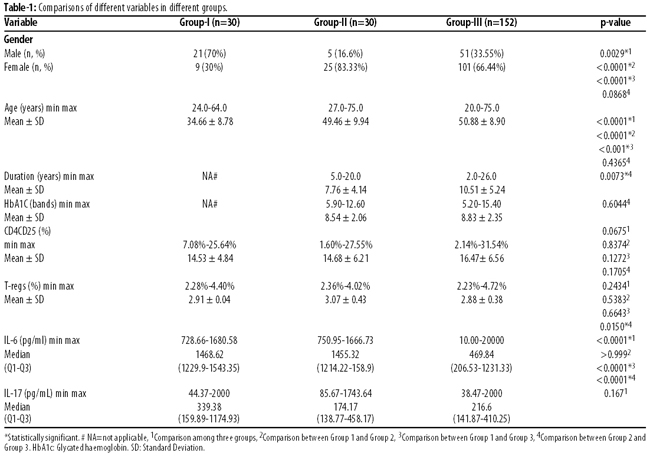
Higher mean age was in Group 3 (50.88±8.9 years) and Group 2 (49.46±9.94 years) compared to Group 1 (34.66±8.78 years). There was significant difference among the three groups, between Group 1 and Group 2, and between Group 1 and Group 3 (p<0.0001 each) and no significant difference between Group 2 and Group 3 (p>0.05).
Longer mean disease duration was in Group 3 (10.51±5.24 years) than Group 2 (7.76±4.14 years) (p=0.007). Highest median ratio of IL-6 was in Group 1 (1468.62) (Q1-Q3: 1229.9-1543.35), followed by Group 2 (1455.32) (Q1-Q3:1214.22-158.9) and Group 3 (469.84) (Q1-Q3: 206.53-1231.33) whereas IL-17 was the highest in Group 1 (339.38) (Q1-Q3: 159.89-1174.93), followed by Group 3 (216.60) (Q1-Q3: 141.87-410.25) and Group 2 (174.17) (Q1-Q3: 138.77-458.17). Higher percentage of Tregs was in Group 2 (3.07±0.43) followed by Group 1 (2.91±0.04) and Group 3 (2.88±0.38) (p=0.015). Significant difference was observed in gender, age, disease duration, level of IL-6 and IL-17 (p<0.05 each), while no difference was found in HbA1c, CD4+CD25+ and Tregs (p>0.05 each).
On using multinomial regression model, significant difference was found in terms of age and female gender (p<=0.001 each) between Group 1 and Group 2. Age, IL-6 and female gender was significantly different between Group 1 and Group 3 (p=0.000, p=0.032 and p=0.03 respectively) (Table-2).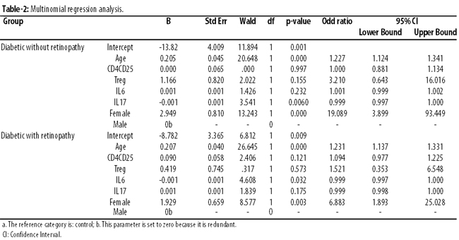
Discussion
The study found a significant difference, on comparison of gender, among the three groups, between Group 1 and Group 2, and between Group 1 and Group 3, but no significant difference between Group 2 and Group 3. This could be so because both groups had more females compared to males. The study is in agreement with earlier ones,5,13 but not with the one that documented higher prevalence of diabetes in males compared to females.3
In the current study, higher mean age was in Group 2 and Group 3 compared to Group 1. On comparison there was significant difference among the groups but no significant difference in terms of age was detected between Group 2 and Group 3. This could be because both groups had diabetic patients and DR might be due to other factors, like environmental.2 The current study is in agreement with several studies,1,2,5,13 while it differs from one study.14
On comparison of HbA1c between Group 2 and Group 3, there was no significant difference in our study, which is in line with some studies,5,13 but another study14 has reported differently. For the current study, diabetic patients were recruited from public hospital/non-governmental organisations (NGOs), and they were from low socio-economic background and their education level was not high which could be the reason for poor diabetes control as suggested earlier.15
Regarding duration of diabetes, our study had significant difference in two groups and it is in agreement with some studies13,14 but others have differed.5,6
On comparison of CD4+CD25+ T cells, no significant difference was observed. There are studies16 in agreement but such studies should not be compared with the current work because none of them involved T2DM patients. One study17 has reported different results in this context. The current study shows that these cells are probably not involved in DR.
Higher percentage of Tregs was in Group 2 compared to Group 3, but there was no significant difference. One study17 is in agreement with the current work, while others12,16 are not. However, none of these studies involved T2DM patients. There are very few studies on Tregs in T1D18 and T2DM19 patients and a possible explanation could be that Treg cells are not associated with DR in T2DM.
In the current study, on comparison of IL-6, there was significant difference among the three groups, between Group 1 and Group 3, and between Group 2 and Group 3, while there was no significant difference between Group 1 and Group 2. Many researchers suggested involvement of IL-6 in eye diseases e.g. uveitis,9 T1D retinopathy and proliferative DR.20 However, none of these studies were in T2DM. In the current work, low IL-6 in DR could be due to their treatment i.e. laser/local eye drops and medicines which may reduce inflammation because a study10 documented suppression of IL-6 reduced inflammation.
In the current study, increase of IL-6 in healthy controls and diabetics without retinopathy could be the normal level of IL-6. Further, individuals with high level of inflammatory markers may get diabetes in later life.7 Although, infection in the preceding two weeks was part of the exclusion criteria, but sub-acute infection could cause low level of IL-6 in DR. Additionally, poorly-controlled diabetics have raised IL-6 and normalisation of plasma glucose causes its decrease.21
Increase of IL-6 in controls could be a protective aspect of IL-6 which was confirmed in non-obese diabetic (NOD) mice who over-express IL-6 and have decreased level of fasting glucose with delayed onset of diabetes.18 Protective aspect of IL-6 polymorphism has been highlighted in Retinopathy and nephropathy of T1D.20 Elevated level of serum IL-6 and its association with raised insulin levels at fasting and after meals have been suggested.21,22
In the current study, on comparison of IL-17 there was no significant difference. In literature there are studies that have highlighted protective aspect of IL-17: it recruits protective INF-g CD4+ T cells into lung, is produced in lung as host response to Klebsiella pneumonia and mycobacterium tuberculosis, neutralisation of IL-17 enhanced eosinophil in asthma, while recombinant IL-17 decreases airway hyperactivity,23 and IL-17 producing cells in oesophageal squamous cell carcinoma have prognostic value.24 However, none of these studies was performed in diabetic patients. IL-17 is initiator of T cell dependent inflammation and causes lupus nephritis and autoantibodies production.25
Some studies8,14 are not in agreement and have documented increase of IL-17 in T2DM, T1D, Behcet disease and antineutrophil cytoplasmic antibody (ANCA) associated vasculitis.
In the current study, age of the participants was not matched and there was a gender bias as majority of the participants of Groups 2 and 3 were females compared to the control group. Patients having diabetes between 5-25 years duration were included in the study, but the duration of diabetes was unequal in the two diabetic groups.
Conclusion
Age, gender, and Treg cells contributed, while CD4+CD25+ T cells, IL-6 and IL-17 did not contribute towards T2DM retinopathy. Differences in CD4+CD25+ T cells, Treg cells, IL-6 and IL-17 could be due to disease manifestations. Functional assays of CD4+CD25+ T cells and more reliable markers of Treg cells i.e. Foxp3 or CD127 should be performed in T2DM DR. Determination of IL-17 in intra vitreous fluid should also be carried out.
Acknowledgement
We are grateful to the Higher Education Commission (HEC) of Pakistan for financial support, to the UHS, Lahore, for providing equipment and facilities, and to the participating subjects.
References
1. Shaw J.E, Sicree R.A., Zimmet P.Z. Global estimates of the prevalence of diabetes for 2010 and 2030. Diab Res Clin Prac. 2010; 87: 4-14.
2. Chhutto MA, Qadar Habib-ur-R, Abro HA, Shaikh MA, Sheikh BA, Sheikh N, et al. Awareness of diabetes mellitus and its complications in diabetic patients. Medi Channel. 2009; 4: 153-6.
3. Qidwai W, Ashfaq T. Imminent epidemic of diabetes mellitus in Pakistan: Issues and challenges for health care providers. JLUMHS. 2010;9:112-3.
4. Singh R, Shaw J, Zimmet P. Epidemiology of childhood type 2 diabetes in the developing world. Pediatr Diabetes 2004;5:154-68.
5. Tam VH1, Lam EP, Chu BC, Tse KK, Fung LM. Incidence and progression of diabetic retinopathy in Hong Kong Chinese with type 2 diabetes mellitus. J Diabetes Complications. 2009;23:185-93.
6. Jamal-u-D, Qureshi MB, Khan AJ, Khan MD, Ahmad K. Prevalence of diabetic retinopathy among individuals screened positive for diabetes in five community-based eye camps in northern Karachi Pakistan. J Ayub Med Coll Abbotabad. 2006;18:40-3.
7. King GL. The role of inflammatory cytokines in diabetes and its complications. J Periodontal 2008;79:1527-34.
8. Honkanen J, Nieminen JK, Gao R., Luopajarvi K, Salo HM, Llonen J, et al. IL-17 Immunity in Human Type 1 Diabetes. J Immunol 2010;185:1959-67.
9. Yoshimura T, Sonoda KH, Ohguro N, Ohsugi Y, Ishibashi T, Cua D.J, et al. Involvement of Th17 cells and the effect of anti-IL-6 therapy in autoimmune uveitis. Rheumatology 2009;48:347-54.
10. Ohta K, Yamagami S, Taylor AW, Streilein JW. IL-6 Antagonizes TGF-beta and abolishes Immune Privilege in Eyes with Endotoxin-Induced Uveitis. Invest Ophthalmol Vis Sci. 2000; 41: 2591-9.
11. Afshan G, Afzal N, Qureshi S. CD4+CD25hi regulatory T cells in healthy males and females mediate gender difference in the prevalence of autoimmune diseases. Clin. Lab. 2012;58:567-71.
12. Hoffmann HJ, Malling TM, Topcu A, Ryder LP, Nielsen KR, Varming K, et al. CD4dim CD25bright Treg Cell Frequencies Above a Standadized Gating Threshold are Similar in Asthmatics and Controls. Intl Soc Anal Cytology; Cytometry Part A. 2007; 71: 371-8.
13. Ahmadani MY, Fawwad A, Basit A, Hydrie Z I. Microalbuminuria Prevalence Study in Hypertensive Patients with Type 2 Diabetes in Pakistan. J Ayub Med Coll Abbottabad. 2008;20:117-20.
14. Zhang X, Saaddine JB, Chou CF, Coteh MF, Cheng YJ, Geiss LS, et al. Prevalence of diabetic retinopathy in the United States, 2005-2008. JAMA 2010;304:649-56.
15. Seeman T, Merkin SS, Crimmins E, Koretz B, Karalamangla A. Education, income and ethnic differences in cumulative biological risk profiles in a national sample of US adults. NHANES III (1988-1994). Soc Sci Med 2008;66:72-87.
16. Sun M, Y.P., Du L., Zhou H., Ren X., Kijlstra A. Contribution of CD4+CD25+ T Cells to the Regression Phase of Experimental Autoimmune Uveoretinitis. Invest Ophthalmol 2010;51:383-9.
17. Radstake TR, Bon LV, Broen J, Wenink M, Santegoets K, Deng Y, et al. Increased Frequency and Compromised Function of T Regulatory Cells in Systemic Sclerosis (SSc) Is Related to a Diminished CD69 and TGF-beta Expression. PLoS ONE. 2009;4: e5981.
18. D\'Alise AM. The defect in T cell regulation in NOD mice is an effect on the T cell effectors. Proc Acad Sci USA. 2008;105:19857-62.
19. Zeng C, Xiaoyun S, Baojun Z, He L, Lianjun Z, Wenjun D, et al. The imbalance of Th17/Th1/Tregs in patients with type 2 diabetes: relationship with metabolic factors and complications. J Mol Med 2012; 90:175-86.
20. Hermann C, Krikovszky D, Fust G, Kovacs M, Korner A, Szabo A, et al. Association between interleukin-6 polymorphism and age-at-onset of type-1 diabetes. Epistatic influences of the tumor necrosis factor-? and interleukin-1? polymorphism. Eur Cytokine Netw. 2005;16:277-81.
21. Mysliwiec M, Balcerska A, Zorena K, Mysliwska J, Lipowski P, Raczynska K. The role of vascular endothelial growth factor alpha and interleukin-6 in pathogenesis of diabetic retinopathy. Diab Res Clin Pract 2008;79: 141-6.
22. Esposito K, Nappo F, Marfella R, Giugliano G, Giugliano F, Ciotola M, et al. Inflammatory cytokine concentrations are acutely increased by hyperglycemia in humans: role of oxidative stress. Circulation. 2002;106: 2067-72.
23. Re Sandra L, Dumoutier L, Couillin I, Vyve CV, Yakoub Y, Uwambayinema F, et al. IL-17A Producing T and Th17 Lymphocytes Mediate Lung Inflammation but not Fibrosis in Experimental Silicosis. J Immunol 2010;184: 6367-77.
24. Lv L, Pan K, Li XD, She KL, Zhao JJ, Wang W, et al. The Accumulation and Prognosis Value of Tumor Infiltrating IL-17 Producing Cells in Esophageal Squamous Cell Carcinoma. PLoS one 201; 6: e18219.
25. Guangfu D, Rengao YE, Wei SH, Shuangxin LI, Tao W, Xiao Y, et al. IL-17 induces autantibody overproduction and peripheral blood mononuclear cell overexpression of IL-6 in lupus nephritis patients. Chin Med J (Engl). 2003;116:543-8.
Related Articles
Journal of the Pakistan Medical Association has agreed to receive and publish manuscripts in accordance with the principles of the following committees:


