

Ahmad Zeeshan Jamil ( Ophthalmology, Rashid Latif Medical College and Hameed Latif Hospital Lahore. )
Muhammad Younis Tahir ( Sahiwal Medical College, Sahiwal. )
Muhammad Hammad Ayub ( Allama Iqbal Medical College, Lahore, Pakistan. )
Khurram Azam Mirza ( HameedLatif Hospital, Lahore, Pakistan. )
February 2015, Volume 65, Issue 2
Original Article

Abstract
Objective: To study the demographic and clinical features of Retinopathy of Prematurity in urban Punjab.
Methods: The cross-sectional study was performed at Hameed Latif Hospital, Lahore, from April 2010 to October 2013 and comprised neonates born with birth weight less than 2000g, gestational age less than 37 weeks, or those who were considered high risk for Retinopathy of Prematurity. Variables recorded included history, birth weight, gestational age, oxygen supplementation, development of Retinopathy of Prematurity, and laser treatment. Data was analysed using SPSS 17.
Results: There were 285 neonates in the study with a mean birth weight of 1280.34 ± 350.43g and mean gestational age being 29.38 ± 3.14 weeks. Overall, 167(58.6%) received supplemental oxygen, 86(30.2%) were anaemic and 44(15.4%) received blood transfusion. Besides, 47(16.5%) premature babies were product of multiple gestation, 34(11.9%) were having respiratory distress, 25(8.8%) had sepsis and received intravenous antibiotics, 70(24.6%) developed Retinopathy of Prematurity, and 22(7.7%) developed threshold disease and received laser treatment at mean gestational age of 32.11±2.53 weeks.
Conclusion: Screening is key to preventing childhood blindness caused by Retinopathy of Prematurity. Prematurity, low birth weight and supplemental oxygen therapy are significant risk factors for the condition.
Keywords: Gestational age, Premature, Retinopathy of Prematurity. (JPMA 65: 156; 2015).
Introduction
Retinopathy of prematurity (ROP) is a vasoproliferative disorder of premature retina.1 It is one of the leading causes of childhood blindness.2 It is estimated that there are about 50,000 children blinded by ROP.3 Prematurity, extreme low birth weight (LBW) and supplemental oxygen therapy are widely accepted risk factors of this blinding disease. But there are many more risk factors that contribute in its pathogenesis. Genetic susceptibility, anaemia, sepsis and mechanical ventilation are attributed to its development.4
As a result of increased survival of premature neonates, ROP is on the rise. Depending on the resources available for screening and treatment of ROP, the number of premature babies developing blinding complications is different in different healthcare systems.5 Pakistan, a developing country, has shown improvement in neonatal care, especially in large cities. With the survival of premature and LBW neonates, there is a rise in ocular morbidity as a sequel of ROP.6,7 Screening of neonates at risk is the key for timely detection and appropriate treatment of this blinding condition.8 Thus not only the child has better life, but also the society has less social and economic burden.
The current study was planned to evaluate the features of premature babies at risk of developing ROP, as well as screening programme at our health facility for the detection of ROP.
Subjects and Methods
The cross-sectional study was conducted from April 2010 to October 2013 at the ophthalmology department of Hameed Latif Hospital, a tertiary care teaching hospital in Lahore, after obtaining institutional approval. Those included were children born with birth weight less than 2000g, gestational age less than 37 weeks, or those who were considered high risk for ROP by a paediatrician. Children who failed to complete the follow-up of ROP screening or treatment were excluded.
All newborns were dilated with 0.5% cyclopentolate and 2.5% phenylephrine. Indirect ophthalmoscopic examination was performed by an ophthalmologist well experienced in dealing with ROP. If ROP was present, it was graded and a management plan was worked out. Clinical findings and the management plan were discussed with the parents concerned. After taking informed parental consent, children having threshold disease were treated with indirect laser retinal photocoagulation. They were followed up at one week, four weeks and eight weeks after laser treatment. Children with no signs of ROP and who showed complete vascularisation of retina were released from follow-up. Newborns who showed ROP but no signs of threshold disease were followed up at 1 week, 2 weeks, 4 weeks and 8 weeks to keep an eye on possible signs of threshold disease and treatment administration accordingly.
Results
There were 285 neonates in the study with a mean birth weight of 1280.34±350.43g and mean gestational age being 29.38 ± 3.14 weeks. There were 171(60%) boys and 114(40%) girls. Overall, 167(58.6%) received supplemental oxygen. Of them, 122(42.8%) babies received it for one week or less, while 45(15.8%) received it for more than a week. Besides, 86(30.2%) premature babies were anaemic, 44(15.4%) received blood transfusion, 22(7.7%) were given ventilator support for one day, 12(4.2%) were given ventilator support for more than a day, 47(16.5%) were product of multiple gestation, 34(11.9%) were having respiratory distress, 25(8.8%) had sepsis and received intravenous antibiotics, 70(24.6%) developed ROP, and 22(7.7%) developed threshold disease and received laser treatment at mean gestational age of 32.11±2.53 weeks (Table).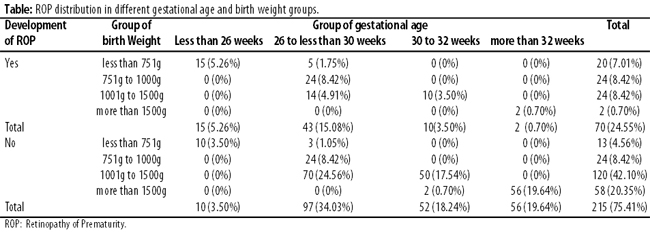
Discussion
According to the study\'s findings, the incidence of ROP was 24.6%. Another Karachi-based study showed the incidence of ROP to be 32.4%.5 One study reported ROP incidence of 34.5% in Iran.9
In our study the mean birth weight and mean gestational age were 1280.34±350.43g and 29.38±3.14 weeks. It has earlier been reported to be 1235±281g and 29.8±2.2 weeks.5 In our study the mean birth weight of infants developing ROP was 1016.5±315.93g and mean gestational age was 27.74±2.36 weeks, and the results are comparable with those reported in literature.10,11
In our study, children born with birth weight less than 2000g, gestational age less than 37 weeks, or those who were considered high risk for ROP by a paediatrician were screened for the development of ROP. Guidelines by the American Academy of Ophthalmology for screening of ROP recommend examination of infants with birth weight of 1500g or less and gestational age of 32 weeks or less and those babies with a birth weight over 1500g and gestational age greater than 32 weeks if recommended by a neonatologist.12
In our study, 70(24.6%) newborns developed ROP and 22(7.7%) developed threshold disease and received laser treatment. Similar results were observed by other studies.13-15 Besides, 68 infants who developed ROP had birth weight less than 1500g and gestational age less than 32 weeks. While there were 2 infants with better birth weight and more gestational age who, developed ROP, but they did not develop threshold disease.
LBW and gestational age are considered to be the most important risk factors for ROP development.15 Other risk factors that we could identify in our study included supplemental oxygen therapy, mechanical ventilation, anaemia, blood transfusion and sepsis.16
Screening criterion in our hospital appears to be reasonable as no infant with ROP was missed. However, since it was a single-centre study, its results cannot be generalised. Different hospitals may have different screening protocols and, as such, our results may not be applicable to other healthcare facilities. In Pakistan, little work is done on ROP17 Moreover, in rural and semi-urban regions of the country, there is little awareness about the disease not only among the general healthcare physician, but also paediatricians and ophthalmologists.2 We emphasise the need of neonatal screening for ROP in high-risk group so that timely treatment may save a child from catastrophic consequences of untreated ROP.
Conclusion
Screening is key to preventing childhood blindness caused by ROP. Prematurity, LBW and supplemental oxygen therapy are significant risk factors for the development of ROP.
References
1. Holmstro¨m G, Wijngaarden P, Coster DJ, Williams KA. Genetic susceptibility to retinopathy of prematurity: theevidence from clinical and experimental animal studies. Br J Ophthalmol 2007;91:1704-8.
2. Hashmi FK, Chaudhry TA, Ahmad K. An evaluation of referral system for retinopathy of prematurity in leading health centers across Karachi, Pakistan. JPMA 2010; 60:840-4.
3. Gilbert C. Retinopathy of prematurity: a global perspective of the epidemics, population of babies at risk and implications for control. Early Hum Dev 2008;84:77-82.
4. Ersan BA, Kir N, AkarcayK, CandanOA, KeskekNS, Demirel A, et al. Epidemiological analysis of retinopathy of prematurity in a referral centre in Turkey.Br J Ophthalmol 2013;97:15-7.
5. Taqui AM, Syed R, Chaudhry TA, Ahmad K, Salat MS. Retinopathy of Prematurity: Frequency and Risk Factors in a Tertiary Care Hospital in Karachi, Pakistan. J Pak Med Assoc 2008; 58:186-90.
6. Akçakaya A, Yaylali S, Akçay G. Screening for retinopathy of prematurity in a tertiary hospital in istanbul: Incidence and risk factors. J Pediatr Ophthalmol Strabismus 2011;15:1-5.
7. Dominico R, Davis K, Davis O. Documenting the NICU design dilemma: Comparative patient progress in open-ward and single family room units. J Perinatol 2011;31:281-8.
8. Hakeem AH, Mohamed GB, Othman MF. Retinopathy of prematurity: A study of prevalence and risk factors. Middle East Afr J Ophthalmol 2012;19:289-94.
9. Karkhaneh R1, Mousavi SZ, Riazi-Esfahani M, Ebrahimzadeh SA, Roohipoor R, Kadivar M, , et al. Incidence and risk factors of retinopathy of prematurity in a tertiary eye hospital in Tehran. Br J Ophthalmol 2008;92:1446-9.
10. Mathew MR, Fern AI, Hill R. Retinopathy of prematurity: are we screening too many babies? Eye (Lond) 2002;16:538-42.
11. Good WV. Early treatment for retinopathy of prematurity Cooperative group. Final results of the early treatment for retinopathy of prematurity (ETROP) randomized trial. Trans Am Ophthalmol Soc 2004;102:233-48.
12. Anon. Section on Ophthalmology American Academy of Pediatrics, American Academy of Ophthalmology, American Association for Pediatric Ophthalmology and Strabismus. Screening examination of premature infants for retinopathy of prematurity. Pediatrics 2006;117:572-6.
13. Gilbert C, Fielder A, Gordillo L, Quinn G, Semiglia R, Visintin P, et al. Characteristics of infants with severe retinopathy of prematurity in countries with low, moderate, and high levels of development: implications for screening programs. Pediatrics 2005; 115: 518-25.
14. Larsson E, Holmstrom G. Screening for retinopathy of prematurity: evaluation and modification of guidelines. Br J Ophthalmol 2002;86:1399-402.
15. Hussain N, Clive J, Bhandari V. Current incidence of retinopathy of prematurity, 1989-1997. Pediatrics 1999;104:e26.
16. Binkhathlan AA, Almahmoud LA, SalehMJ, Srungeri S. Retinopathy of prematurity in Saudi Arabia: incidence, risk factors, and the applicability of current screening criteria. Br J Ophthalmol 2008;92;167-9.
17. Naz S, Dawar Jadoon MZ, Saeed N. Retinopathy of prematurity: frequency measures In two tertiary care hospitals. J Med Sci 2011; 19: 148- 51.
Related Articles
Journal of the Pakistan Medical Association has agreed to receive and publish manuscripts in accordance with the principles of the following committees:


