

Rukhsana Tariq ( Department of Radiology, Advanced Radiology Clinic, Karachi. )
Shehla Hasnain ( Department of Radiology, Advanced Radiology Clinic, Karachi. )
Maria Tariq Siddiqui ( Medical Student, University of Karachi. )
Rashid Ahmed ( Department of Radiology, Advanced Radiology Clinic, Karachi. )
March 2014, Volume 64, Issue 3
Case Reports

Abstract
Aggressive angiomyxoma is a rare, myofibroblastic tumour, of pelvi-perineum of young women. It is a slow growing, low grade neoplasm with a high risk of recurrence following initial resection. Aggressive angiomyxoma is often clinically misdiagnosed because of its variable presentation as a soft tissue mass of the vulva, perianal region, buttock or pelvis. It displays translevator extension with growth around the perineal structures. Fewer than 150 cases have been reported in the literature since 1983. Imaging is important to determine the extent of the lesion. We present layered configuration of the mass on ultrasound and Magnetic Resonance Imaging in a 40 years old woman with a left pelvi- perineal mass since 5 years. Histapathology after excision gave a diagnosis of Aggressive Angiomyxoma.
Keywords: Aggressive angiomyxoma, Ultrasound, Magnetic Resonance Imaging.
Introduction
Aggressive angiomyxoma (AAM) is the most typical primary tumour of the ischio-rectal fossa in the pelviperineum. It is a rare, locally infiltrative, mesenchymal tumour of young women. It was first described by Steeper and Rosai in 1983.1 AAM is a slow-growing low grade neoplasm. It is often clinically misdiagnosed as a lipoma on account of its variable presentation as a soft mass. Initial surgical resection is unsuccessful, as the anatomical extent is frequently not perceived. There is a high risk of local recurrence after many years. Imaging is important to determine the extent of the lesion and surgical approach. We describe the ultrasound and MRI findings in a 40-years old woman with a left sided pelvi-perineal mass. It proved to be an AAM on histopathology.
Case Report
A 40-years-old lady presented to our facility with a soft tissue mass located in the lower left pelvi-perineal and medial gluteal region for the last 5 years. Mild pain and perineal discomfort were also reported. Clinically, a soft tissue bulky mass was located on the medial aspect of the left perineum. The bulge increased in size on coughing. The overlying skin was intact with normal colour. It was soft, well defined, lobulated, and freely mobile. Baseline laboratory investigations including haematological profile, urine detailed report and X-ray chest were normal. The clinical diagnosis was a perineal malignant fibrous histiocytoma and dermatofibrosarcoma protruberans.
Transvaginal ultrasound revealed an elongated, soft tissue mass in the left hemi pelvi-perineum with contralateral displacement of the midline structures. It measured 11.8 x 6.0cms and it was reducible on probe manouvre. It demonstrated mixed echogenicity, lamellated appearance with alternating layers of hypo and hyperechoic tissues (Figure-1a).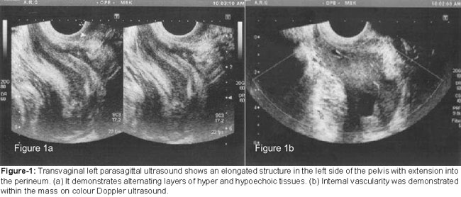
Layered sonographic features have not been reported in the literature. Colour Doppler demonstrated internal vascularity (Figure-1b). The differential diagnosis after ultrasound examination included liposarcoma, haemangiopericytoma and leiomyosarcoma.
Magnetic Resonance Imaging (MRI) revealed a large well circumscribed mass lesion in the left pelvi-perineum. It demonstrated translevator extension along the left ischio-rectal fossa into the subcutaneous tissues of the left hemiperineum. The perilesional fat planes were intact. The mass displaced the urinary bladder, uterus and the rectum to the right without infiltrating them. The mass demonstrated, isointensity to the muscle on T1W images (Figure-2a).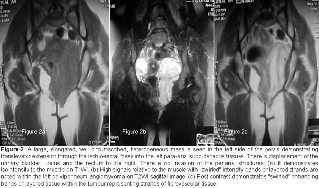
On T2W images high signal intensity relative to the muscle with "swirled" low signals intensity bands or layered strands within the hyperintense tumour was noted. (Fig 2b) The mass appeared as high signal intensity on fat saturated images. Avid heterogeneous enhancement with a whorled configuration representing fibrovascular tissue was present on postcontrast images (Figure-2c).
Image guided fine needle aspiration biopsy was performed using a 16 gauge fine needle. Two passes were made. Histopathology, revealed a neoplastic lesion composed of scattered spindle and stellate shaped cells with ill defined cytoplasm. Variable sized thin and thick walled vascular channels against a myxoid stroma rich in collagen fibers was seen. Mitotic activity was insignificant. It was reported as an AAM. The patient was referred for surgical resection and was later lost to follow up.
Discussion
AAM is a slow growing, infiltrative, low grade mesenchymal tumour displaying unusal growth pattern and exclusively involves the lower pelvis and perineum. The tumour was first reported as a new clinical pathology by Steeper and Rosai in 1983.1 Male to female ratio is 1:6. The age distribution is from second to the eigth decade with a peak incidence in reproductive age group.2 In females it may present as a labial or vulval mass, perineal hernia, vaginal or an endometrial polyp. It is soft and compressible. Generally, large in size at presentation it is greater than 10cm.3 It causes discomfort or pressure effect on adjacent pelvic organs. They have a tendency to deviate rather than infiltrate the local structures including vagina, urethra, bladder and the rectum. It displays unusual growth pattern of translevator extension with growth around perineal structures without invasion.4
Pathologically, it is a lobular, poorly encapsulated, gelatinous, rubbery mass with gray bluish appearance. Microscopically, the lesion is composed of stellate and spindle shaped neoplastic cells embedded in collagenous and hyaluronic acid containing stroma with loose myxoid background. The lesion is vascular with thickened or hyalinized vessels. Nuclear atypia and mitosis are absent. Immunohistochemistry is positive for vimentin, desmin, actin, CD 34 and Factor VIIIa. Estrogen and progesterone receptors may be positive.4 On haematoxylin and eosin staining, vessels of varying caliber scattered haphazardly through the parenchyma with few mitotic figures is considered a hallmark of AAM.
On Ultrasound the mass has generally been mentioned as hypoechoeic and cystic.4 Our patient however presented with an elongated mass with a lamellated appearance of hyper and hypoechoeic tissues alternating with each other. The lamellated configuration and reducibility of mass with probe manouvre and increase in size on coughing have not been reported earlier. On MRI the tumour is iso to low-signals to the muscle on T1W-images, swirled hyperintensity is seen on T2W-images3 attributable to the myxomatous content of the tumour. A whorled pattern of signal intensity on T2W images, fat-suppressed T2W-images and post contrast T1W-images has been reported as typical features of aggressive angiomyxoma.5 Contrast enhancement reflects inherent vascularity related to fibrovascular stroma. The tumour displaces and grows around adjacent structures.3 These features were demonstrable in our patient and are highly characteristic and distinctive.
Differential diagnosis includes lipomas, myxoid peripheral nerve sheath tumours, pelvic fibromatosis and angiomyofibroblastomas. Malignant tumours simulating AAM include mixed mesodermal tumours, malignant fibrous histiocytomas, botyroid pseudosarcoma, embryonal rhabdomyosarcomas squamous cell carcinomas and extramucosal anal adenocarcinomas. Misdiagnosis is seen in 82% of the cases .
Lipomas have high signals on T1W images with signal drop out on fat-suppressed images. Plexiform neurofibromas may affect pudendal nerve and are symmetrically arranged within the pelvis. They are low on T1WI and high on T2WI with intense postcontrast enhancement. Pelvic fibromatosis are large masses with low signals. Angiomyofibroblastomas are small and involve superficial parts of the vulva. Rhabdomyosarcomas occur in younger patients. Squamous cell carcinomas demonstrate invasion. Malignant fibrous histiocytoma demonstrate low signals on T1WI and high signals on T2WI with postcontrast enhancement. Extramucosal anal adenocarcinomas are typically located in an area where anal glands are present. The characteristic "swirling" is not seen in any of these malignant tumours on MRI.
The histopathological diagnosis can be established after surgical resection or via a biopsy. The mainstay of management includes surgery with wide local excision. Since most tumours are large, infiltrative, and merge with adjacent soft tissues with loss of fat plane without invasion of like bladder and rectum, considerable morbidity may result from wide excision. Hence in such situations, patients are observed periodically with clinical and radiological correlation. Radiotherapy has been disappointing but as most AAM express estrogen and progesterone receptors, hormonal treatment with a gonadotrophin-releasing hormone agonist has been applied for small AAM, in addition to adjuvant therapy for residual tumours.6
Multidisciplinary approach is opted to avoid high risk of multiple local recurrences. To decrease the likelihood, preoperative angiographic embolization, preoperative external beam irradiation and intraoperative electron beam radiotherapy have been applied. The risk for distant metastasis is rare. Two cases of distant metastasis have been reported. The first includes nodal and peritoneal metastasis and the other case reports lung metastasis.7
In our case, the limitation of the study was a lost to follow up of the patient and no further information could be obtained after the initial imaging and biopsy. Hence, a follow-up with regard to the course of the disease, and whether she underwent surgery or not, or was there a recurrence post surgically or a development of a distant metastasis could not be attained.
Conclusion
AAM is a rare tumour with distinct imaging appearance. It affects the pelvi- perineum of women. Whorled or swirled appearance retaining enhancing layered appearance on postcontrast images on MRI is characteristic. MRI accurately evaluates translevator extension and growth around pelvic organs. MRI allows planning of the best surgical route. This is crucial in order to prevent recurrences related to inadequate resection and residual tumours.
References
1. Steeper TA, Rosai J. Aggressive angiomyxoma of the female pelvis and perineum. Report of nine cases a distinctive type of gynecologic soft-tissue neoplasm. Am J Surg Pathol 1983; 7: 463-75.
2. Magitibay PM, Salmon Z, Keeney GL, Podratz KC. Aggressive angiomyxoma of the female pelvis and perineum: a case series. Int J Gynecol Cancer 2006; 16: 396-401.
3. Sinha R, Verma R. Case 106: aggressive angiomyxoma. Radiology 2007; 242: 625-7.
4. Evis S, Jo H. Aggressive Angiomyxoma, Vulva. In: Hedvig H, editor. Diagnostic Imaging Gynaecology. Manitoba, Canada Amirsys: Elsevier Saunders, 2007; 16-9.
5. Jeyadevan NN, Sohaib SA, Thomas JM, Jeyarajah A, Shepherd JH, Fischer C. Imaging features of aggressive angiomyxoma. Clin Radiol 2003; 58: 157-62.
6. Shinohara N, Nonomura K, Ishikawa S, Seki H, Koyanagi T. Medical management of recurrent aggressive angiomyxoma with gonadotropin-releasing hormone agonist. Int J Urol 2004; 11: 432-5.
7. Geng J, Cao B, Wang L. Aggressive Angiomyxoma: an Unusual Presentation. Korean J Radiol 2012; 13: 90-3.
Journal of the Pakistan Medical Association has agreed to receive and publish manuscripts in accordance with the principles of the following committees:


