

Kiran Gujrati ( Department of Pediatrics and Child Health, The Aga Khan University Hospital, Karachi. )
Arshalooz Jamila Rahman ( Department of Pediatrics and Child Health, The Aga Khan University Hospital, Karachi. )
Gulsher ( Department of Pediatrics and Child Health, The Aga Khan University Hospital, Karachi. )
March 2014, Volume 64, Issue 3
Case Reports

Introduction
Congenital chloride diarrhoea (CLD) is an autosomal recessive disease, reported mainly in Finland.The infrequency with which it is encountered makes its diagnosis a challenge. Furthermore,due to un-availability of stool electrolyte test in Pakistan, diagnosis of CLD is most often missed or misdiagnosed. The case of a 16-month-old male child is described who was treated as Bartter Syndrome since the age of 9 months, later diagnosed to have CLD with the support of history and some routine laboratory work.
Case Report
A sixteen-month-old male child of consanguineous parents,who was being treated for Bartter syndrome in another hospital since the age of 9 months, presented in our hospital with complaints of 15-20 daily episodes of watery diarrhoea since 1 week. History revealed evidence of polyhydramnios and dilated small bowel loops on ultrasound, premature birth and no passage of meconium. There had been two admissions for jaundice within the first month of life. He had never passed a normal stool since birth and the content of stool was always confused with that of urine. He had multiple admissions due to moderate to severe dehydration which were managed as acute gastroenteritis. Diagnosis of Bartter syndrome was established based on serum electrolytes. Treatment involved spironolactone, indomethacine, and potassium and no other electrolyte supplementation was given. Obstetric history of the mother revealed one missed abortion at three months of gestational age and death of a seven-day-old female child who had the similar antenatal ultrasound findings. At the time of admission to the hospital, the boy weighed 7.9kg (percentile below 5), with length 71cm (percentile below 5), heart rate 150beats/min, respiratory rate 32breaths/min, blood pressure 103/55mm Hg, temperature 37°C. On examination; the child was severely dehydrated and had diaper dermatitis. Other systemic examinations were unremarkable. Initial laboratory workup showed hypochloraemia (plasma chlorine-90mmol/L), hypokalaemia (plasma potassium-3.1mmol/L), and hyponatraemia (sodium 131 mmol/L). Since blood pH and partial pressure of carbondioxide changes rapidly in crying infants,1 whereas changes in serum bicarbonate concentration occurs much more slowly and its level provide equivalent information to arterial blood gases,2 only serum bicarbonate level was done which showed metabolic alkalosis (venous blood bicarbonate-30.8mmol/L); aldosterone level (2.4ng/dl) and renin (6.1ng/ml/hr). Stool pH was 5.0 (normal: 7-7.5). Other stool microscopy was normal. As spot urine electrolytes results can be compared with adequate accuracy with 24-hr urinary investigation,3 therefore spot urine samples were taken which showed sodium <10mmol/L (less than 10mmol/L-extra renal depletion while greater than 10-renal, adrenal, syndrome of inappropriate antidiuretic hormone [SIADH]), K: 16mmol/L (Normal: 25-100mmol/L), chlorine <15mmol/L (less than 25mmol/L-extra renal loss while greater than 40mEq/L-renal loss). Stool for reducing substance and fat globules was negative. Ultrasound KUB (kidney, ureters, and bladder) was normal. Coeliac serology was also negative. Sweat test could not be performed successfully due to lack of production of sweat, therefore cystic fibrosis (CF) was excluded on the basis of history, absence of faecal fat and negative Delta-508 mutation (only available test for CF mutation). Stool electrolytes could not be obtained as the test was not available in Pakistan. Spironolactone and indomethacine was discontinued. Oral cholestyramine and butyrate in the form of pineapple essence was started. Oral substitution with sodium chloride and potassium chloride was continued. Patient is now being followed as an outpatient. In the 4 month followup, stool frequency and hospital admissions have significantly decreased and he has also gained around 2kg weight.
Discussion
In 1945, Darrow and Gamble described the first cases of congenital chloride diarrhoea (CLD) under the name of congenital alkalosis with diarrhoea. Later, familial enrichment of the cases proved the autosomal recessive inheritance of the disease, which was called CLD.4 The condition is characterised by loss of faecal chloride and osmotic diarrhoea owning to primary defect in the active transport of Cl/HCO3 in the distal ileum and colon manifesting as hypochloraemia.5 In addition, absolute loss of sodium and potassium due to increase quantity of stools results in hyponatraemia and hypokalaemia. Also, acidity of intestinal contents hampers sodium absorption and aggravates hyponatraemia.5 Inadequate secretion of HCO3 in the ileum and colon and excessive loss of H+ through the kidneys leads to alkalosis. Hyperaldosteronism occurs as a compensatory mechanism to conserve sodium. Onset of CLD is intrauterine, leading to watery diarrhoea and polyhydramnios. Prenatal scans show dilated bowel loops.6 Birth is generally two weeks premature. There is no passage of meconium due to watery, 'urine like' diarrhoea.5 As the disease is rare, there is a possibility of misdiagnoses. Watery diarrhoea may be easily confused with urine. As both CLD and Bartter syndrome are associated with hypokalaemic metabolic alkalosis, this led to the diagnosis of Bartter syndrome in our patient. Aldosterone and renin levels in this case were within normal ranges which could be due to previous treatment with indomethacin.7 Differentiation of both is possible due to different site of defect in both the conditions. There is increased stool chloride in CLD while chloride is lost in urine in Bartter syndrome.This difference becomes more distinctive in untreated CLD when urine becomes chloride free due to severe dehydration and intestinal loss of chloride,4 making urine chloride measurement an important investigation. CLD is then confirmed by faecal electrolytes. A faecal chloride concentration exceeding the sum of the sodium and potassium concentrations suggests the diagnosis of CLD. Although clinical picture and high faecal chloride are the basis for the diagnosis in CLD, searching for the disease-causing SLC26A3 mutations is possible.4 Thus so far, over 30 mutations have been reported.4 CLD must be considered in the differential diagnosis in children with a tendency to chronic dehydration, failure to thrive, slow growth, and hypokalaemic and hypochloraemic metabolic alkalosis. As in this case, even in the absence of stool electrolyte test, detailed medical history including prenatal and early postnatal findings, synchronous serum and urinary chloride measurements helped in the diagnosis of CLD (Table).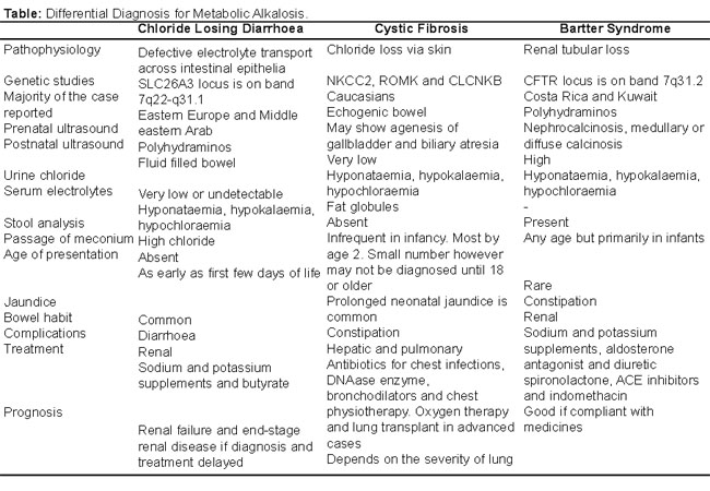
Condition is usually fatal if left untreated. Salt replacement with sodium chloride (NaCl) and potassium chloride(KCl) has no significant effect on chronic diarrhoea but allows maintenance of serum electrolyte and acid base balance with normal physical and mental growth. Studies have shown oral butyrate to have beneficial effects in CLD.8 As the diarrhoea persists throughout life, patients gradually learn to cope with the condition.
Conclusion
Metabolic alkalosis in a child with diarrhoea should always be looked carefully with differentials like cystic fibrosis and Barters Syndrome however a meticulous history with focus to prenatal history may help in cornering chloride losing diarrhoea from others.
References
1. Ronald E. Kleinman. Renal tubular acidosis. Manual of Pediatric Practice. 2nd ed. United Kingdom: Saunders (W.B.) Co Ltd, 2002; pp 789-90.
2. Martin MJ, FitzSullivan E, Salim A, Berne TV, Towfigh S. Use of serum bicarbonate measurement in place of arterial base deficit in the surgical intensive care unit. Arch Surg 2005; 140: 745-51.
3. Mann SJ, Gerber LM. Estimation of 24-hour sodium excretion from spot urine samples. J Clin Hypertens (Greenwich) 2010; 12: 178-80.
4. Odul E, Afi RT, Dalgiç B, Wedenoja S. Congenital chloride diarrhea misdiagnosed as bartter syndrome. Turk J Gastroenterol 2011; 22: 321-3.
5. Parikh B, Khubchandani, Amdekar YK, Ugra D, Patel A, Nardekar J. Congenital chloride diarrhea. Indian Pediatr 1993; 30: 811-3.
6. Cuillier, F, Alessandri JL, Kauffman E, Heiser M. Congenital chloride diarrhea. (Online) 2012 (Cited 2012 Jun 11). Available from URL: http://www.sonoworld.com/TheFetus/page.aspx?id=1901.
7. Mayatepek E, Seyberth HW, Nützenadel W. Effects of indomethacin in congenital chloride diarrhea. J Pediatr Gastroenterol Nutr 1992; 14: 319-22.
8. Canani R, Gianluca T, Cirillo P, Castaldo G, Salvatore F, Cardillo G, et al. Butyrate as an Effective Treatment of Congenital Chloride Diarrhea. Gastroenterol 2004; 127: 630-4.
9. ALAbbad A. Hypochloremic alkalosis workup. (Online) 2012 Mar 30 last updated (Cited 2012 Aug 12). Available from URL: http://emedicine.medscape.com/article/945263-overview.
10. Mersin SS, Ramelli GP, Laux-End R, Bianchetti MG. Urinary chloride excretion distinguishes between renal and ex-trarenal metabolic alkalosis. Eur J Pediatr 1995; 154: 979-82.
Journal of the Pakistan Medical Association has agreed to receive and publish manuscripts in accordance with the principles of the following committees:


