

Farrukh Azmi ( Public Health Division, National Institute of Health, Islamabad. )
Jamuila Iqbal ( Public Health Division, National Institute of Health, Islamabad. )
Nasreen Nomani ( Public Health Division, National Institute of Health, Islamabad. )
Zahid Ishaque ( Public Health Division, National Institute of Health, Islamabad. )
Jamshaid Iqbal ( Public Health Division, National Institute of Health, Islamabad. )
Mohammad Abdur Rab ( Public Health Division, National Institute of Health, Islamabad. )
June 1987, Volume 37, Issue 6
Original Article

Abstract
Counter immuno electrophoresis was done on concentrated urine samples of 166 children with lower respiratory tract infection for the detection of Streptococcus pneumoniae and haemophilus influenzae type b antigens. About 10¼ cases showed the presence of Strept. Pneumoniae Polysaccharide antigen and1.8% H. influenzae antigen. No growth was seen on blood culture in 91.8% while 0.8% showed Strep. pneumoniae. Bacterial antigen detection can be used as an adjunct to routine diagnostic tests in patients
with lower respiratory tract infection (JPMA 37: 148,1987).
INTRODUCTION
Acute respiratory tract infection (A.R.I) in infants and young children is an important cause of morbidity and mortality in developing cotrntries. Every year there are over 3 million deaths in developing countries due to pneumonia alone1. In Pakistan as in other developing countries diarrhoea, malnutrition and acute respiratory tract infections are the most common causes of death in young children. These three conditions often exist together and may potentiate each other’s lethal effects2.
In acute upper respiratory tract infections little or no effort is made to look for the etiological agent. Adequate facilities For bacteriological and virological examination at most places are rarely available or, if available, the delay encountered in obtaining results discourages the use of these facilities.
Alternative methods of diagnosis such as ELISA, latex agglutination, co-agglutination and counter immuno electrophoresis3,5 for strept. Pneumoniae and H. influenzae developed recently, have shown encouraging results. These techniques have been used for both antigen and antibody detection5. They are sensitive, specific and easy to perform.
The aim of this study was to assess the value of C.I.E. for the detection of Strept. pnewnoniae and H. influenzae antigen in urine of patients with acute respiratory tract infection.
MATERIAL AND METHODS
PATIENTS
One hundred and sixtysix children under 4 years of age, with symptoms and signs suggestive of lower respiratory tract infections were included in the study. Complete history and physical examination was recorded by a physician.
MICROBIOLOGICAL STUDIES
Throat swabs were taken in transport medium brought under cold conditions to the laboratory and processed by standard culture method6.
BLOOD CULTURE
Two ml blood was taken in 20 ml heart infusion broth and was cultured as described7.
URINE SAMPLES FOR ANTIGEN DETECTION
Urine samples were collected using argyle paediatric urine collector (Sherwood Medical, St. Louis M.O. 63103 U.S.A). The samples were transported in cold to the laboratory and frozen at-20°Ctill processed i.e. 30-100 days.
Urine samples thawed on the day of the test were concentrated using alcohol precipitation technique8. Ten ml urine was centrifuged at 250 g for 10 minutes; to 5 ml, supernatant was added in or to 15 ml 95% cold ethanol, it was vortexed and incubated at 4°C for 60 minutes. The sample was centrifuged at 400 g for 20 minutes, supernatant was discarded and precipitate air dried; to the dried sediment 0.25 ml normal or 0.851 saline was added. The mixture was centrifuged at 400 g for 10 minutes and then used for the test.
COUNTER IMMUNO ELECFROPHORESIS (C.I.E)
C.I.E. was performed as described by Ingram et al9 with slight modifications as follows: 1% Agarose in 0.05 M sodium barbital buffer pH. 8.6 was used to prepare plates. 3 mm wells 3mm apart were punched. All the specimens were tested against pneumococcal omni serum (Statens Serum Institute Copenhagen Denmark) and H. influenzae type b antiserum (Bostid). Positive and negative controls were included with each plate.
Electrophoresis, 20 mA/plate, was performed for one hour. The plates were read immediately and after overnight incubation at 4°C. The plates were also washed and stained with amido black.
RESULTS
A total of 166 children with lower respiratory tract were included in the study. Their ages ranged from 15 days to 4 years; male to female ratio was 2.3 :1. Seventynine percent cases came from urban areas and most belonged to the low-middle socio economic group (Figure 1).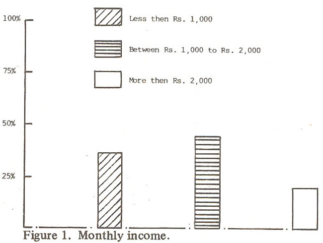
Commonest presenting features were fever (99%), cough (97%), vomiting (43%) and diarrhea (30%). About 62% children had already taken various broad spectrum antibiotics before seeking hospital admission and only 30% cases had not taken any antibiotic. Eight percent cases took unknown drugs for respiratory tract infection. Radiography of the chest was done in 78% cases. Lesions included patchy consolidation (44%), hyper inflation (18%), lobar consolidation (11%) and pleural effusion and pneumothorax in 1% cases each. Radiology was normal in 3% cases. Blood culture was done in 123 cases.. No growth was obtained in 96%, 2% grew citro bactor and 1% each streptococcus pneumoniae and klebsiella,pneurnoniae.
Throat swabs were sent for bacteriological examination in 70% cases. Normal flora were seen in 72%, Kiebsiella pneumoniae in 24%, E. Coli in 3% and Strept. pneumomae in 1% cases. On the basis of history, physical examination and X-ray chest, clinical diagnosis of broncho pneurnoniae, acute bronchiotitis and lobar pneumoniae was made in most of the cases.
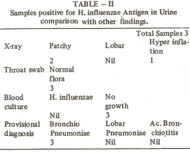
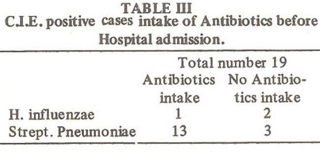
Counterimmuno electrophoresis was performed on all 166 samples. Eleven percent revealed the presence of either strept. pneumoniae or H influenzae antigen in the urine. A breakdown of samples showing the presence of H. influenzae and strept. pneumoniae antigen and their comparison with clinical diagnosis, X-ray findings, blood culture and throat swabs is given in Tables I, II and III.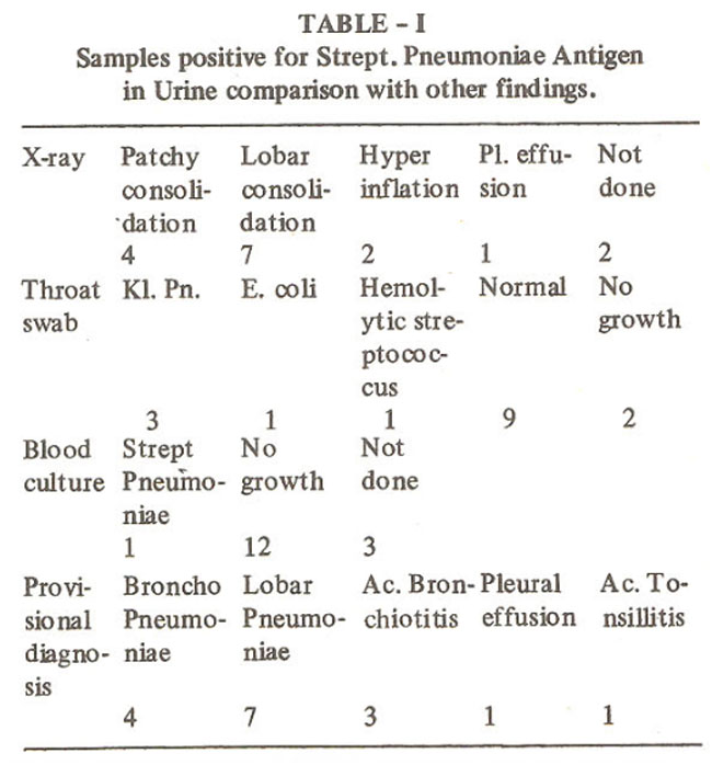
DISCUSSION
Antigen detection for the diagnosis of many bacterial and parasitic diseases has been used with great success in the past.10 The usual methods for diagnosis in respiratory tract infection are blood Count, nasopharyngeal Swab,sputum examination, blood culture and X-ray of the chest. Though X-ray chest helps in confirming the clinical diagnosis of Pneumonia, but the etiological diagnosis is established by bacteriological. virological and serological studies. In certain situations such as associated pleural effusion or pneumothorax, the X-ray findings often become difficult to interpret. Although blood cultures were dose in 116 cases in this study. 91% of the blood cultures failed to reveal any growth. One of the reasons is that most of the patients were already taking antibiotics before hospital attendance. Kahn et al11 were able to culture Strept. Pneumoniae from 35% of blood, nasopharyngeal and sputum samples of their ARL. patients. This study also suggests that the blood culture is not the method of choice under our conditions. Sputum can be used for looking at the etiological agent but in young children, debilitated and elderly patients it is often difficult to obtain.
Incidence of respiratory and gastro-intestinal infections in the developing countries does not vary greatly from developed countries2. There is, however, a big difference in mortality ‘being very high in developing countries. It could be due to the fact that diarrhoea, malnutrition and acute respiratory tract infection coexist in many children. In the present study 43% of the children had accompanying vomiting and 30% had diarrhoea along with lower respiratory tract infection. An added advantage in such a study is to look at the etiology of acute respiratory tract infections which could explain regional and national differences in disease pattern and mortality rates. The delay in identifying causative organism and institution of appropriate chemotherapy accounts for higher mortality in our clinical, social milieu.
Over 79% of the patients attending the hospital belonged to lower and middle income group families. There is a need to develop simple and inexpensive methods of diagnosis. Antigen detection technique offers a promising alternative. Counter-immunoelectrophoresis, although difficult to set up, is quick, reliable and quite objective. In this study C.I.E. was able to demonstrate the presence of S.trept. Pneumoniae antigen in 9% and H influenzae antigens in 2% patients, whereas blood culture failed to reveal the presence of Strept. pneumoniae and H. influenzae in all but one case. It has been demonstrated by several investigators that there is a prolonged presence of pneumococcal antigen in the patient’s urine and serum12. It was suggested by studies on rodent models that pneumococcal polysaccharides is highly resistant to degradation, therefore metabolism occurs slowly and these antigens can be detected for long periods13 Many investigators have found that the prolonged antigen detection is not affected by antibiotic intake8. Antigen detection can be a very useful diagnostic tool after antibiotics have been started. Urine samples can reveal positive results as late as 9-12 days after the onset of pneumococcal pneumonia14. Nineteen samples revealed the presence of Hinfluenzae and Strept. pneumoniae antigen, out of these 16 were taking antibiotics. The sensitivity of C.I.E. for the detection of Strept.pneumoniae antigen has been studied by Cerosaletti et al15 , wh found that the lower limits for detecting type 3 purified capsular poly-saccharide by C.I.E. was 15.6 ng/ml.
Counterimmuno electrophoresis and other antigens detection techniques i.e. ELISA, latex agglutination and coagglutination can be used as an adjunct to routine diagnostic methods. Latex agglutination and coagglutination do not need any specialized equipment, but they are more difficult to interpret and latex agglutination can be more inconclusive15. There is a need to evaluate these recent immunodiagnostic techniques under our conditions and incorporate them into routine diagnostic procedures.
ACKNOWLEDGEMENT
Financial support for this study was provided by Board on Science and Technology for International Development (BOSTID) U.S.A.
REFERENCES
1. Grant, G.P. The state of world’s children. Newyork, Oxford University Press, 1984.
2. Denny, F.W. and Loda, F.A. Acute respiratory infections are the leading cause of death in children in developing countries. Am. J. Trop. Med. Hyg., 1986; 35 : 1.
3. Edwards, E.A. and Coonrod, J.D. Coagglutination and counter-immunoelectrophoresis for detection of pneumococcal antigens in the sputum of pneumonia patients. J. Clin Microbiol., 1980; 11:488.
4. Ingram, D.L. Pearson, A.W. and Occhuti, A.R. Detection of bacterial antigens in body fluids with Weilcogen Haemophilus influenzae b. Streptococcus pneumoniae, and Niseria meningitidis (ACYW 135) latex agglutination tests. J. Clin Microbiol. 1983;18: 1119.
5. Messina, J.P., Hickox, P.G., Lepow, M.L., Pollara, B. and Venezia, R.A. Modification of a direct enzymelinked iinmunosorbent assay for the detection of iinmunoglobuiln G and M antibodies to pneumococcal capsular polysaccharide. J. Clin. Microbiol., 1985;21:390.
6. Lenette, E.H. ed. Manual of clinical microbiology. Washington, American Society for Microbiology, 1985, p. 80.
7. Lenette, E.H. ed. Manual of clinical microbiology. Washington, American Society for Microbiology, 1985, p. 75.
8. Coonrod, J.D. and Rytel, M.W. Detection of type-specific pneumococcal antigens by counterimmunoelectrophoresis. II. Etiologic diagnosis of pneumococcal pneumonia. J. Lab. Clin Med., 1973; 81:778.
9. Ingram, D.L., Anderson, P. and Smith, D.H. Countercurrent immuno-electrophoresis in the diagnosis of systemic diseases caused by Hemophilus influenzae type b. J. Pediatr. 1972; 81:1156.
10. Rytel, M.W. Counterimmunoelectrophoresis in diagnosis of infectious diseases. Hosp. Pract., 1975; 10:75.
11. Kalin, M. and Linderug, AA. Diagnosis of pneumococcal pneumonia; a comparison between microscopic examination of expectorate, antigen detection and cultural procedures Scand. J. Infect. Dis., 1983; 15 :247.
12. Coonrod, J.D. and Drennan, D.P. Pneumococcal pneumonia; capsular polyseccharide antigenemia and antibody responses. Ann. Intern. Med., 1976; 84:254.
13. Coonrod, J.D. Evidence of breakdown of pneuinococcal polysaccharides m vivo. Proc. Soc. Exp. Biol. Med., 1979; 162 :249.
14. Tugwell, P. and Greenwood, B.M. Pneumococcal antigen in lobar pneumonia. J. Clin. PathoL, 1975; 28 :118.
15. Cerosaletti, K.M., Roghmann, M.C. ant Bently, D.W. Comparison of latex agglutination and counter immuno electrophoresis for detection of pneuinococcal antigen in elderly pneumonia patients. J. Clin. Microbiol., 1985; 22:553.
Journal of the Pakistan Medical Association has agreed to receive and publish manuscripts in accordance with the principles of the following committees:


