

S.M.W. Jafri ( Department of Medicine, The Aga Khan Medical College, Hospital, Karachi. )
H.A. Shah ( Department of Medicine, The Aga Khan Medical College, Hospital, Karachi. )
H. Sheikh ( Department of Medicine, The Aga Khan Medical College, Hospital, Karachi. )
September 1989, Volume 39, Issue 9
Case Reports

Primary biliary cirrhosis is worldwide in its distribution with a low prevalence of 3.7 - 14 cases per 100,000 population. The disease is rarely diagnosed in the tropics. The incidence and prevalence is not known but if a thorough search is made for the diagnosis of hepatitis B Surface Antigen negative cirrhosis the diagnosis of this condition would probably increase. The following case report clearly demonstrates this:
Case Report:
A 52 year old female presented in May 1988 to the Aga Khan University Hospital with the coinplaints of high grade fever accompanied by rigors eighteen months ago which lasted for about two to three weeks. This fever recurred however and the patient was never afebrile over the following months. She received treatment irregularly and intermittently for this. This was followed by a feeling of weakness, weight loss, generalised body aches and difficulty in moving around the house for three to four months prior to admission. She also gave a four thy history of progressive abdominal distension. She was diagnosed to be hypertensive for 3 months; for this she was taking Lasix and Adalat. Her menopause occurred eight years ago. In her personal history there was nothing significant. There was no history of liver ailments in the family and she did not havejaundice in the past. On examination she was pale, mildly jaundiced, afebrile and drowsy with evidence of vasculitic rash on both legs (Figure 1).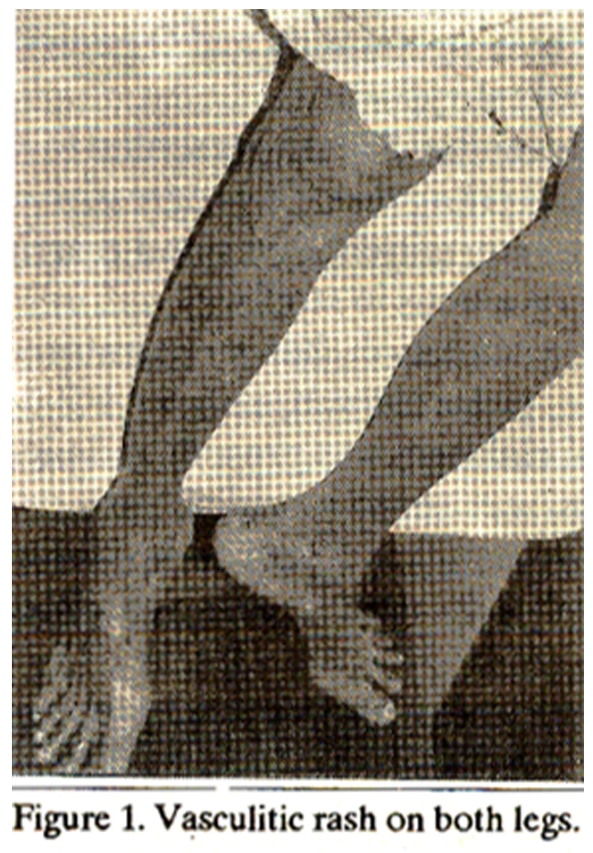
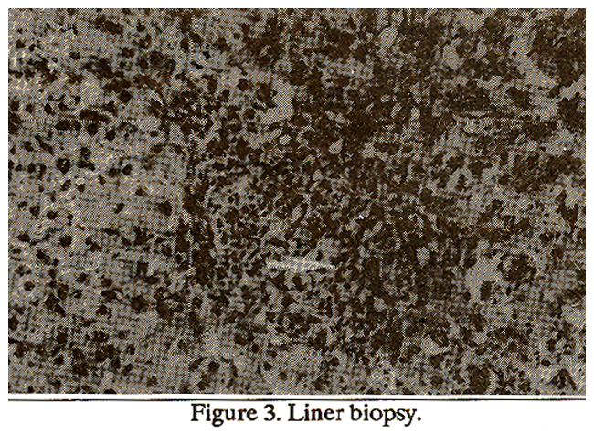
Her pulse was 80/mm, blood pressure was 180/100mm Hg in supine position. JVP was normal, there was slight pedal edema, heart sounds were normal, systolic ejection murmur (Grade 2/6) was heard over the precordium. Abdomen was distended, non tender with tense ascites and hence no abdominal viscera were palpable. Bowel sounds were audible. Chest and nervous system examinations were within normal limits. There was no encephalopathy. Her drug history revealed that she was taking Isordil, Lasbc, Adalat and Disprin. Following investigations were done during this admission. Hemoglobin 8.7 g/dl, HCT 25.2, WBC 5.0 x 10⁹/I, platelets 278 x 109/I, retics 4.3%, ESR 110, BUN 31 mgfdl, serum creatinine 13 mg/dl, sodium 122 mEq/l, potassium 4.2 niEq/l, chloride 91 mEq/l, bicarbonate 16.9 mEq/I, SGOT 57 I.U.L, LDH 347 I.U.L., CFK 27 I.U.L, plasma bilirubin 031 mg/dl, clotting screen was normal. Serum C3 = 0364 mg/I (0.70 - 1.80), serum C4 = 0.44 gm/I (0.15 - 0.70). Ascitic tap revealed ascitic fluid protein 754mg/dl, glucose 125 mg/dl, WBC 356/cmm with polys 25%, lymphos 75%, RBC 10/cmm. Aerobic and anaerobic acid fast bacilli cultures were negative. Plasma cholesterol 122 mg%, plasma triglycerides 117 mg%, serum iron 51.2 ug/dl, TIBC 1832 ug/di. Saturation of transferrin 27.9(20 - 45). Anti-nuclear antibody + + + (homogenous), anti smooth muscle antibody negative, anti mitochondrial antibody + +. The above tests were done with enzyme immunoassay technique. Hepatitis B surface antigen, hepatills surface antibody and hepatitis core antibody (IGG) were negative. Abdominal ultrasound was suggestive of cirrhosis, there was marked ascites and splenomegaly. Stool examinations times 3 for direct microscopyrevealed nothing significant. Urine occasional granular casts. RBC 18/HPF, 4 pus cells/HPF, occult blood +ve. On the basis of the above presentation and laboratory investigations it was believed her primary diagnosis was PBC (primary biliary cirrhosis, with decompensation. She was continued on adalat, isordil, angised and spironolactone with 20 mg of frusenude were added. A liver biopsy was planned but had to be postponed because the patient developed severe chest pain about the time of procedure. She recovered satisfactorily from her initial presenting complaints of fever, lethargy, abdominal distension and weakness. She was diichargedwitb a follow-up appointment in the clinic. In July 1988 she presented again with bradycardia, her pulse of 40/min which was regular, ECG revealed prolonged PR interval, junctional rhythm and peaked ‘T’ waves. Her serum potassium was found to be 8.0 mEq/I, indeed a complication of aldactone therapy; plus as she was feeling nauseated, she was taking nothing but fruit juices. She was treated with calcium gluconate, bicarbonate, insulin dextrose and lasix. Her serum potassium came down to 5 mEq/1 and her bradycardia disappeared. ECU now revealed normal sinus rhythm. Her liver biopsy was done during this admission which confirmed the initial diagnosis of primary biliary cirrhosis (Figure 2 & 3).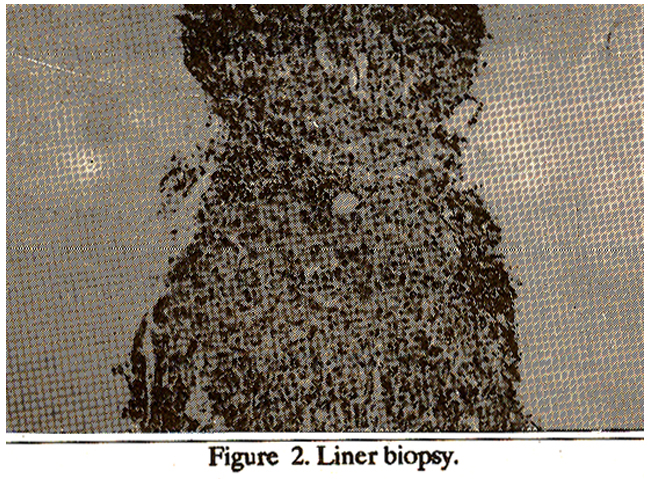
She was started on tablets D-penicilimine 125 mg dailywhicb was later increased to 125 mg twice a day. In October88 she was seen in the clinic once again where her initial vascutitic rash on the legs was still persistent but this time it was itchy. Ascites also recurred and the dose of diuretics had to be increased. She was noticed to have proteinuria and D penicilimine was consequently stopped. In November 88, she presented to emergency room at AKUH again with progressive abdominal distension, intermittent fever and diarrhoea without blood or mucus in stools. On examination, her temperature was 140/90 mm Hg, digital clubbing was present and pigmentation of the legs was seen, vasculitis rash was still the same. Abdomen was found to be distended, umbilicus everted, ascites was considerable but generally the abdomen was non- tender. Liver edge was palpable 3-4 cm below costal margin. Bowel sounds were audible. Ascitic tap was done and revealed WBC count 308/cmm, polys 6%, lymphocytes 94%. RBC licmm. Ascitic fluid protein 13 gms%, glucose 120 mg%. Her Hbwas 9.7gm/dI, WBC 7.5 x 10⁹/litre with normal differential count, platelets 129 x 109/litre, ESR 112 mm/1st hour. She was treated as a case of gastroenteritis complicating spontaneous bacterial peritonitis and was started on gentamycin, ampicillin and flagyl intravenously. Her temperature came down in three days time. Stool cultures grew Salmonella type B. She was subsequently discharged home to be seen in the clinic. The ascitic fluid cell count was not diagnostic of spontaneous bacterial peritonitis however it was clinically thought as a possibility and she was treated as such.
DISCUSSION
Primary biliary cirrhosis, a chronic progressive disease of liver (often fatal), is characterisedby autoimmune destruction of intra hepaticbite ducts, portal inflammation and scarring with ultimate result being cirrhosis and liver failure. The patient is more often a middle aged female and presents with fatigue, itching or unexplained bepatosplenomegaly. Biochemical tests of liver function typically reveal cholestatic pattern. Gamma glutamyl transpeptidase is also increased. A positive antimitochondrial antibody test makes the diagnosis almost certain. Patency of bile ducts can be ascertained by abdominal ultrasound, computerized tomography scanning or ERCP. Liver biopsy provides histological proof and helps staging the disease which is prognostically important.1 Primary biliary cirrhosis occurs worldwide and in all races aceounting for 0.6 to 2 percent of deaths from cirrhosis worldwide. Genetic factors are im portant in the production of the disorder although no simple recessive or dominant pattern has been described. Familial occurrences are known and unaffected family members have more often than normal population, shown to have immunoligic abnormalities e.g. circulating antibodies. 2,3 An association between primary biliary cirrhosis and HLADR8 has been suggested. 4 Our patient was diagnosed as primary biliary cirrhosis on the basis of her clinical presentation, female sex with laboratory evidence of positive ANA and antimitochondrial antibody and positive. liver biopsy. Antimitochondrial antibody occurs in 95% of cases of PBC. There is no definite treatment of PBC. D- Penidilimine, because of its associated side effects and evidence of no beneficial effect on survival and histological progression of disease, is no longer favoured. Cochicine has shown some benefit. Corticosteroid therapy is not recommended in PBC. However liver transplantation using cyclosporin and prednisolone as immuno-suppressive has shown promising results.
REFERENCES
1. Hamlyn, AN. and Sherlock, S. The epidemiology of primary biliary cirrhosis; a survey of mortalityin England and Wales. Gut, 1974; 15:473.
2. Galbraith,R.M., Smith,M., MacKenzie, R.M.;Tee, D.E., Doniach, D. and Williams, R. High prevalence of seroimmunologic abnormalities in relatives of patients with active chronic hepatitis or primaiy biliaty cirrhosis. N. Engl. J. Med., 1974; 290:63.
3. Miller, KB., Sepersky, R.A., Brown, KM., Goldberg, MJ. and Kaplan, M.M. Genetic abnormalities of immunoregulation in primary biliary cirrhosis. Am. J. Med., 1983; 75:75.
4. Gores, GJ., Moore, S.B., Fisher, L.D., et al. Primary biliazy cirrhosis: associations with major histocompatibility complex class II antigens. Gastroenterology, 1986; 90:1728. abstract.
Journal of the Pakistan Medical Association has agreed to receive and publish manuscripts in accordance with the principles of the following committees:


