

Iftikhar Ahmad ( Department of E.N.T. and Head and Neck Surgery, Khyber Medical College, Peshawar. )
M. Rafiq Khan ( Department of E.N.T. and Head and Neck Surgery, Khyber Medical College, Peshawar. )
Abdullah Jan ( Department of E.N.T. and Head and Neck Surgery, Khyber Medical College, Peshawar. )
Abdur Rasheed ( Department of E.N.T. and Head and Neck Surgery, Khyber Medical College, Peshawar. )
May 1990, Volume 40, Issue 5
Original Article

ABSTRACT
A technique for repairing in one stage, wide complete palatal cleft of congenital origin with obvious shortening of the soft palate, Is described. Mucoperiosteal flaps were obtained from vomer bone and palate while muco-muscular flap from posterior pharyngeal wall by means of a simple technique. A layer closure was used to fill in the palatat defect. This technique was used successfully in 24 out of 25 cases, with a mean follow-up of 2 years. When fully healed, the palate attained a normal shape and function (JPMA 40:105, 1990).
INTRODUCTION
Congenital defects in the palate are encountered frequently in E.N.T. practice. These defects, if not repaired, lead to speech, otologic, cosmetic and potential psychological problems1. Complete palatal cleft of the secondary palate is the one which extends to the anterior edge of the palate behind the incisive foramen2,3. They are always in the midline and the vomer is joined to neither part of the palatal bone. In the surgical repair of post alveolar clefts of the palate, the goal is separation of oronasal cavities, improvement of respiration, deglutition, oronasal hygiene, good anatomic relations and provision of a mechanism for normal speech4. Over the years a number of palatal reconstructive techniques have been evolved. Major techniques are Von Langenbeck Bipedicle Flap palatoplasty, Veau Pedicle Flap Technique, and Wardill and Kilner four flap palatoplasty5. The above mentioned techniques usually do not succeed in repairing wide complete palatal clefts with shortening of the palate in a single stage operation, leading post-operatively to either fistula formation or VeloPharyngeal incompetence, requiring some secondary operative procedure. A planned multi- stage procedure is usually used for repair of such defects6. However, repeated surgical intervention in these cases can lead to growth defects of the upper jaw. There is considerable evidence that early palatal surgery is responsible for inhibition of facial growth7, palato-plasties are not performed between 2 and 2.5 years of age8. The desirability of using one-stage operation for repair in wide complete palatal clefts with resultant minimal postoperative complications led us to begin using simultaneously vomerine, palatal and pharyngeal flaps for reconstruction. This article describes a single step technique for repairing wide complete palatal clefts with obvious shortening of the soft palate.
PATIENTS AND METHODS
Twenty five patients with wide complete palatal clefts of congenital origin with short soft palates were included in this study. Their ages ranged from 2to9 years. All the cases were operated in one step by using vomerine, palatal and pharyngeal flaps for repair. Palatal flaps were elevated by the Veau’s method9. On the inner surface of hamulus the muscles of the soft palate were mobilized. The nasal mucuosae were next elevated from the palatal bones. Vomerine flaps were elevated sub-periosteally and sutured to the edge of nasal layers10 (Figure 1).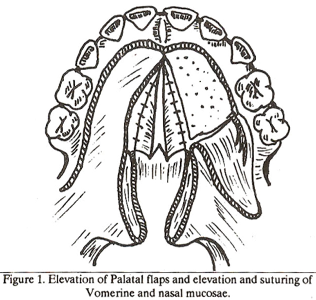
A superiorlybased pharyngeal flap was formed11 . The flap was turned up and sutured into anterior part of the nasal layer of soft palate. The donor site was closed after mobilization of the wound edges (Figure 2).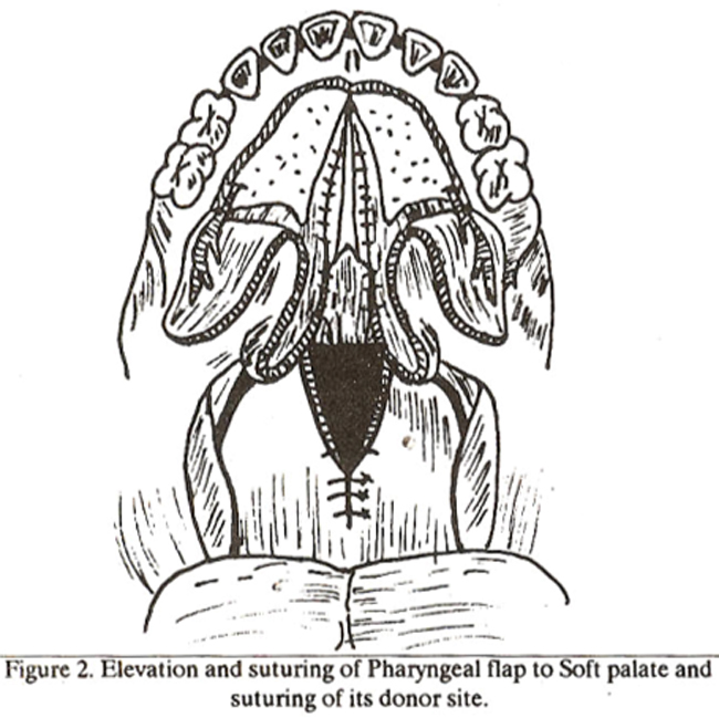
The muscles of the soft palate were approximated with deep sutures. The oral mucosal layer over the full length of palate was then closed with interrupted 4x0 black silk. The lateral releasing incisions were packed locsely for 5 days. A pledget was sewn in for a week to achieve apposition of the palatal flaps to vomerine flaps and to the bone of the hard palate. A feeding tube was introduced to put the palate at rest for a week. The silk stitches were removed from the palatal mucosa after two weeks and the patient advised palatal exercises for a month.
RESULTS
Patients were followed up post operatively. A mean follow up of 2 years was achieved with a range of 1-4 years. Success was defined as an intact and anatomically correct palate with sufficient length and movements, no escape of air through the nose during speech, no retardation Of maxillofacial growth and development. An anatomically correct palate was obtained in 24 cases (Figure-3 a,b).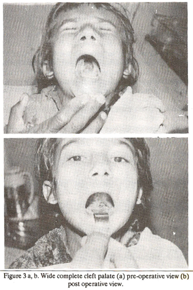
There was a failure in 1 case because of post-operative infection with breakdown of the repair line and pharyngeal flap. The palate healed with a resultant velopharyngeal incompetence that was repaired in a second stage operation. One case demonstrated a pin hole fistula over the reconstructed area that gradually healed with granulation tissue leaving an intact palate. Improvement in velopharyngeal incompetence or insufficiency was achieved completely in 24 cases and partially in one case. The speech was improved to a variable extent in all cases. Growth of the upper jaw was unaffected by palatal reconstruction in all cases to date.
DISCUSSION
This technique was well suited to the repair of wide complete palatal clefts with obvious shortening of the soft palate. The lateral elliptical defects over the palatal bone healed by granulation and provided a hard palate with normal appearing mucosa. The soft palate and the uvula appeared normal and the pharyngeal flap was not directly visible. The donor site at the base of the pharyngeal flap, where primary closure was not possible, healed bygranulation tissue. Restriction of movements of the superior constrictor muscle did not occur after formation of the cranially based pharyngeal flap. The flap provided an organic soft tissue obturator for the velopharyngeal sphincter12, sufficient room being left laterally on each side for access to the nasal cavity. Complete retropharyngeal closure depends on lateral wall movement medially to contact the flap, thus closing off the lateral airways. A physiological method of closure was maintained by the freely mobile posterior part of the soft palate. In Von Langenbeck Bipedicle flap palato-plasty anterior fixation of the palatal flap prevents lengthening of the soft palate, 10 the Veau procedure or Wardill-Kilner technique is used, therefore in cases where lengthening of the palate is necessary5. However, in 1960 Calnan13 reported that the palatal lengthening achieved by the Veau procedure and Wardill-Kilner operation is not sustained. The frequency of velopharyngeal insufficiency after repair by these techniques varies from 20-40%14,15 . In our series it was 4.0% only. In wide complete palatal clefts repaired by the Von Langenbeck, Veau or Wardill-Kilner technique, suitable flaps cannot be formed which allow closing of the palates. There is always a great danger of non-union with persisting fistulae5. Surgical treatment of the fistulae represents a considerable problem. In our surgical technique persisting fistulae were prevented by double closure in the region of the hard palate permitting use of the vomerine mucous membrane.
REFERENCES
1. Schultz, LW. Correct time, sequence and technique for closure of cleft lip and palate. J. Int. Coll. Surg., 1963; 39: 585.
2. Kernaban, D.A. and Stark. R.B. A new classification for cleft lip and palate. Plast. Reconatr. Surg., 1958; fl: 435.
3. Harkins, CS., Berlin, A., Harding, R.L, Longacre, J.J., Snodgrasse, R.M. A classification of cleft lip and cleft palate. Plast. Reconstr. Surg., 1962; 29: 31.
4. Lewis, G.K. Recent trends in surgical treatment of the cleft palate. J. Int., Coll. Surg, 1956;20:478
5. Undsay. W.K. The end results of cleft palate surgery and management, in long, term results in plastic and reconstructive surgery. Edited by RM. Goldwyn. Boston, Little Brown. 1980, p. 5&
6. Schuchardt, K. and Pfeifer, 0. Primary and secondary operation for cleft palate. J. Int. CoIL Surg., 1962; 38: 237.
7. Kremenak, CR. and Searls, J.C Experimental manipulation of midfacial growth; a synthesis of five years of research at the IOWA Maxillofacial growth laboratory. 3. Dent. Rea., 1971; 50: 1488.
8. Bernstein, L The effect of timing of cleft palate operations on subsequent growth of the maxilla, Laryngoscope, 1968; 78: 1510.
9. Veau, V. The treatment of cleft palate by operation. Proc. R. Soc. Med., 1927; 20: 1916.
10. Stellmach, R. Surgery of the common anomalies of the face, in bead and neck surgery. Edited by H.H. Naumann, Stuttgart. Georg Thieme Verlag. 1980, p. 28.
11. Lore, J.M. Jr. An atlas of head and neck surgery. 2nd ed. Philadelphia, Saunders, 1973; p.534.
12. Shprintzen, R.J.. Lewin, M.l., Croft, C.B., Daniller, A.I., Argamaso, LV., Ship, A.G. and Strauch, B. A comprehensive study of pharyngeal flap surgery . Tailor made flaps. Cleft Palate 3., 1979; 16 : 46.
13. Calnan, J.S Cleft palate lengthening of the soft palate following the V-Y repair. Br. 3. Plast. Surg., 1960; 13 : 243.
14. Krause, CJ., Tharp, R.P. and Moms, H.L A comparative study of results of the von Langenbeck and V-Y pusbback palatoplastics Cleft-Palate .1., 1976; 13: 11.
15. Bernstein, L Treatment of velopharyngeal incompetence. Arch. Otolaryngol., 1967; 85: 67.
Journal of the Pakistan Medical Association has agreed to receive and publish manuscripts in accordance with the principles of the following committees:


