

Emine Kucukates ( Laboratory Microbiology and Clinical Microbiology, Cerrahpasa Medical Faculty, Istanbul University, Istanbul, Turkey. )
Nazmi Gultekin ( Department of Cardiology, Institute of Cardiology, Cerrahpasa Medical Faculty, Istanbul University, Istanbul, Turkey. )
Yasar Bagdatli ( Department of Mirobiology and Clinical Microbiology, Cerrahpasa Medical Faculty, Istanbul University, Istanbul, Turkey. )
September 2013, Volume 63, Issue 9
Original Article

Abstract
Objective: To determine the features of patients with active infective endocarditis, and to identify causative microorganisms in blood and/or valve cultures.
Methods: The retrospective study was conducted at the Surgical Intensive Care Unit of Istanbul University Cardiology Institute, Istanbul, Turkey, and comprised 22 patients with clinical evidence of the affliction who underwent operation between January 2001 and December 2010. Modified Duke Criteria was used for the diagnosis of the condition. Frequencies and percentages were calculated.
Results: The mean age of the patients was 46.68±18 years, and 12 (54.5%) were male. Native valve endocarditis was present in 13 (59.1%) cases and prosthetic valve endocarditis was present in 9 (40.9%). Rheumatic heart disease was the commonest underlying heart disease (n=12; 54.5%). None of patient was intravenous drug abuser and positive for human immunodeficiency virus. Operative mortality was 18% (n=4). Vegetation and abscess formation were detected in 19 (86.3%) cases. Blood culture positivity was 50% (n=11). Staphylococci was the most common causative micro-organism (n=4; 36.3%), followed by Streptococci (n=3; 27.3%), and 1 (9.1%) each for Enterococci, Brucella species, Candida albicans and Stenotrophomonas maltophilia. Positive cultures from vegetations and tissue retrieved at the operation was not detected.
Conclusion: Active IE remains a severe disease affecting the young population in our study and rheumatic heart disease continues to be the most underlying heart disease. Culture negativity was found to be very high and prior antibiotic therapy may be the prime reason.
Keywords: Infective endocarditis, Blood culture, Microorganism, Treatment, Mortality. (JPMA 63: 1163; 2013).
Introduction
Active infective endocarditis (IE) is lethal if not aggressively treated with antibiotics, combined or not with surgery. Developments in antibacterial therapy, clinical microbiology, cardiac imaging and cardiac surgery have revolutionised its diagnosis and prognosis. Despite improvements in healthcare, the incidence of diseases has not changed over the past two decades. The apparent paradox results from a progressive change in risk factors for IE.1 Overall mortality remains elevated, ranging from 21% to 50% over the past three decades, with an operative mortality of 5-30% in large recently published series.2 Therefore, active IE seems to be the most serious problem in valvular heart disease, in spite of appropriate antibiotic treatment and improved surgical techniques, hospital mortality is higher than expected and the incidence of recurrence is still high.3 Bacteria isolation from valvular tissue is not always possible; healed endocarditis, broad-spectrum perioperative antibiotic therapy, systemic hypothermia, haemodilution and cold cardioplegia reduce the possibility of germ isolation from the infected tissue. Nevertheles, it is still unclear whether bacteria isolation from the infected tissue and sometimes from blood and specific postoperative antibiotic therapy may improve long-term prognosis of IE.3
The aim of this study was to definite the features of patients with active IE who were operated upon and identification of causative micro-organism in blood and/or valve cultures.
Material and Methods
We retrospectively analysed 22 patients with active IE who were operated upon between January 2001 and December 2010 in the Surgical Intensive Care Unit of Istanbul University Cardiology Institute. The patients had undergone microbiological examination of blood cultures and all excised material at valvular surgery procedures, regardless of the etiology of the valvular disease. The diagnosis of active IE was based on clinical, microbiological and echocardiographic findings. Cases of active IE were evaluated according to the Modified Duke Criteria.4
From each case of active IE, the following demographic and clinical variables were extracted; year of occurence, patient’s age and gender, underlying heart disease, length of surgical intensive care unit (SICU) admission, symptoms at presentation, duration of symptoms, fever of 38.5ºC and higher, new heart murmur, intravenous drug abuser, Anti-human immunodeficiency virus (HIV) test, antibiotics prescribed before and after the diagnosis, surgical treatment, complications and outcome. Other investigations were done as and where required. Transthoracic and transesophageal echocardiography results were obtained from reports in patients\' medical records.
Blood cultures were performed routinely in all patients who were operated upon with valvular disease and fever. At least three blood cultures were taken in all patients by three separate vein punctures at least one hour apart. Fungal blood cultures were done when thought necessary.5
Microbiological data included the number of positive blood cultures, the number of positive blood cultures drawn per patient and micro-organisms isolated from blood cultures and from all other available fluid or tissue samples (especially valve and/or vegetation obtained from surgery). For the blood cultures, the Bactec 9050 blood culture instrument (Becton Dickinson, Baltimore, Md. USA) was used. All specimens were inoculated on blood agar, endo agar (Oxoid, Unipath Ltd, Basingstoke, UK) and Mueller Hinton Broth MHB) (Oxoid); all plates were incubated for 24 h at 35ºC aerobically. Gram staining was performed on positive culture in order to identify the organism likely to be present. All microorganisms were identified using standard micro-biological procedures.6
Being a descriptive study, only percentages were calculated.
Results
Of the 22 cases there were 12 (54.54%) men and 10 (45.45%) women; overall mean age was 46.68±18 years. There were 10 (45%) patients under 40 years (range: 19-39 years); the others were above 40 years of age (range: 40-73 years). Native valve endocarditis (NVE) was present in 13 (59.1%) cases and prosthetic valve endocarditis (PVE) in 9 (40.9%). The aortic valve was infected in 7 (31.8%) cases, the mitral valve in 13 (59.1%); and multiple valves (aortic and mitral valve) in 2 (9.1%).
Rheumatic heart disease was the commonest underlying disease (n=12; 54.54%) (Table-1).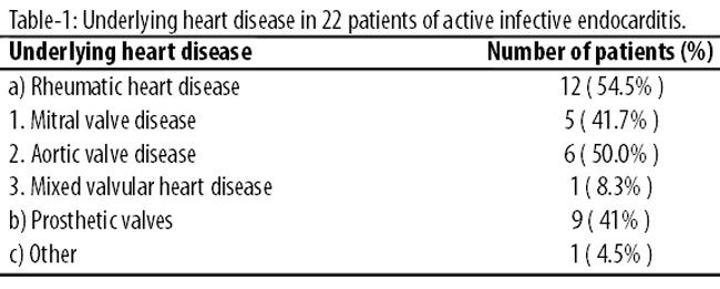
Mechanical prosthetic valves were included in 9 (41% ) patients. The side of anatomic involvements was as follows: mitral valve in 8 (36%) cases and mitral valve and aortic valve in 1 (4.5%) case. One (4.5%) case was also operated for mitral valve and coronary artery bypass grafting (CABG). The characteristics of the patients with active IE were noted (Table-2).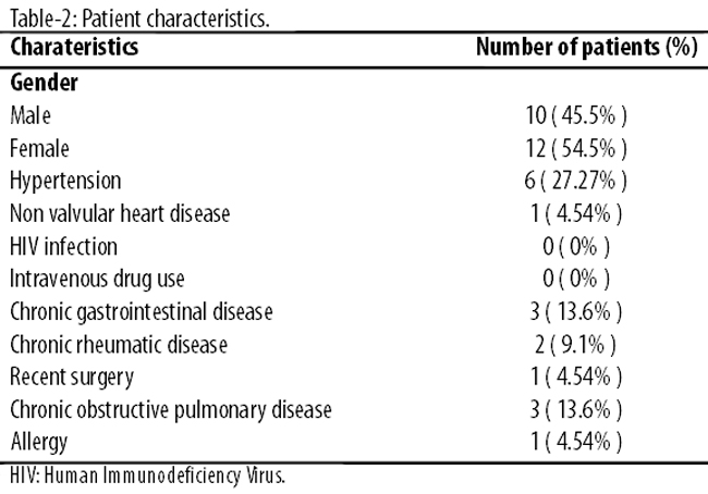
In these patients symptoms were present before the diagnosis of active IE was established. The presenting symptoms of active IE were separately noted (Table-3).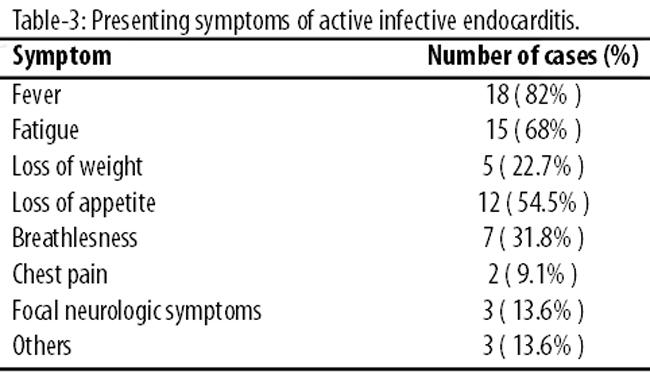
Incidence of pre-operative fever, fatigue and NVE were significantly higher in patients with positive blood cultures. There were no HIV-positive patients, and no intravenous drug abuser.
All patients underwent transthoracic echocardiography (TTE) and transesophageal echocardiography (TEE). Vegetation and abscess formation were detected in 19 (86.3%) cases. No isolates were identified from native and prosthetic valve materials. All isolates were identified from blood cultures. No patients had underlying immunosuppressive condition.
Blood cultures were positive in 11 (50%) cases (Table-4).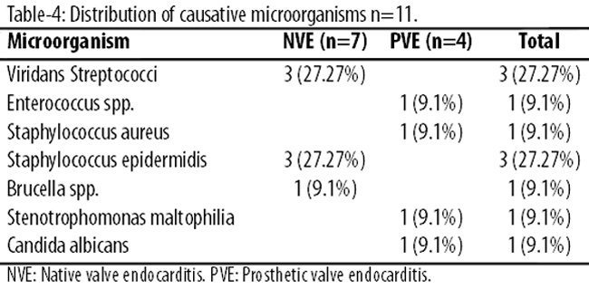
Staphylococci was the most frequent microorganisms (n=4; 36.3%). Of these 4 cases, 3 (75%) were identified Staphylococcus epidermidis (all cases; native mitral valve) and 1 (25%) case was isolated Staphylococcus aureus (prosthetic mitral valve). The other isolated microorganisms were Streptococci (n=3; 27.3%), and 1 (9.1%) Enterococci, Brucella species (native aortic valve), Candida albicans, and Stenotrophomonas maltophilia each. In the 3 patients, Viridans Streptococci (all cases;native aortic valve) was isolated. Enterococci was identified as a cause of active IE from prosthetic mitral valve. Stenotrophomonas maltophilia was isolated from prosthetic mitral valve. Another case had fungal endocarditis from prosthetic mitral valve (Candida albicans).
All patients received at least 4 weeks of post-operative multi-drug antibiotic treatment and all underwent operation before the completion of antibiotic treatment. Crystalline penicilin with or without aminoglycosides (gentamicin/amikacin/tobramicin) was the most commonly used antibiotic and used in 14 (64%) cases. Other antibiotics used were beta-lactams 6 (27%), vancomycin 4 (18%), teicoplanin 3 (13%) and fluroquinolones 4 (18%).
Hospital mortality occured in 4 (18%) patients, including 1 (25%) with NVE (multiple valves; aort and mitral valve and blood and valve culture negative case) and 3 (75%) patients with PVE (all cases; mitral valve and culture positive). The main reasons for death were septic multi-organ failure in 3 (75%) patients and cardiac failure in 1 (25%) patient.
Discussion
IE remains a frequent disease with constantly changing aspects and highly variable clinical picture, partly due to an increased frequency of intravenous drug users; a more frequent use of invasive procedures involving intravenous devices, HIV-positive patients, heart surgery with prosthetic valves and pacemaker implantation.2 In our patients, there were no HIV positivity and history of intravenous drug abuse.
In our study, rheumatic valvular disease remained the common underlying heart disease. Our patients were younger, was consistent with previously published studies from developing countries.7-9 Most of the studies in developed countries have demonstrated a gradual increase in the mean age of IE patients.4,10
Our study also confirmed many clinical features of endocarditis occuring in other parts of the world. For example, most of our cases involved native valves and the mitral valve was the most common affected.11,12 Fever was the most prevalent symptom in our study (82%), followed by fatigue (68%). The lack of fever should not exclude a diagnosis of IE in a patient with suggestive clinical features.12,13
TTE and TEE play a critical role in the decision-making process, especially when surgical treatment is contemplated. Because IE is considered a medical and surgical disease, and considering that the current rate of surgery is about 50%, echocardiography has definite value in pre-operative diagnosis and surgical planning, intra-operative confirmation of lesions and quality of repair or replacement before and after cardiopulmonary bypass, and post-operative assessment. Use of TTE and TEE in patients with clinically suspected IE explains the higher incidence of vegetation in our study and were similar to the other studies.14,15 Echocardiography had contributed to establishing the diagnosis of active IE in our patients. Vegetations were detected in 86.3% of our cases.
Many studies reported operative mortality between 5-30% in large recently published series.2,16 In our study, mortality was 18% among patients. Deaths in all patients were directly attributed to active IE if patients died from either sepsis or recognised major complications.
In our study, 50% of patients with active IE were blood culture positive. Blood cultures have been found to be positive in more than 80% patients with IE in European countries.7,17 Prior antibiotic therapy may be the important reason for high culture negativity (50%) in this study.
A significant proportion of culture-negative patients had received antibiotics therapy before admission to our unit (81.8%). In several studies, the incidence of negative blood cultures ranges were 20-60% of cases who had undergone surgical treatment.3,8 Culture-negative disease is often associated with antibiotic consumption within the previous 2 weeks. IE might also be due to fastidious (example; Haemophilus aphrophius, Adinopacillus actinomycetemcomitans, Cardiobacterium hominis, Eikenella corroclens and Kingella kingae [HACEK] group micro-organisms) or intracellular pathogens (example; Chlamydia spp.) that are not easily detected by standard culture conditions. Many factors such as healed endocarditis, low virulence germs, use of broad-spectrum antibiotic and poor techniques of blood sampling may account for the lack of identification of micro-organisms from the blood. Positive cultures from vegetations and tissue retrieved at the operation was not found in our unit. Failure in detecting micro-organisms from vegetations and explanted materials may be caused by healed endocarditis, use of broad-spectrum antibiotics, attitude to prime oxygenator with crystalloid cardioplegia that may cause an osmotic and physical shock to the bacteria still living in the vegetations.4,18
In recent years, staphylococci have overtaken streptococci as the most frequent causes of native and prosthetic valve endocarditis.3,7,9,17,19-22 In this study, staphylococci were the most common pathogen. All of staphylococci were methicillin sensible. Staphylococcus epidermidis was identified from native mitral valve of the three patients. Staphylococcus aureus was isolated from prosthetic mitral valve of the patient. Viridans streptococci was identified as second most frequent from native aortic valve of three patients. Enterococcus species was isolated as a cause of active IE from prosthetic mitral valve of patients who died post-operatively.
Brucella infective endocarditis is an uncommon, but life-threatening complication of brucellosis. The aortic valve is the most commonly affected cardiac valve. Hence, it is an important cause of morbidity, especially in countries of the Mediterranean basin and of the Middle East. Several studies reported brucella endocarditis from aortic and mitral valves.23,24 We detected brucella endocarditis in a patient of native aortic valve and he was treated successfully.
Stenotrophomonas maltophilia is a nosocomial pathogen resistant to multiple antibiotics. It is a rare cause of endocarditis and only 21 previous cases of S. maltophilia endocarditis have been reported in literature.25 Of these, 11 cases had prosthetic valve endocarditis and the others native valve endocarditis. S. maltophilia endocarditis carries high mortality and morbidity. The prosthetic valves are commonly involved than the native valve.25 In our study, S. maltophilia was identified as a cause of active IE from a prosthetic mitral valve of patient who died post-operatively.
Candida species are an uncommon cause of infective endocarditis. Fungi are responsible for 1.8% to 10% of cases of prosthetic valve endocarditis. Candida albicans is the most common species and stressed the difficulty encountered in making the diagnosis of Candida endocarditis.26,27 In our unit, Candida albicans was isolated as a cause of fungal prosthetic mitral valve endocarditis who died post-operatively.
Conclusion
Infective endocarditis remains a severe disease affecting a young people in our unit. Rheumatic heart disease continues to be the major underlying heart disease. Survival was higher in patients with native valve than those with prosthetic valve involvement. Isolation of causative micro-organism is an essential part of diagnosis of IE for an appropriate antimicrobial therapy. Identification of the pathogen in culture-negative disease depend on special procedures. In such situaton, The other diagnostic tests such as serology, agglutination, indirect fluorescence, ELISA, complement fixation and polymerase chain reaction (PCR) can be used for definitive diagnosis. Culture-negative IE is a problem because unusual pathogens might not respond to empirical treatment. Molecular results can remain positive even after long-term anti-microbial therapy.
References
1. Moreillon P, Que Y. Infective endocarditis. Lancet 2004; 363: 139-49.
2. Mouly S, Ruimy R, Launay O, Arnoult F, Brochet H, Trouillet JL, et al. The changing clinical aspects of infective endocarditis: descriptive review of 90 episodes in a French teaching hospital and risk factors for death. J Infect 2002; 45: 246-56.
3. Renzulli A, Carozza A, Marra C, Romano GP, Ismeno G, De Feo M, et al. Are blood and valve cultures predictive for long-term outcome following surgery for infective endocarditis? Eur J Cardiothorac Surg 2000; 17: 228-33.
4. Li JS, Sexton DJ, Mick N, Nettles R, Fowler VG Jr, Ryan T, et al. Proposed modifications to the Duke criteria for the diagnosis of infective endocarditis. Clin Infect Dis 2000; 30: 633-8.
5. Rubeinstein E, Lang R. Fungal endocarditis. Eur Heart J 1995; 16 (Suppl B): 84-9.
6. Carroll KC, Weinstein MP. Manual and automated systems for detection and identification of microorganisms. In: Murray PR, Baron EJ, Jorgensen JH, Landry ML, Pfaller MA, (eds.). Manual of Clinical Microbiology. 9th ed. Washington DC; American Society for Microbiology; 2007; pp 192-217.
7. Roca B, Marco JM. Presentation and outcome of infective endocarditis in Spain: a retrospective study. Int J Infect Dis 2007; 11: 198-203.
8. Jalal S, Khan KA, Alai MS, Jan V, Iqbal T, Tramboo NA, et al. Clinical spectrum of infective endocarditis: 15 years experience. Indian Heart J 1998; 50: 516-9.
9. Garg N, Kandpal B, Garg N, Tewari S, Kapoor A, Goel P, et al. Characteristics of infective endocarditis in a developing country-clinical profile and outcome in 192 Indian patients, 1992-2001. Int J Cardiol 2005; 98: 253-60.
10. Di Salvo G, Habib G, Pergola V, Avierinos JF, Philip E, Casalta JP, et al. Echocardiography predicts embolic events in infective endocarditis. J Am Coll Cardiol 2001; 37: 1069-76.
11. Hoen B, Alla F, Selton-Suty C, Beguinot I, Bouvet A, Briançon S, et al. . Changing profile of infective endocarditis: results of a 1-year survey in France. JAMA 2002; 288: 75-81.
12. Netzer RO, Zollinger E, Seiler C, Cerny A. Infective endocarditis: clinical spectrum, presentation and outcome. An analysis of 212 cases 1980-1995. Heart 2000; 84: 25-30.
13. Fefer P, Raveh D, Rudensky B, Schlesinger Y, Yinnon AM. Changing epidemiology of infective endocarditis: a retrospective survey of 108 cases, 1990-1999. Eur J Clin Microbiol Infect Dis 2002; 21: 432-7.
14. Van Hal SJ, Mathur G, Kelly J, Aronis C, Cranney GB, Jones PD. The role of transthoracic echocardiography in excluding left sided infective endocarditis in Staphylococcus aureus bacteraemia. J Infect 2005; 51: 218-21.
15. Mestres CA, Fita G, Azqueta M, Miró JM. Role of echocardiogram in decision making for surgery in endocarditis. Curr Infect Dis Rep 2010; 12: 321-8.
16. Sekido M, Takano T, Takayama M, Hayakawa H. Survey of infective endocarditis in the last 10 years: analysis of clinical, microbiological and therapeutic features. J Cardiol 1999; 33: 209-15.
17. Pazdernik M, Baddour LM, Pelouch R. Infective endocarditis in the Czech Republic: eight years of experience at one of the country\'s largest medical centers. J Heart Valve Dis 2009; 18: 395-300.
18. Van den Berg L. Effect of temperature on growth and activity of a methanogenic culture utilising acetate. Can J Microbiol 1977; 23: 898-902.
19. Cicalini S, Puro V, Angeletti C, Chinello P, Macrì G, Petrosillo N. Profile of infective endocarditis in a referral hospital over the last 24 years. J Infect 2006; 52: 140-6.
20. Nunes MC, Gelape CL, Ferrari TC. Profile of infective endocarditis at a tertiary care center in Brazil during a seven-year period: prognostic factors and in-hospital outcome. Int J Infect Dis 2010; 14: e394-8.
21. Lupis F, Giordano S, Pampinella D, Scarlata F, Romano A. Infective endocarditis: review of 36 cases. Infez Med 2009; 17: 159-63.
22. Lee JH, Burner KD, Fealey ME, Edwards WD, Tazelaar HD, Orszulak TA, et al. Prosthetic valve endocarditis: clinicopathological correlates in 122 surgical specimens from 116 patients (1985-2004). Cardiovasc Pathol 2011; 20: 26-35.
23. Hadjinikolaou L, Triposkiadis F, Zairis M, Chlapoutakis E, Spyrou P. Successful management of Brucella mellitensis endocarditis with combined medical and surgical approach. Eur J Cardiothorac Surg 2001; 19: 806-10.
24. Sasmazel A, Baysal A, Fedakar A, Bugra O, Ozkokeli M, Büyükbayrak F, et al. Treatment of Brucella endocarditis: 15 years of clinical and surgical experience. Ann Thorac Surg 2010; 89: 1432-6.
25. Mehta NJ, Khan IA, Mehta RN, Gulati A. Stenotrophomonas maltophilia endocarditis of prosthetic aortic valve: report of a case and review of literature. Heart Lung 2000; 29: 351-5.
26. Falcone M, Barzaghi N, Carosi G, Grossi P, Minoli L, Ravasio V, et al. Candida infective endocarditis: report of 15 cases from a prospective multicenter study. Medicine (Baltimore) 2009; 88: 160-8.
27. Talvard M, Paranon S, Dulac Y, Mansir T, Kreitmann B, Acar P. Candida albicans endocarditis after pulmonary artery banding. Arch Pediatr 2009; 16: 1146-9.
Journal of the Pakistan Medical Association has agreed to receive and publish manuscripts in accordance with the principles of the following committees:


