

Leili Hafizi ( Department of Obstetrics and Gynecology, Imam Reza Hospital, Mashhad, Iran. )
Zahra Mirfeizi ( Department of Rheumatology, Imam Reza Hospital, Mashhad, Iran. )
Nastaran Razmjoo ( Faculty of Nursing and Midwifery, Mashhad University of Medical Sciences, Mashhad, Iran. )
Maliheh Keshvari ( Department of Urology, Fellowship of Urogynecology, Imam Reza Hospital, Mashhad, Iran. )
Atefeh Jabbari ( Faculty of Nursing and Midwifery, Mashhad University of Medical Sciences, Mashhad, Iran. )
Hami Ashraf ( Women Health Research Center, Imam Reza Hospital, Faculty of Medicine, Mashhad, Iran. )
Fatemeh Yousefi ( Women Health Research Center, Imam Reza Hospital, Faculty of Medicine, Mashhad, Iran. )
September 2013, Volume 63, Issue 9
Original Article

Abstract
Objective: To determine whether joint hypermobility is associated with pelvic organ prolapse.
Methods: The case-control study was conducted from January to April 2011 and comprised 30 women with pelvic organ prolapse, stage > II and 30 controls with stages 0 and I with similar age and parity. They were recruited from the gynaecology clinic at Imam Reza Hospital in Mashhad, Iran. The condition was evaluated by a quantification system and, for the purposes of this study, pelvic organ prolapse was defined as stage > II. All the subjects were examined in the dorsal lithotomic position with an empty bladder. A separate investigator evaluated each subject for joint hypermobility by using Beighton score which was calculated by doing five simple manoeuvres. SPSS 11.5 was used for data analysis.
Results: The mean age of the 30 cases was 35.40±6.39 years, while for the controls it was 35.36±5.9 years. Overall clinical joint hypermobility was found in 24 of the 60 (40%) subjects. There were no significant difference in the prevalence of joint hypermobility between the two groups. The prevalence of hypermobility in the cases was 36.7% (n=11) versus 43.3% (n=13) in the controls (p= 0.59). The prevalence of cystocele in subjects with joint hypermobility was 41.7% (n=10) versus 38.9% (n=14), (p <0.83); rectocele 33.3% (n=8) versus 41.7% (n=15), (p <0.73) women with normal joint mobility. No Significant differences were found between the groups with regard to other markers of connective tissue weakness such as the presence of varicose veins (p<0.37), easy bruising (p<0.43) and observed striae (p<0.42).
Conclusion: Joint hypermobility was not associated with pelvic organ prolapse in the study population. Further studies involving more patients with pelvic organ prolapse are recommended.
Keywords: Pelvic organ prolapse, Joint instability, Connective tissue diseases. (JPMA 63: 1152; 2013).
Introduction
Pelvic organ prolapse (POP) is a global health problem, and is believed to affect adult women of all ages and decrease their quality of life (QoL) considerably.1 Despite the high incidence of POP, little is known about its underlying pathophysiology. The etiology of POP is considered to be multifactorial based on the integrated life span model.2 It can be assumed that the development of POP includes predisposing factors, inciting factors and intervening factors.3 Unfortunately, only a minority of these risk factors can easily be prevented.
One possible cause of genital prolapse is an inherent weakness or laxity of the connective tissue of pelvic support structures.4 Joint hypermobility is a very common finding in conditions of known connective tissue abnormalities, such as the Ehlers-Danlos syndrome, and is considered a reliable marker for altered connective tissue.5 There is little evidence on prolapse and joint hypermobility in different populations.3,5-9 But some findings suggest that local, rather than systemic, alterations in biomechanical skin properties are associated with POP.10
The purpose of this study was to determine whether joint hypermobility, a clinical marker for connective tissue abnormalities, is associated with POP in northeastern Iranian women.
Patients and Methods
The case-control study comprised women aged 18 to 49 years attending gynecological clinics at the Imam Reza Hospital, Mashhad University of Medical Sciences, Mashhad, Iran, between January and April 2011. The sample size was calculated on the basisof prevalence data of joint hypermobility among Turkish women6 due to similarities in the socioeconomic and genetic profile of the two countries. With a power of 80 and alpha (type 1 error) of 0.05, the sample size was calculated through PASS software to be 29 in each group. The study included 30 female patients with POP (cystocele, rectocele, desensus uteri, prolapsus uteri, vaginal vault prolapse). After approval by the Institutional Ethics Committee, informed written consent was obtained from all the participants who were given a brief description of the research by their physician before a pelvic organ prolapse quantification (POP-Q) examination for staging was performed.11
Women with no delivery during the last year were included in the study. The exclusion criteria comprised: menopause, previous POP surgery, neurological disorders, pelvic cancer and mass, psychiatric disorders and history of joint problems (systemic lupus erythematosus, rheumatoid arthritis, Ehlers-Danlos syndrome or any fracture near or in a joint).
All the subjects were matched on age and parity, and stratified by the degree of prolapse. For POP-Q evaluation, all the subjects were examined by one physician in the dorsal lithotomic position with an empty bladder. POP was defined as stage >2.12 Determining the Beighton score is essential for making the diagnosis of joint hypermobility and is calculated by doing simple manoeuvres. A Beighton score of 4 or more is considered indicative of joint laxity.3,4,6,13 All the nine tests performed in our study have been recommended by the British Society of Rheumatology and have been tested for reliability.14
The tests include passive extension of each five fingers, passive apposition of each thumb to the forearm, hyperextension of each elbow, hyperextension of each knee and trunk flexion to allow the palms to lie flat on the floor.
The hypermobility was independently examined by a separate researcher blinded to the results of the physical examination.
Other markers of connective tissue weakness such as varicose veins, easy bruising and striae were also assessed.3 Varicose veins and a tendency to get bruised easily were assessed through a questionnaire. The presence of striae was investigated during clinical examination.
Each woman completed the detailed questionnaire regarding her medical and gynaecological history, including age, parity, body mass index (BMI), obstetrical factors, smoking, heavy work, physical activity, family history of pelvic floor disorders, socioeconomic status, contraception, and oestrogen replacement therapy.
Goniometer was used for accurate joint angle measurement.
Women were defined as postmenopausal if they had gone 12 months without a menstral period. The BMI was calculated from the measured weight and self-reported height. High socioeconomic status was assessed through the questionnaire and defined as having an income of 10 million Rials per month (about 10,000 USD) or more and an education at the university level or higher. Current smoking was assessed through the questionnaire. If a woman reported smoking, she was asked how many cigarettes she smoked per day.
To be classified as doing heavy occupational work, three variables in the questionnaire were needed to be present. The jobs were classified regarding physical exertion requirements as light, medium and heavy: self-report of occupation as physically heavy; lifting more than 20 heavy lifts per week; and working in a standing position for more than 50% of the labour time. If one or two factors were present, the job was classified as medium, and if none was present, the job was classified as light.
A positive family history of pelvic floor disorders was assessed through the questionnaire, asking: "Has your mother or grandmother experienced pelvic floor disorders" (\'Yes\', \'do not know\', \'no\').
The question about exercises pattern was: "How much average time do you spend in the activities such as walking, jogging, bicycling, swimming, gymnastics, ball-playing during a week" The answers were categorised into (1) less than 2 hours, (2) 2-5 hours, or (3) more than 5 hours.
A semi-structured interview addressed the recall of obstetric factors. Birth weight and the number and types of birth were registered.
Varicose veins and a tendency to get bruised easily were assessed through the questionnaire. The presence of striae was observed during clinical examination. Beighton\'s scoring system was used to assess joint mobility, and hypermobility was defined as four or more positive tests out of nine.
Statistical analysis was performed on SPSS version 11.5. Independent two samples T test was used to compare the mean age of the groups. Differences between the case and the control groups were evaluated by K2 test and one-way analysis of variance (ANOVA). P values <0.05 was considered to be significant.
Results
The age of the 60 participants ranged between 24 and 49 years.
In the control group, 13 (43.3%) women were classified as stage 0 and 17 (56.7%) as stage I on POP-Q. In the POP group, 29 (96.7%) women had stage II, and 1 (3.3%) was stage III on POP-Q. Besides, 24 (80%) patients in the POP group had prolapse in one vaginal compartment, whereas 15 (50%) had prolapse in two compartments (Table-1).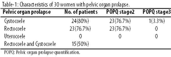
The mean age of the cases was 35.40±6.39 years and 35.36±5.9 years in the controls. The two groups were similar in age, BMI, parity (p < 0.98; 0.99; 0.34) (Table-2).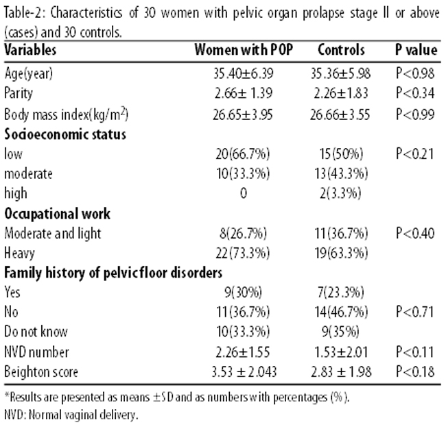
Women in the two groups had a median parity of two (range 0 to 7).
There was no significant difference in the mean number of caesarean section (1.01±0.18 versus 0.83±0.15; p<0.33) and vaginal delivery (1.55±0.28 versus 2.01±3.67; p<0.11) between the groups. However, the number of patients with positive history of caesarean section was significantly lower (n=8; 26.7% versus n=16; 53.3%; p<0.035) and vaginal delivery (n=26; 86.7% versus n=16; 53.3%; p<0.005) was significantly higher in the POP group.
The prevalence of cystocoele in subjects with joint hypermobility was (10 of 24 [41.7%] versus 14 of 36 [38.9%], p= 0.83); rectocoele (8 of 24 [33.3%] versus 15 of 36 [41.7%]; p=0.73) compared to women with normal joint mobility (Table-3).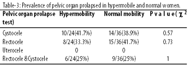
The mean Beighton score was 3.53±2.0.43 in the case group, and 2.83±1.98 in the control (p<0.18). No significant difference was found between the two groups in Beigton score.
The prevalence of joint hypermobility in the POP group was 36.7% (n=11) versus 43.3% (n=13) in healthy women (p<0.59). No significant differences were found between the groups regarding other markers of connective tissue weakness such as the presence of varicose veins (p=0.37), easy bruising (p=0.43) and observed striae (p=0.42) (Table-4).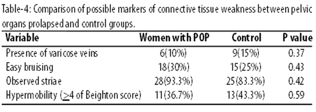
Discussion
The findings demonstrated that the prevalence of joint hypermobility was 40% in the study population. There were no significant differences in joint hypermobility among women with POP, compared to the controls.
These findings contradict prior research that suggests women with POP are more likely to have a joint hypermobility and systemic collagen or elastin disorder.4
We found no other studies that compared hypermobility in women between 18-45 years of age regarding POP. One study reported no association between hypermobility and pelvic organ mobility in a study on nullipar young women.15 Similar results are found in a study that characterised and compared pelvic floor muscle function and pelvic organ descent in high-impact, frequent intense training athletic and non-athletic women. There was no correlation between Beighton score and biometric indices of hiatal size or pelvic organ mobility in that study.16
Another one found that women with clinically hypermobile joints were more likely to have POP than women without hypermobile joints.5 Joint laxity prevalence was 36% in prolapsus patients that was similar with ours (36.7% in the prolapsed group), but there are significant differences between the two studies. In the other study joint laxity was evaluated by using Carter-Wilkinson criteria, and POP was examined in the standing position and non-straining position for cystocoele, rectocoele and uterine prolapse However, Beigton score and POPQ system was used in our study, which is an efficinet system that allows researchers to report findings in a standardised fashion.6
One study examined 76 Iraqi women with genital prolapse and found that 66% had clinical joint hypermobility.7 The control group had an 18% prevalence of joint hypermobility. In Turkey, a study found 53.8% of the prolapsus women to have joint laxity which was higher than our results.6 This may be explained by the variations of hypermobility prevalence among some racial groups.17 The average age of our subjects was 35 years whereas it was 54 in the Turkish study. Both the Iraqi and Turkish studies showed that the prevalence of joint hypermobility was higher in the POP group which is in contrtast with our results.6,7
Varicose veins, striae, diastasis recti abdominis and easy bruising are markers of systemic alterations in connective tissue, and these factors have not been the focus of previous POP surveys. A recently published study assessed these markers and showed differences amongst women with POP and controls with regard to bruising and varicose veins; the latter being strongly associated with POP.3 The study was in agreement with our own, showing no significant relationship between joint hypermobility and POP among women with Ehlers-Danlos syndrome.18
In another study a positive association between joint hypermobility and recurrent POP was found. It suggested connective tissue abnormalities as an underlying factor for POP by studying type I and III collagen metabolism products.4
A recently study compared histological structure of POP and healthy individuals and reported that underlying elastin and collagen structures were equivalent in the two groups.10 The results suggest that POP is a local, rather than systemic connective tissue disorder. Biomechanical tissue quality, pelvic floor muscle deficits, and defects in pelvic floor connective tissues are etiologies for pelvic organ prolapse.
Though each of these theories provides some insight to the evolution of POP, no theory adequately explains all cases. More likely, a combination of these forces work together to cause POP. It is nesessary that further understanding of the pathogenesis of POP is achieved to improve the QOL for these women.
One of the most important limitations in our study was the sample size. The clinical diagnosis of ligament laxity was also one of the limitations. The focus was on ensuring that all physical examinations were performed by just one physician to prevent highly probable measurement bias. Because of our clinic settings, (presence of the physician on just two days per week and high diversity in the refereed patients in our clinic) most of the patients did not meet the inclusion criteria. In the absence of enough literature, we calculated the sample size on the basis of a study in Turkey,6 which has similar socio-economic conditions as Iran. We did all we could to make age and other effective variables similar between the two groups.
Conclusion
Joint hypermobility was not found to be associated with genital prolapse. Further studies involving younger patients with POP are recommended. However, molecular studies are also required to determine the genetic basis of POP.
Acknowledgment
We are grateful to all those who participated in data collection and statistical analysis.
References
1. Kerkhof MH, Hendriks L, Brölmann HA. Changes in connective tissue in patients with pelvic organ prolapsed - a review of the current literature. Int Urogynecol J Pelvic Floor Dysfunct 2009; 20: 461-74.
2. DeLancey JO, Kane Low L, Miller JM, Patel DA, Tumbarello JA. Graphic integration of causal factors of pelvic floor disorders: an integrated life span model. Am J Obstet Gynecol. 2008; 199: 610.e1-5.
3. Braekken IH, Majida M, Ellström Engh M, Holme IM, Bø K. Pelvic floor function is independently associated with pelvic organ prolapse. BJOG 2009; 116: 1706-14.
4. Knuuti E, Kauppila S, Kotila V, Risteli J, Nissi R. Genitourinary prolapse and joint hypermobility are associated with altered type I and III collagen metabolism. Arch Gynecol Obstet 2011; 283: 1081-5.
5. Norton PA, Baker JE, Sharp HC, Warenski JC. Genitourinary prolapse and joint hypermobility in women. Obstet Gynecol 1995; 85: 225-8.
6. Aydeniz A, Dikensoy E , Cebesoy B, Altinda O, Gürsoy S, Balat O. The relation between genitourinary prolapse and joint hypermobility in Turkish women. Arch Gynecol Obstet 2010; 281: 301-4.
7. Al-Rawi ZS, Al-Rawi ZT. Joint hypermobility in women with genital prolapse. Lancet 1982; 1: 1439-41.
8. Bai SW, Choe BH, Kim JY, Park KH. Pelvic organ prolapse and connective tissue abnormalities in Korean women. J Reprod Med 2002; 47: 231-4.
9. Lammers K, Lince SL, Spath MA, van Kempen LC, Hendriks JC, Vierhout ME, et al. Pelvic organ prolapse and collagen-associated disorders. Int Urogynecol J 2012; 23: 313-9.
10. Epstein LB, Graham CA, Heit MH. Systemic and vaginal biomechanical properties of women with normal vaginal support and pelvic organ prolapse. Am J Obstet Gynecol 2007; 197: 165. e1-6.
11. Bump RC, Mattiasson A, Bø K, Brubaker LP, DeLancey JO, Klarskov P, et al. The standardization of terminology of female pelvic organ prolapse and pelvic floor dysfunction. Am J Obstet Gynecol. 1996; 175: 10-7.
12. Weber AM, Abrams P, Brubaker L, Cundiff G, Davis G, Dmochowski RR, et al. The standardization of terminology for researchers in female pelvic floor disorders. Int Urogynecol J Pelvic Floor Dysfunct 2001; 12: 178-86.
13. Beighton P, Solomon L, Soskolne CL. Articular mobility in an African population. Ann Rheum Dis 1973; 32: 413-8.
14. Remvig L, Jensen DV, Ward RC. Are diagnostic criteria for general joint hypermobility and benign joint hypermobility syndrome based on reproducible and valid tests. A review of the literature. J Rheumatol 2007; 34: 798-803.
15. Dietz HP, Eldridge A, Grace M, Clarke B. Pelvic organ descent in young nulligravid women. Am J Obstet Gynecol 2004; 191: 95-9.
16. Kruger JA, Dietz HP, Murphy BA. Pelvic floor function in elite nulliparous athletes. Ultrasound Obstet Gynecol 2007; 30: 81-5.
17. Hakim AJ, Sahota A. Joint hypermobility and skin elasticity: the hereditary disorders of connective tissue. Clin Dermatol 2006; 24: 521-33.
18. McIntosh LJ, Mallett VT, Frahm JD, Richardson DA, Evans MI. Gynecologic disorders in women with Ehlers-Danlos syndrome. J Soc Gynecol Invest 1995; 2: 559-64.
Journal of the Pakistan Medical Association has agreed to receive and publish manuscripts in accordance with the principles of the following committees:


