

Cem Ungor ( Department of Oral and Maxillofacial Surgery, Faculty of Dentistry, Karadeniz Technical University, Turkey. )
Anil Guven ( Department of Oral and Maxillofacial Surgery, Faculty of Dentistry, Ankara University, Turkey. )
Timur Songur ( Department of Oral and Maxillofacial Surgery, Faculty of Dentistry, Ankara University, Turkey. )
Ezher Dayisoylu ( Department of Oral and Maxillofacial Surgery, Faculty of Dentistry, Karadeniz Technical University, Turkey. )
Hakan Kurt ( Department of Oral and Maxillofacial Radiology, Faculty of Dentistry, Ankara University, Turkey. )
Emre Tosun ( Department of Oral and Maxillofacial Surgery, Faculty of Dentistry, Karadeniz Technical University, Turkey. )
Figen Cizmeci Senel ( Department of Oral and Maxillofacial Surgery, Faculty of Dentistry, Karadeniz Technical University, Turkey. )
September 2013, Volume 63, Issue 9
Original Article

Abstract
Objective: To determine the relative success of two different bone grafting material - putty and powder forms of De-mineralised Bone Matrix (DBM) - used in sinus lift procedure.
Methods: The retrospective study was conducted at the Department of Oral and Maxillofacial Surgery, Ankara University, Ankara, Turkey, and comprised data related to the patients referred for bilateral maxillary sinus augmentation between 2007 and 2010. During the period, 48 endoosseous implants were placed concurrently with the sinus augmentation in 12 patients. Marginal bone loss around the implants was measured at the time of loading, 12 and 30 months after the treatment. SPSS 11.5 was used for data analysis.
Results: Of the 12 patients, 8 (66.6%) were females and 4 (33.3%) were males. All implants osseointegrated in both the putty and powder groups well without any significant clinical finding. The average volume of marginal bone resorption at implants for the putty side was 0.43±0.22 mm, 0.8±0.33 mm and 1.12±0.49 mm at prosthetic loading, 12-month and 30-month follow-up, respectively. For the powder side, the corresponding numbers were 0.48±0.32 mm, 0.82±0.46 mm and 1.24±0.57 mm. No statistically significant difference in bone loss between the two groups was observed (p >0.05).
Conclusion: Both putty and powder forms of de-mineralised Bone Matrix showed satisfactory results and there was no significant difference in marginal bone loss around dental implants and survival rates.
Keywords: Sinus lift, Bone graft, Dental implant, De-mineralised Bone Matrix. (JPMA 63: 1124; 2013).
Introduction
Rehabilitation of edentulous posterior maxilla with dental implants is a challenging problem in oral and maxillofacial surgery due to alveolar resorption and excessive pneumatisation of maxillary sinus that follows teeth extraction. In cases of inadequate bone volume, sinus augmentation procedure with bone grafts is considered effective for endosseous implant survival. Sinus augmentation has become a routine procedure to accommodate maxillary bone to the needs of the endosseous implant.1,2
There are various types of graft material used in the sinus augmentation procedure. Autogenous bone grafts are considered the gold standard for graft materials3 but potential complications emerging at the donor site and prolonged operation time may accompany the procedure.1,4 Therefore, alternative approaches have been devised to overcome these disadvantages by using bone substitutes such as allografts, tricalciumphosphate, hydroxyapatite, glass ceramic and xenografts.
Over the years de-mineralised bone matrix (DBM) has been frequently used for bone grafting. DBM was first described in 1889 by Senn and used in oral and maxillofacial surgery in 1975.5,6 A study showed that DBM could be successfully used in cases of small defects. DBM contains active proteins such as bone morphogenetic protein (BMP), transforming growth factor-beta (TGF-b), osteogenin, insulin-like growth factor, and fibroblast growth factor, which are mostly regarded as members of the TGF-b superfamily.7 In recent years, several studies have demonstrated the success of DBM for reconstructive maxillofacial surgery, and sinus augmentation is used in various graft sizes and forms.2-4,8-10
Marginal bone loss is considered to be an important criteria for evaluating the success of dental implants. It can range from the loss of marginal bone to the complete failure of the implant and dramatically decreases after the first year. Hypotheses for the causes of crestal bone loss include the reflection of the periosteum during surgery, preperation of the implant osteotomy, level of the microgap between the fixture and osteotomy, bacterial invasion, the establishment of a biological width, the implant crest module design and occlusal overload.11
There have been studies comparing the types of DBM based on particulate size, survival of implants and operation time.4-8 In these studies, there were no significant difference in terms of implant success during the loading time, but the putty form was found to be more successful than the powder form for the ease of application and operation time. Also, optimal bone induction was found with DBM particle sizes of 250 to 500µm. On the other hand, marginal bone resorption and implant success between the putty and powder forms has not been evaluated. The aim of this retrospective study was to compare the dental implant survival rate and the marginal bone resorption around dental implants placed following sinus augmentation with putty and powder forms of DBM.
Materials and Methods
The retrospective study comprised patients referred to the Department of Oral and Maxillofacial Surgery at Ankara University, Ankara, Turkey, for bilateral maxillary sinus augmentation between 2007 and 2010. All the patients were American Society of Anaesthesiology (ASA) I with no systemic disease and had good oral hygiene regarding plaque and gingival status. Before the procedure, the anatomy and pathology of the sinuses were evaluated using panaromic view and Water\'s view. According to Cawood-Hawell\'s classification,12 Class V and VI cases were included in the study. Patients who had residual bone height less than 2mm were excluded. The other exclusion criteria were sinus pathologies, systemic diseases, smoking habits, alcohol consumption and poor oral hygiene. All patients underwent bilateral sinus surgery and the residual bone height of the edentulous sites for implant placement was measured. The study was conducted with the approval of the Ankara University Faculty of Dentistry Research Ethics Committee. Two most preferred forms of DBM — putty and powder — were applied for sinus augmentation during the same session.
A total of 24 sinus lifts were performed and 48 dental implants (Nucleoss Implants, Izmir, Turkey) measuring 3.4 to 5.0 mm in width and 12 to 14 mm in length were placed concurrently with sinus augmentation. Four implants each were inserted in grafted regions of 12 patients. In all patients, the left side was grafted with DBM putty form (DynaGraft Keystone Dental, Burlington, Massachusetts) and the right side was grafted with DBM powder form (Pacific Coast Tissue Bank, Los Angeles, California). None of the implants were loaded before a minimum of 6 months from the date of first surgery. Implant success was evaluated based on the clinical and radiologic criterias13,14 that included: absence of mobility; absence of persistent subjective complaints (pain, foreign body sensation and/or dysaesthesia); absence of a continuous radiolucency around the implant; and marginal bone level changes in the first year implant insertion less than 1-1.5mm and the ongoing annual bone loss less than 0.2mm.
All operations were performed under local anaesthesia, using articaine hydrochloride (Ultracaine D-S Forte®Aventis). With a mid-crestal incision and vertical releasing incisions, a mucoperiosteal flap was elevated to expose the sinus wall. An osteotomy was carried out on the lateral wall of the maxilla with a round bur. The schneiderian membrane was detached from the sinus wall to create space for the placement of the graft materials. Implant sockets were then prepared and the dental implants were placed. The left and right maxillary sinuses were augmented in the same session in each patient. The left sinuses were augmented with DBM putty form and the right sinuses were augmented with DBM powder form after a minimum of 30 min rehydration process in 0.9% Saline solution. After the graft had been placed, the flap was re-positioned and sutured with 3/0 silk suture. Antibiotic (Augmentin 625mg, Glaxo Smith Kline, Britian) and analgesic (Apranax Fort 550 mg, Abdi Ibrahim, Turkey) therapy was administrated 1 hour before surgery and for 5 days following the surgery. Chlorohexidine gluconate 0.12% mouthwash was used twice daily for 2 weeks. The patients were advised to have a soft diet and to avoid sneezing till suture removal.
A rigid cross-arch bar was used with bite-registration material and Rinn XCP (Dentsply , Des Plaines, IL) rod and ring were firmly attached to the bar and placed in contact with the X-ray cone. Marginal bone loss around mesial and distal side of the implants were measured (in mm) at the time of loading, after 12 and 24 months of loading by a digital plot (Figure-1).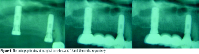
For measurements purposes, 2 visible and easily localised reference points were selected at the junction point between the implant and prosthetic restoration. A straight line was traced joining the 2 reference points (Figure-2).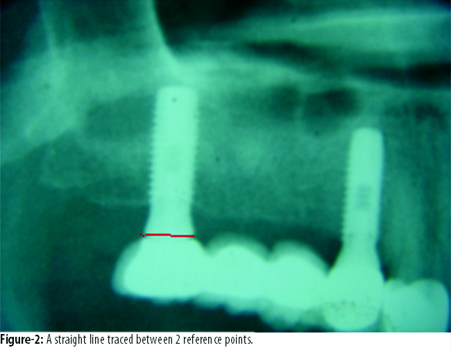
The marginal bone resorption was determined by measuring between this line and the highest crestal bone point around the implant.
The study was approved by the Research Ethics Committee of the Ankara University, Faculty of Dentistry.
Statistical analyses were performed with SPSS 11.5. Two-way repeated measures analysis of variance (ANOVA) and Fisher\'s LSD tests were used for between and within group comparisons. A p value less than 0.05 was considered significant.
Results
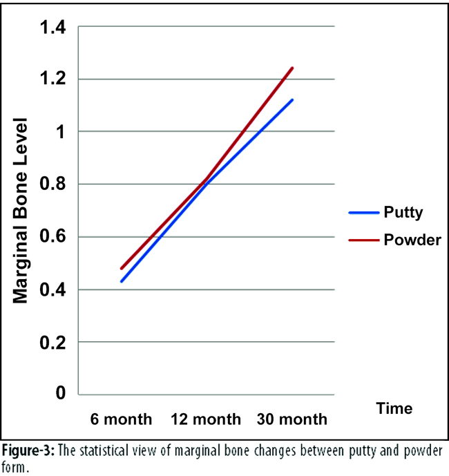
Of the 12 patients, 8 (66.6%) were females and 4 (33.3%) were males. The overall age range of the patients was 29-64 years. All implants wtih complete upper prothesis had a 100% survival rate at the point of final observation. All implants osteointegrated sucessfully in grafted sides and showed successful results. The average volume of marginal bone resorption at implants for the putty side was 0.43±0.22 mm, 0.8±0.33 mm and 1.12±0.49 mm at prosthetic loading, 12-month and 30-month follow-up, respectively. Significant differences were found in the putty side between 6-12, 6-30 and 12-30 months (p<0.001). The average volume of marginal bone resorption at implants for the powder side was 0.48±0.32 mm, 0.82±0.46 mm and 1.24±0.57 mm at prosthetic loading, 12-month and 30-month follow-up, respectively. Also, significant differences were found in the powder side between 6-12, 6-30 and 12-30 months (p<0.001). The average marginal bone loss around the implants was detected as 0.45±0.27, 0.80±0.40 and 1.21±0.53 at prosthetic loading, 12-month and 30-month follow-up, respectively No statistically significant differences were observed in marginal bone loss around the implants between the powder and the putty groups at 6 months (p=0.620), 12 months (p=0.843) and 30 months (0.488). Repeated measurements of two-way ANOVA and Fisher\'s LSD test revealed no significant interactions between the putty and the powder sides (p=0.338) (Table, Figure-3).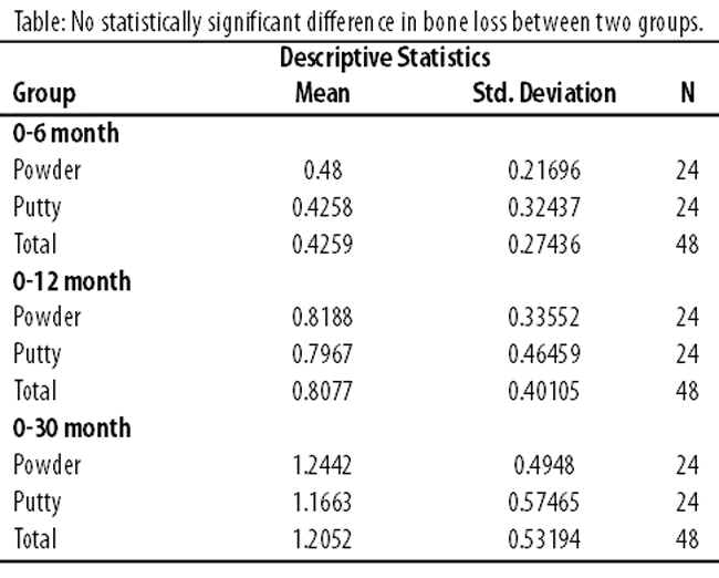
Discussion
Techniques to augment the sinus floor in combination with several grafting materials are commonly used to restore adequate volumes for implantation. As a general rule, grafting techniques should be as simple, less invasive, complication-free and with the shortest healing time as possible.15
The present study compared the survival rate and marginal bone levels of dental implants inserted into grafted maxillary sinus with powder and putty forms of DBM. DBM powder is the most osteoinductive form as it possesses the maximum surface area for interaction with target cells at the graft site.9 However, difficulties with handling, its tendency to migrate from the graft sites, and lack of stability after surgery are clinical problems associated with the use of powdered and particulated forms of DBM.8,9,16
Various carrier materials such as glycerol, fibrin sealent, hyaluronic acid, lecithin, polyorthoester, polyethyleneoxide-polybutylene terephthalate (PEO-PBT) copolymer, polylactic-co-glycolic acid (PLGA) and Poloxamer 407 have been used to facilitate the handling of DBM powder.9 In the present study, DynaGraft-D™ was used in one side of the patients as a paste form of DBM for sinus graft material. DynaGraft-D™ is a unique graft substitute composed of a high content of human DBM in a reverse phase medium provided by Poloxamer 407, and is designed to promote natural bone formation.8
Different results have been demonstrated by several studies regarding marginal bone loss around dental implants in maxillary sinus grafted with various graft materials. One such study evaluated radiological bone resorption around dental implants placed in grafted sinuses after up to 4 years of function on 18 cases.17 Twenty-six sinus augmentations were performed with alloplastic or xenogenic grafting materials on these 18 patients, and 37 implants were inserted. The change in marginal bone level around the implants was found at the mesial and distal side to be 1mm and 1.1mm during the first year after the abutment connection, followed by an annual loss of 0.1mm and 0.2 mm, respectively. In our study, similar results were found. The average marginal bone loss around dental implants at sinus sites grafted with both putty and powder form of DBM was 0.45, 0.80 and 1.21 at prosthetic loading, 12-month and 30-month follow-up, respectively
Another study inserted 38 implants in 16 sinuses together with simultaneous sinus lifting, and evaluated survival rate and marginal bone resorption volume around the implants after follow-up periods of 1-7 years. The reported survival rate of these implants was 92.1% with 3 failed implants and the marginal bone loss evaluated at final observation was 1.35-1.87mm.18 Another clinical study evaluated the sinus bone graft resorption and marginal bone loss around the implants.19 The study compared xenograft and a minimal amount of autogenous bone (Group 1) versus a minimal amount of autogenous bone and equal amounts of allograft and xenograft (Group 2) in sinus augmentation. The average marginal bone loss 1 year after prosthodontic loading and after 20.8 months\' follow-up was found to be 0.6 mm and 0.7 mm, respectively, in Group I. A 93.9% success rate was observed for Group I, with 3 implants showing bone resorption of >1.5mm within 1 year of loading. For Group II, the average marginal bone loss 1 year after prosthodontic loading and after 19.7 months\' follow-up was 0.7 mm and 1.0 mm, respectively. An 83.3% success rate was observed for Group II, with 4 implants showing bone resorption of >1.5 mm within 1 year of loading. But it concluded that mixed grafting with DBM for maxillary sinus bone grafting had no significant short-term merit regarding bone healing and stability of implants compared with anorganic bovine bone alone.
A retrospective study evaluated the survivability and success of single implants placed simultaneously during direct sinus lifts with particulate and paste forms of DBM and evaluated the effect of paste form on time management and membrane integrity during the procedure.8 There were no significant differences between the two forms of DBM when survivability of the implants were judged. But the operation time was significantly decreased in the paste form group.8 In our study, no significant difference was found between two forms of DBM according to survival rates and marginal bone levels. Also, operation time was not compared between the 2 groups, but in our experience, the paste form was applied more easily than the powder form.
A study has reported the effect of paste form of DBM on sinus floor augmentation using clinical parameters.3 Sinus augmentation procedures were performed on 5 cases and after an average of 6 months\' healing period, all implants showed favourable osseointegration and final restorations were completed. The study reported that the paste form of DBM was clinically useful for the increase of bone volume in sinus augmentation, because of its favourable behaviour towards new bone formation and easy handling. Another study showed satisfactory results of maxillary sinus lift with paste form of DBM after 9 months\' healing period.2 In our study, both putty and powder forms demonstrated satisfactory results. There were no significant differences between the two groups.
A retrospective study reported a total of 282 dental implants with sinus augmentation and recorded that the cumulative survival rates of these implants were 95.6% and 100% for autogenous and bovine bone material, respectively. The marginal bone resorption were shown as 1.8mm mesially and 2.1mm distally at the 1-year follow-up, and 1.4mm mesially and 1.5mm distally at the 2-year follow-up.20 Studies have also shown that delayed implant positioning resulted in a mean marginal bone loss three times greater than that of simultaneous implant positioning.20,21 In the current study, all implants were performed with simultaneous sinus lifting and demonstrated acceptable results in line with literature.20,21
Conclusion
Both putty and powder forms of DBM showed satisfactory results and had no significant difference at marginal bone loss around dental implants and survival rates according to long-term followup. As the current study was limited by the small number of patients, more long-term studies with larger groups of patients are required before its findings could be generalized.
Disclosure
There was no financial assistance from any source for this study.
References
1. Merkx MA, Maltha JC, Stoelinga PJ. Assessment of the value of anorganic bone additives in sinus floor augmentation: a review of clinical reports. Int J Oral Maxillofac Surg 2003; 32: 1-6.
2. Won YH, Kim SG, Oh JS, Lim SC. Clinical evaluation of demineralized bone allograft for sinus lifts in humans: a clinical and histologic study. Implant Dent 2011; 20: 460-4.
3. Sohn DS, Bae MS, Choi BJ, An KM, Shin HI. Efficacy of demineralized bone matrix paste for maxillary sinus augmentation: a histologic and clinical study in humans. Oral Surg Oral Med Oral Pathol Oral Radiol Endod 2009; 108: e30-5.
4. Groeneveld EH, van den Bergh JP, Holzmann P, ten Bruggenkate CM, Tuinzing DB, Burger EH. Mineralization processes in demineralized bone matrix grafts in human maxillary sinus floor elevations. J Biomed Mater Res 1999; 48: 393-402.
5. Block JE, Poser J. Does xenogeneic demineralized bone matrix have clinical utility as a bone graft substitute? Med Hypotheses 1995; 45: 27-32.
6. Libin BM, Ward HL, Fishman L. Decalcified, lyophilized bone allografts for use in human periodontal defects. J Periodontol 1975; 46: 51-6.
7. Gross RH. The use of bone grafts and bone graft substitutes in pediatric orthopaedics: an overview. J Pediatr Orthop 2012; 32: 100-5.
8. Irinakis T. Efficacy of injectable demineralized bone matrix as graft material during sinus elevation surgery with simultaneous implant placement in the posterior maxilla: clinical evaluation of 49 sinuses. J Oral Maxillofac Surg 2011; 69: 134-41.
9. Tian M, Yang Z, Kuwahara K, Nimni ME, Wan C, Han B. Delivery of demineralized bone matrix powder using a thermogelling chitosan carrier. Acta Biomater 2012; 8: 753-62.
10. Eryilmaz T, Ozmen S, Lortlar N, Omeroglu S, Ayhan S, Celebi C. Feasibility of demineralized bone matrix for craniomaxillofacial contour restoration. J Craniofac Surg 2011; 22: 1888-92.
11. Koh RU, Oh T-J, Rudek I, Neiva GF, Misch CE, Rothman ED, et al. Hard and soft tissue changes after crestal and subcrestal immediate implant placement. J Periodontol 2011; 82: 1112-20.
12. Cawood JI, Howell RA. A classification of the edentulous jaws. Int J Oral Maxillofac Surg 1988; 17: 232-6.
13. Buser D, Ingimarsson S, Dula K, Lussi A, Hirt HP, Belser UC. Long-term stability of osseointegrated implants in augmented bone: a 5-year prospective study in partially edentulous patients. Int J Periodontics Restorative Dent 2002; 22: 109-17.
14. Albrektsson T, Zarb G, Worthington P, Eriksson AR. The long-term efficacy of currently used dental implants: a review and proposed criteria of success. Int J Oral Maxillofac Implants 1986; 1: 11-25.
15. Esposito M, Grusovin MG, Kwan S, Worthington HV, Coulthard P. Interventions for replacing missing teeth: bone augmentation techniques for dental implant treatment. Cochrane Database Syst Rev 2008; CD003607.
16. Jang CH, Park H, Cho YB, Song CH. Mastoid obliteration using a hyaluronic acid gel to deliver a mesenchymal stem cells-loaded demineralized bone matrix: an experimental study. Int J Pediatr Otorhinolaryngol 2008; 72: 1627-32.
17. Maiorana C, Sigurtà D, Mirandola A, Garlini G, Santoro F. Bone resorption around dental implants placed in grafted sinuses: clinical and radiologic follow-up after up to 4 years. Int J Oral Maxillofac Implants 2005; 20: 261-6.
18. Simion M, Fontana F, Rasperini G, Maiorana C. Long-term evaluation of osseointegrated implants placed in sites augmented with sinus floor elevation associated with vertical ridge augmentation: a retrospective study of 38 consecutive implants with 1- to 7-year follow-up. Int J Periodontics Restorative Dent 2004; 24: 208-21.
19. Kim YK, Yun PY, Kim SG, Kim BS, Ong JL. Evaluation of sinus bone resorption and marginal bone loss after sinus bone grafting and implant placement. Oral Surg Oral Med Oral Pathol Oral Radiol Endod 2009; 107: e21-8
20. Sbordone L, Levin L, Guidetti F, Sbordone C, Glikman A, Schwartz-Arad D. Apical and marginal bone alterations around implants in maxillary sinus augmentation grafted with autogenous bone or bovine bone material and simultaneous or delayed dental implant positioning. Clin Oral Implants Res 2011; 22: 485-91.
21. Herzberg R, Dolev E, Schwartz-Arad D. Implant marginal bone loss in maxillary sinus grafts. Int J Oral Maxillofac Implants 2006; 21: 103-10.
Journal of the Pakistan Medical Association has agreed to receive and publish manuscripts in accordance with the principles of the following committees:


