

Mohammad Raheel Nawaz Jajja ( Department of Surgery, The Aga Khan University, Karachi. )
Zain Feroze Cawasji ( Department of Surgery, The Aga Khan University, Karachi. )
Sehyr Imran ( Department of Surgery, The Aga Khan University, Karachi. )
Muhammad Shahjahan Khan ( Department of Surgery, The Aga Khan University, Karachi. )
Salila Shoaib Hashmi ( Department of Surgery, The Aga Khan University, Karachi. )
Tahir Shafi Khan ( Department of Surgery, The Aga Khan University, Karachi. )
Alina Ghani ( Department of Surgery, The Aga Khan University, Karachi. )
September 2013, Volume 63, Issue 9
Original Article

Abstract
Objective: To identify the frequency of different types of oral clefts and presence of known risk factors among patients.
Methods: The retrospective review of 292 patients, presenting with oral clefts between 1992 and 2011, was conducted at the Aga Khan University Hospital, Karachi. A pre-designed questionnaire was used to collect details, including demographics, type of cleft, presence of known risk factors, surgical details, and follow-up visits. SPSS 16 was used for data analysis. Chi-square test and analysis of variance was used: whenever applicable.
Results: Of the total, 168 (57-53%) patients had cleft lip with or without cleft palate, and 124 (42.5%) had cleft palate alone. The most common defect was left-sided complete cleft lip and palate and midline incomplete cleft palate in the two groups respectively. Consanguinity among the parents was found to be the most common risk factor (n=50; 17.1%). Median age of repair was 4 months for cleft lip and 10 months for cleft palate in the first group. For the other group, the median age of primary repair was 13 months. First-week follow-up after surgery was 50% (n=84) for the lip repair, and 65% (n=81) for palate repair.
Conclusion: Our review revealed that most patients had cleft lip with or without cleft palate (CL/P). The most common risk factor was consanguinity among parents. Delay in seeking care, low follow-up rates after surgical repair of the anomaly and lack of involvement of speech therapist and orthodontist was observed.
Keywords: Oral clefts, Cleft palate, Surgical repair. (JPMA 63: 1098; 2013).
Introduction
Craniofacial anomalies comprise a significant component of human birth defects.1 Oral clefts are the second most common entity in this group.2
Two major categories of oral clefts based on differences in the embryological pathogenesis have been recognised: Cleft lip with or without cleft palate (CL/P) and Cleft palate alone (CP).3 Epidemiological studies have revealed the incidence to be 1 in 700 around the world with marked geographical and ethnic differences.3 Asian populations have the highest frequency (1 in 500) with the Caucasian population as intermediate and African population having the lowest (1 in 2500).4
Oral clefts have a complex and multi-faceted genetic and environmental etiology. Apart from being a part of well-recognised syndromes, numerous risk factors have been elucidated in the disease causal pathway. Maternal smoking, alcohol consumption, anti-epileptic drugs usage, maternal (toxoplasmosis, rubella, cytomegalovirus, herpes simplex TORCH) infections, nutritional deficiencies (especially folic acid) during pregnancy, and consanguineous marriage between the parents are the most quoted risk factors.5-7
Children with oral clefts have a higher morbidity and mortality throughout their lives compared to the unaffected children. They require a multi-disciplinary approach for primary repair and further management.1,7 A delay in seeking health-care for these children can lead to detrimental outcomes. In addition to impairment of appearance, their speech and hearing capabilities may also be affected. The aim of this study was to identify the incidence of different types of oral clefts among patients with orofacial anomalies, their association with other congenital malformations and presence of associated risk factors in these patients.
Patients and Methods
The retrospective review of 329 patients who had presented with oral clefts between 1992 and 2011 with subsequent surgical management was carried out at Aga Khan University Hospital (AKUH), Karachi. Incomplete records were excluded. Data collection was started after receiving approval from the institutional Ethics Review Committee.
A pre-designed questionnaire was used to collect data. The questionnaire had three sections for data collection. The first part included age at presentation, gender, birth-weight and gestational age at birth. The second section collected details about the specific type of cleft. The third section recorded identifiable risk factors, surgical details, and subsequent follow-up visits.
Data was analysed using SPSS version 16.0. Analysis included descriptive statistics. Frequencies and percentages as well as means and standard deviation were calculated and tabulated.
The study period was divided in three different 6-year time periods i.e. 1992-1998, 1999-2004 and 2005-2011. Median age at presentation and at the time of repair, consanguinity, frequencies of follow-up after surgery, visits to cardiologist, speech therapist and orthodontist were calculated for these time periods separately. Chi-square and analysis of variance (ANOVA) was used where appropriate.
Results
Of the initial 329 patients, cases of 292 (88.75%) with complete records were included in the study.
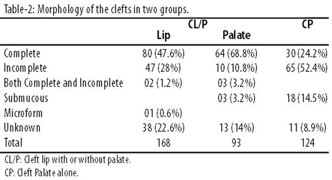
Of them, 168 (57.5%) had CL/P; 93 (55.4%) of these had a cleft palate as well. Besides, 124(42.5%) patients had CP alone (Tables-1 and 2).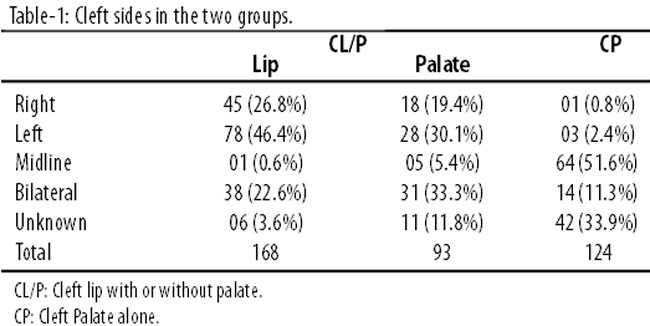
Overall, there were 155 (53.1%) males and 137 (46.9%) females. There was a male predominance in the CL/P group and female predominance in the CP group; the male-to-female ratio being 1.47:1 and 1:1.25 in the two groups respectively.
Age at presentation ranged from birth to 35 years (median-5 months) for the CL/P group, and birth to 29 years (median-12 months) for those with CP. The most common age of presentation was within the first month of life for both groups.
Out of the total, 37 (12.7%) patients were born at the AKUH, while the rest were outside referrals. The mean birth weight was 6.56±1.28 lbs and 6.73±1.34 lbs for the CL/P and CP groups respectively; 3(1.8%) patients in the CL/P and 2 (1.6%) patients in the CP groups were born pre-term.
Overall, 8(2.7%) patients had a syndromic presentation of Pierre Robin sequence; 17(5.8%) had other morphological abnormalities that included hypertelorism, bat ears, talipes, hypospadias and thumb hyperplasia. Congenital heart defects (CHD) were found in 26(8.9%) cases. The most common heart defect was found to be patent foramen ovale (n=8; 2.7%). Other defects included atrial and ventricular septal defect, patent ductus arteriosus (n=4; 1.36% each) and tetralogy of fallot (n=3; 1.8%).
Consanguinity among parents was found in a total of 50 (17.1%) cases and it was the most common identifiable risk factor. It was followed by positive family history in 36 (12.3%) cases. Maternal infection during pregnancy was positive in 5 (1.7%) cases; maternal anti-epileptic drug use was found in 2 (0.7%) cases. No case for maternal smoking or alcohol use or vitamin A or radiation exposure during pregnancy was identified.
Out of 120 CL/P patients 86(71.4%), and out of 108 CP patients, 94(87.1%) had primary lip repair surgery done at the AKUH. The median age of primary repair in the CL/P group was 4 months for cleft lip cases and 10 months for the cases that also had a cleft of the palate. The median age of primary repair in the CP group was 13 months. Nine (7.5%) patients in the CL/P group and 6(5.6%) in the CP group had to undergo revision surgery.
Patients with CL/P undergoing lip repair surgery had a follow-up rate of 49.4% (n=83) at 1 week and 19.6% (n=33) at 6 weeks. Following palate repair 64.5% (n=60) patients were followed up at 1 week and 18.3% (n=17) and 3.2% (n=3) followed up at 2.5 years and 5 years respectively. In the CP group, 65.3% (n=81) patients were followed up 1 week after palate repair, with 15.3% (n=19) and 5.6% (n=7) at 2.5 and 5 years respectively.
There were 36 (20%) cases with post-operative complications that included breathing problems, bleeding, wound dehiscence, fever, infections and palatal fistula. 1 (0.5%) death was recorded, which was attributed to anaesthesia complications.
Overall, 2177(4.3%) patients were referred to a speech therapist for evaluation; only 36(16.6%) actually followed the advice.
Trends of study variables over a 19-year study period were recorded (Table-3).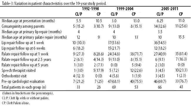
It showed certain variations over the years. For instance, consanguinity in parents among the CL/P and CP groups increased, but this increase was not found to be statistically significant (p= 0.47 and 0.54 respectively). The median age at presentation and at primary repairs for both the groups did not show any statistically significant variation. However, 1st week follow-up after primary lip and primary palate repair did show a significant change over the years with considerably better follow-ups in the later years (p<0.03 and 0.001). Speech therapist and orthodontist visits remained very low over the observed time period and did not show any statistically significant variation. Pre-operative consultation with a paediatric cardiologist witnessed a statistically significant improvement for CL/P and CP groups (p<0.02 and 0.04 respectively).
Discussion
Oral clefts are the most common craniofacial anomalies, occurring in 1 in every 700 births.3 Mongols have the highest reported prevalence3 and Asians are considered to be at most risk, with rates of oral clefts ranging between 1.20 and 1.18 per 1000 live births.8,9 A single-city study conducted in a different province of Pakistan found the mean incidence to be 1.46 per 1000 live births.10
The etiology of oral clefts is complex, involving interactions of numerous genetic, environmental and pre-natal nutrition factors. Our finding of first-degree consanguinity among parents of patients being the most common identifiable risk factor matched the results of another study from Pakistan.11 Our data further showed an increasing trend, though statistically insignificant, in parental consanguinity over the observed time period. Orofacial morphogenesis is controlled by multiple genes. This underlines the role that parental consanguinity and family history may play in the pathogenesis.12 Animal experimental trials have also proven a multi-factorial threshold model of inheritance for oral clefts. The recurrence risk seems to increase with degree relation and extent of anatomical severity in the affected relation.13
There has been a strong observational evidence linking environmental risk factors such as maternal smoking, alcohol use, poor nutrition, viral infections and teratogen use in early pregnancy to incidence of oral clefts.4,7,14-16 We could not elucidate poor maternal nutrition as a risk factor due to the retrospective study design, but it can possibly account for significant contribution in our population. Recall bias cannot be ruled out for low number of viral illnesses during pregnancy. Lack of proper prenatal care may contribute to cases of viral illnesses going undiagnosed and, therefore, may have been under-reported.
The overall male dominance for orofacial anomalies found in our study is in conjunction with several studies world-wide.7,17 A left-sided cleft lip is known to be the most common morphology.18
Syndromic clefts are now recognised as a different entity with 55% of cleft cases being associated with a congenital syndromic.19 CL/P is a known feature of more than 200 syndromes and CP is associated with more than 400 syndromes.7 We found 8 cases of Pierre Robin Sequence in which oral cleft is a well-known feature. This is probably not the true prevalence of the actual syndromic clefts as many of these syndromes end up in stillbirths, neonatal or infant deaths.
The association between CHD and orofacial anomalies is explained by embryological development of face and heart by neural crest cell precursors.20 Cardiac anomalies, mostly of atrioventricular septum, were found in 8.9% patients. Another study done at our centre revealed that CHD were the most commonly associated anomalies with oral clefts.21
Management of oral clefts needs a multi-disciplinary effort with involvement of a plastic surgeon, paediatrician, orthodontist and speech therapist. A long-term follow-up is needed to assess the functional development of the palate, and need of a revision surgery and/or speech therapy. At AKUH, a definite protocol for the multi-disciplinary management of oral clefts is followed. The child is referred to a plastic surgery clinic by a neonatologist/paediatrician. Depending on the health status, cleft lip surgery is planned at 4 months of age and cleft palate repair is planned at 9 months of age. Pre-op evaluation by a paediatric cardiologist is mandatory, including a 3D echocardiography to rule out CHD. Early palate repair (6-9 months) is considered a standard approach all over the world, with a better functional outcome.18,22 We use Millard\'s procedure for cleft lip repair and Veau-Wardill-Kilner procedure and Langenback palatoplasty for palate repairs.
Follow-up after cleft lip repair is at 1 and 6 weeks after surgery and at 1 week, 2.5 and 5 years after cleft palate. If there is a need, an orthodontist or speech therapist referral is suggested at the follow up visits to the surgeon. Despite a well-defined protocol in-line with the international guidelines, our review revealed a deficient follow-up.
Over our study duration, we did note a statistically significant improvement in the initial follow-up visit after a primary lip or palate repair surgery over the years. However, the subsequent follow-up rate remained poor. There was no improvement in the rate of orthodontists and speech therapist visits among these patients. This aspect of the oral cleft management needs further emphasis in future management protocols. Pre-operative cardiology consultations improved from less than 25% initially to around 70% towards the end of the study period. This can be explained by the introduction of the oral cleft protocol at the AKUH in 1996 that recognised a pre-op cardiology evaluation as essential.
Conclusion
Lack of awareness with regards to complexity of oral clefts and limited capacity to afford multi-disciplinary management may contribute to sub-optimal care of children with oral clefts. Importance of a timely surgical intervention with proper subsequent management needs to be emphasised to basic health-care givers. Simultaneously, efforts also need to be geared towards primary prevention. A decrease in consanguinity coupled with better maternal health may decrease the incidence of orofacial anomalies, and help reduce the burden of problems associated with it.
References
1. Strauss RP. The organization and delivery of craniofacial health services: the state of the art. Cleft Palate Craniofac J 1999; 36: 189-95.
2. Strong EB, Buckmiller LM. Management of the cleft palate. Facial Plast Surg Clin North Am 2001; 9: 15-25.
3. World Health Organization. Global Registry and Database on Craniofacial Anomalies: Report of a WHO Registry Meeting on Craniofacial Anomalies (2001: Bauru, Brazil). Geneva, Switzerland: WHO; 2001.
4. Weingärtner J, Lotz K, Fanghänel J, Gedrange T, Bienengräber V, Proff P. Induction and prevention of cleft lip, alveolus and palate and neural tube defects with special consideration of B vitamins and the methylation cycle. J Orofac Orthop 2007; 68: 266-77.
5. Grewal J, Carmichael SL, Ma C, Lammer EJ, Shaw GM. Maternal periconceptional smoking and alcohol consumption and risk for select congenital anomalies. Birth Defects Res A Clin Mol Teratol 2008; 82: 519-26.
6. Murray JC. Gene/environment causes of cleft lip and/or palate. Clin Genet 2002; 61: 248-56.
7. Mossey PA, Little J, Munger RG, Dixon MJ, Shaw WC. Cleft lip and palate. Lancet 2009; 374: 1773-85.
8. Cooper ME, Ratay JS, Marazita ML. Asian oral-facial cleft birth prevalence. Cleft Palate Craniofac J 2006; 43: 580-9.
9. Reddy SG, Reddy RR, Bronkhorst EM, Prasad R, Ettema AM, Sailer HF, et al. Incidence of cleft lip and palate in the state of Andhra Pradesh, South India. Indian J Plast Surg 2010; 43: 184-9.
10. Elahi MM, Jackson IT, Elahi O, Khan AH, Mubarak F, Tariq GB, et al. Epidemiology of cleft lip and cleft palate in Pakistan. Plast Reconst Surg 2004; 113: 1548-55.
11. Natsume N, Kawai T, Ogi N, Yoshida W. Maternal risk factors in cleft lip and palate: case control study.Br J Oral Maxillofac Surg 2000; 38: 23-5.
12. Romitti PA, Sun L, Honein MA, Reefhuis J, Correa A, Rasmussen SA. Maternal periconceptional alcohol consumption and risk of orofacial clefts. Am J Epidemiol 2007; 166: 775-85.
13. Shaw GM, Lammer EJ, Wasserman CR, O\'Malley CD, Tolarova MM. Risks of orofacial clefts in children born to women using multivitamins containing folic acid periconceptionally. Lancet 1995; 346: 393-6.
14. Zafar H, Khan FA, Farooq MU, Niazi NA, Zafar F, Ali M, et al. Prevalence of cleft lip and palate in cousin marriages. Ann King Edward Med Uni 2006; 12: 99-100.
15. Aljohar A, Ravichandran K, Subhani S. Pattern of cleft lip and palate in hospital-based population in Saudi Arabia: retrospective study. Cleft Palate Craniofac J 2008; 45: 592-6.
16. Grosen D, Chevrier C, Skytthe A, Bille C, Mølsted K, Sivertsen A, et al. A cohort study of recurrence patterns among more than 54,000 relatives of oral cleft cases in Denmark: support for the multifactorial threshold model of inheritance. J Med Genet 2010; 47: 162-8.
17. Memon GA, Memon R, Kumar M. A 21 years perspective of cleft lip and palate and outcome of surgical procedures at Jamshoro, Pakistan. J Liaquat Uni Med Health Sci 2003; 2: 3-6.
18. Bicknell S, McFadden LR, Curran JB. Frequency of pharyngoplasty after primary repair of cleft palate. J Can Dent Assoc 2002; 68: 688-92.
19. Calzolari E, Bianchi F, Rubini M, Ritvanen A, Neville AJ; EUROCAT working group. Epidemiology of cleft palate in Europe: implications for genetic research. Cleft Palate Craniofac J 2004; 41: 244-9.
20. Barbosa MM, Rocha CM, Katina T, Cladas M, Codorniz A, Medeiros C. Prevalence of congenital heart diseases in oral cleft patients. Pediatr Cardiol 2003; 24: 369-74.
21. Shafi T, Khan MR, Atiq M. Congenital heart disease and associated malformations in children with cleft lip and palate in Pakistan. Br J Plastic Surgery 2003; 56: 106-9.
22. Ysunza A, Pamplona MC, Quiroz J, Yudovich M, Molina F, González S, et al. Maxillary growth in patients with complete cleft lip and palate, operated on around 4-6 months of age. Int J Pediatr Otorhinolaryngol 2010; 74: 482-5.
Journal of the Pakistan Medical Association has agreed to receive and publish manuscripts in accordance with the principles of the following committees:


