

Zahoor Ahmed ( Department of Thoracic and Vascular Surgery, Mafraq Hospital, Abu-Dhabi, United Arab Emirates. )
Zahoor Mohyuddin ( Department of Thoracic and Vascular Surgery, Mafraq Hospital, Abu-Dhabi, United Arab Emirates. )
July 1998, Volume 48, Issue 7
Case Reports

Introduction
Congenital bilateral diaphragnmtic cventmuon is an extremely rare anomaly. The reported incidence of congenital diaplaragmatic defects varies from in 2200 to I in 5000 live births1,2. The surgical correction is, technically, a simple procedure but parioperative care is quite complicated. Pre-natal diagnosis is often difficult. The birth of a distressed neonate should alert the physician to the possibility of the presence of this anomaly as the method of resuscitation adopted to improve the condition, usually by face mask in emergencies, can in fact w’orsen the situation.
Case No.1
A 3945 grams male baby was born by an elective cesarean section because of breech and cephalopelvic disproportion at 39 weeks gestation. There was neither any history of hydramnios or drug use during the pregnancy nor birth of abnormal siblings before. Abortions, stillbirths or neonatal deaths had not been documented, either, in the family. Apgar score was 4 at one minute. The resuscitation by suctioning and oxygen by face mask did not help so the neonate had to be intubated temporarily which improved his condition. Repeat examination showed persistent tachypuoca (respiratory rate 92/minute) with diminished air entry on both sides on chest auscultalion. Abdomen and heart were however normal clinically. Chest X—ray done soon after the birth (figure IA)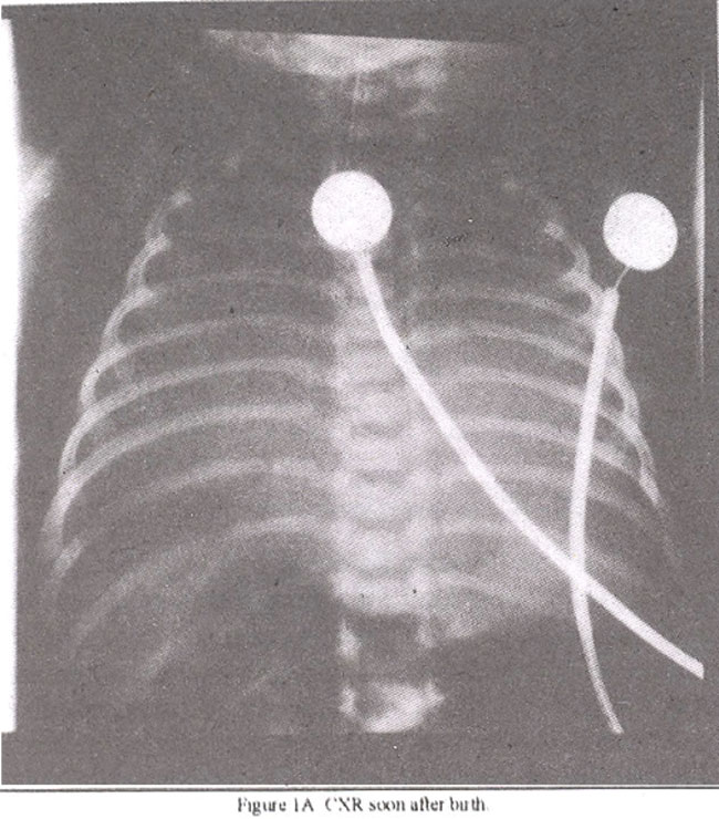
revealed poor aeration of both lungs. Blood gas analysis showed pH of 7,28, PCO2 of 66 mmHg and PO2 of 36 mmHg. The neonate had tobc reIntubated and managed by Continuous Positive Airway Pressure (CPAP) with Fl02 of 0.4. This stabilized the condition and his blood gasses reverted to normal (pH 7.46, PCO2-36, PO2-87).
The repeat chest x-ray (figure 1 B)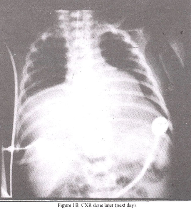
showed good expansion of both upper lobes but a big opacity became well delineated in right lowerzoncwhich was thought to be pushing the heart to the left side leading to the distortion of its left border. Anechocardiogram was done which revealed a normal heart without any demonstrable connection between it and the right lower zone opacity. On ultrasound examination of the chest right diaphragm was found normal in its posterior part but it could not be traced anteriorly. The mass itself had liver like appearance. This virtually confirmed the presence of right diaphragmatic defect. So right thoracotomy was performed whIchconfirmcd diaphragmatic eventrationwIth liver lying in the right chest and causing compressIon atelectasis of lower and middle lobes of the right lung. The heart was found pushed to the left side. The collapsed right lower and middle lobes expanded nicely on inflating the lung. The liver was pushed down to its normal position and the repair was carried out using 2 mm thick Gore-Tex patch since the edges of the diaphragm could not be brought together owing to the large size of the defect. Postoperatively the neonate required assisted ventilation for three days. His spontaneous breathing was normal and he maintained blood gasses within normal range. He also started feeding well. Postoperative chest x-ray (Figure IC)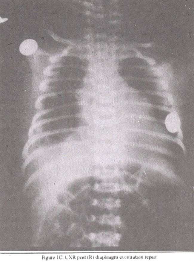
showed complete expansion of the right lung but the lump noticed earlier over the left border of the heart had failed to disappear. This gave rise to the suspicion about the presence of left diaphragmatic defect as well. Ultrasound examination of the chest was repeated but this demonstrated normal intact left diaphragm. Combined liverand lung isotope scans, however, showed elevation of left lobe of liver into the left chest suggesting presence of left diaphragmatic defect.
As the baby was gaining weight and doing well it was decided to postpone the left diaphragmatic repair until the baby had grown well. Three months later left thoracotomy was performed and a small eventration was found. Left lung itself looked nonnal. The defect was repaired with ease. Immediate postoperative chest x- my (Figure ID)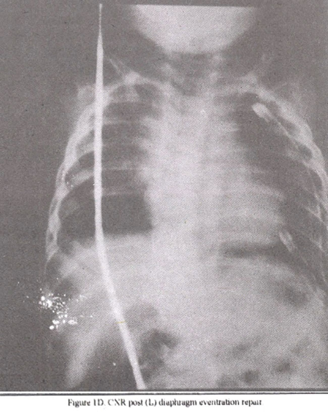
showed completely expanded both lungs. Both diaphragms could be seen in entirety and the lump over the left border of the heart had disappeared. This child could be followed up for about 5 years after surgery and was found to be an active child but he suffered from recurrent attacks of respiratory tract infections.
Case No. 2
A 2350 grams female was born by normal vaginal delivery at 39 weeks gestation. The neonate looked dysmature and was cyanosed. The mother had not taken any dmgs known to result in this anomaly nor did she suffer from Hydramnios or had any abortions or stillbirths before. The Apgar score was I at five minutes. The condition improved by resuscitation with oxygen by face mask in the delivery room. Re-examination of the neonate revealed respiratory rate of 60/minute. Air entry was found diminished on both sides. Heart and abdomen were normal clinically. Chest x-ray (Figure 2A)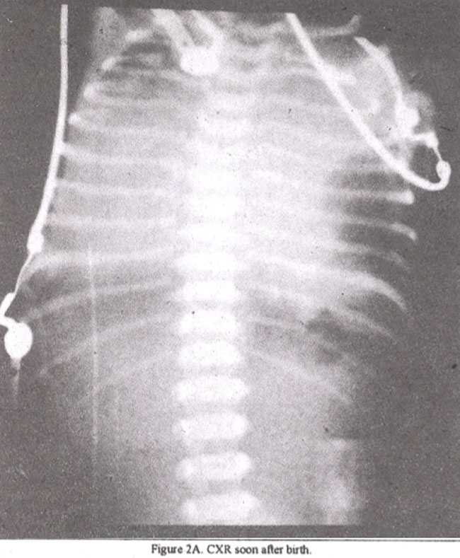
showed haziness on both sides with distended stomach lying in the left chest signifying the presence of left diaph.ragmatic defect. The hazines of right lung field was thought to be due to compression of the right lung by the shifted mediastinurn. The initial blood gas analysIs showed pH of 7.30, PCO2 of 55 mmHg and PO2 58 mmHg. The condition worsened rapidly. The breathing became labored with maiked retraction of suprasternal notch and intercostal spaces. Repeat blood gas analysis showed further deterioration with pH drop to 7.22, PCO2 elevation to 78 mmHg and PO2 drop to 45 mmHg. She was immediately intubated and maintained on a ventilator. This resulted in stabilization of the condition of the neonate with reversal of the blood gasses to normal values. On the second day of life she underwent left thoracotomy. Most of the left diaphragm was thin and flimsy but had a good muscular rim at the periphery. Left lung itself looked smallerin size. The repair of eventration was accomplished using 2mm thick Gore-Tex patch as the edges of the defect could not be approximated without tension. The immediate post-operative chest x-ay (Figure 2B)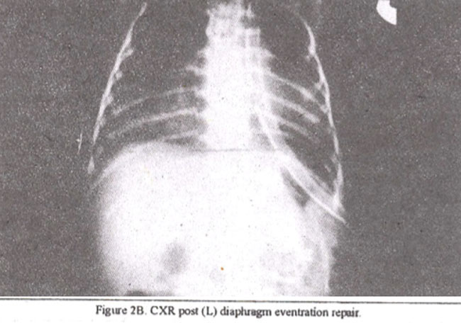
showed complete expansion of both lungs. The right lung expansion was achieved as the mediastinum shifted back to its normal position thus relieving pressure off the collapsed right lung. The neonate was doing fine but the chest x-ray repeated on 3rd postoperative day revealed bowel gas pattern (Figure 2C)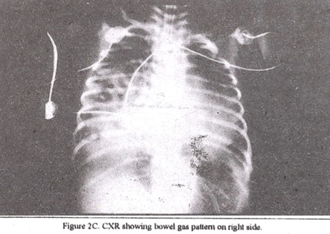
on the right side suggesting presence of right diaphgragmatic defect as well. The delayed protrusion of the small bowel into right chest can probably be explained by the fact that the right diaphragmatic defect being small had got shielded by the compressed right lung and shifted mediastinal structures. The moment the pressure had been taken off by mediastinal shifting back to its normal position, the small right diaphragmatic defect became vulnerable to increased abdominal pressure generated as a result of relocation of herniated viscera into peritoneal cavity during repair of left diaphragrnatic defect and so it led to small bowel protrusion.
Right thoracotomy was performed. Right diaphragm was found thin in the centerbut the size of the defect was much smaller than the left side. The small bowel loops were relocated into the abdominal cavity and repair was done. The postoperative chest x-ray (Figure 2D)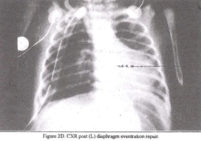
showed complete expansion of both lungs. She needed ventilatoxy support for about a week and was then weaned off. Her spontaneous breathing after coming off the ventilator was good and was able to maintain the blood gasses within normal range. Now she is 8 years old and quite an active and healthy child.
Discussion
Congenital bilateral diaphgragmatic eventration is quite a rare anomaly. The exact etiology is unknown but a high proportion of cases have been found to have chroinosomal disorders such as Trisomy 18.13, or 153. No chromosomal analysis could be done on ourcases. Prenatal cytomcgalovirus infection4 Vitamin A deficiency5. thalidomide6, quinine7. nitrophen8 and phcnmetrazinc9 are the other drugs known to induce this anomaly. Hydramnios10 is found in 20% of the pregnancies ending in the birth of a neonate with this anomaly.
The diaphragmatic defect results in protrusion of viscera into the chest. The continued compression of the developing lung by the herniated viscera during fetal life results in pulmonary hypoplasia11 which is responsible for the respiratozy distress during postnatal life, The degree of pulmonary hypoplasia is dependent upon the timing and the extent of the encroachment of the pleural cavity space by the herniated viscera and the earlier and the greater is the herniation in magnitude the worse would be the outcome. In our case No. 1, the liver did get displaced upwards into the right chest leading to compression of right lower and middle lobes but these readily expanded to full size on inflation. Left lung in case No. 2 was smaller but it did not lead to any respiratory difficult)’ post-operatively and normal expansion was achieved. Congenital diaphragmatic defects can be diagnosed prenatally, but with difficult) by ultrasonography, arnniography and computerized tomography. However, if detected prenatally the delivery of such babies can be undertaken in centers well equipped to resuscitate such neonates. This anomaly does not cause any distress to the fetus but the problem starts soon after the birth. Any neonate who develops respiratory distress soon after birth should be suspected to have this anomaly. One has to be very skillful in immediate resuscitation of a distressed neonate. The ventilation by face mask, usually a first instinct in such emergencies, has to be avoided. Since this could result in distention of the herniated stomach which could aggravate the respiratory distress further by compressing the hypoplastic lung as happened in our case no.2. The ideal method is intubation and ventilation with lowest airway pressures consistent with adequate ventilation. The development of respiratory acidosis with hypercapnia and hypoxia must be avoided as this would trigger off the reactive pulmonary hypertension so one should err on the side of over ventilation rather than underventilation. The resolution of the developed pulmonary hypertension is very difficult as fewer neonates respond to the vasoditators like Tolazoline, POE and PGD2 so the aim should be its prevention. Nifedipine, however, has been reported to give better response12. The neonates who continue to show hypoxaemia despite the use of drugs and adequate ventilatory measures are managed by ECMO13 which is essentially a cardiopulmonary bypass and is employed to treat pulmonary hypertension.
From surgical angle congenital diaphragmatic eventration is relatively a simpler defect to repair but the outcome is rather dependent upon the management of associated physiologic derangements. Initially increased intra-abdominal pressure generated by the relocation of viscera into a smaller unstretched abdominal cavity may compronuse the diaphragmatic function but this soon gets resolved as the peritoneal cavity acconunodates. In those cases where it results in embarrassed ventilation or vena caval obstruction with resultant hypotension a temporary ventral hernia can be created to overeome inunediate difficulty.
Postoperative detenoration inthe pulmonary function is also a well recognized phenomenon and occurs due to the development of pulmonary hypertension which is physiologically akin to persistent fetal circulation. A significant contribution in downhill course could also result from the barotrauma caused by overenthusiaslic ventilation In an attempt to expand the fragile hypoplastic lungs, The deciding factor is the degree of pulmonary hypoptasia which mainIfests itself as respiratory distress, The mortality used to be very high14 but proper preoperative stabilization of the distressed neonate and delaying the repair has been reported to give better results in terms of survival than hurrying for the early operative intervention15, However, early manifestation of dyspnoea, pH of less than 7, PO2 of less than 80 iumHg and PCO2 of more than 50mmHg arc bad prognostic indices, Recurrent chest infection and gastrocsophageal reflux are the most common long term complications16.
References
1. Booker PD "Congenital diaphragmatie hernia in children" Arch. Dis. Child., 1981;56: 253 -257.
2. Herisson MR, de.Lorimer AA. “Congenital diaphragmatie hemia’ Surg. Clin. North Am., 1981;61:1023-1035.
3. Wexler HA, Poole CS "Neonatal diaphragmatic dyafunction" Am J Roenigenol., 1976;127:617-619.
4. Becroft DM0 "Prenatal cytomogalovirus infaction and muscular deflciency (Eventration) of the diephgragm", J. Pediatr., 1979;94:74-75.
S. Andersen DH. "Bffect of diet during pregnancy upon the incidence of congenital hereditary diaphgragmatitic hernia in the rat’ Am 3. Pathol, 1992:25:163-185.
6. Hobolth N."Dugs and congenital abnormalities’ Lancet, 1994;11:1333-1334.
7. Kurmmel, TM, Greenfield Li and Kirkpatrick BV. ‘Alveolar-Arterial oxygen gradients versus the neonatal pulmonary insufficiency index for prediction of mortality in ECMO candidates." J. Pediatr. Surg, 1984;19:380-384.
8. lritan I. "Experimental study on pathogenesis and embryogenesis of congenital diaphragmatic hernia.’ AnaL. Embryol, 1984;169:133-139.
9. Pokomy WD, McGill CW and Harberg Fi. "Morgagni hernias during infancy: Presentations and associated anomalies’. J. Pediatr. Sung., 1984;19:394-397.
10. Butler N and Claireaux AE" Congenital diaphragmatic hernias as a cause of porinatal mortality’. Lancet, 1962;1:639-663.
11. Conde J, Mendoza R, Rafel li ut al. ‘Congenital bilateral posterolateral and anterior diaphragmatic defocts." J Podiatr Surg., 1979;14:185-186.
12. Burguet A, Menget A, Formentin C et al, ‘Efficacy of Nifedipine on respiratory hypoxaemia associated with diaphragmatic hernia in the newborn infant’ Annals De Pediatr., 1989;36:557-559.
13. O\\\' Rourke PP. Lillehei CW, Crone RK et al. \\\'The effect of extracorporeal memberane oxygenation on the survival of neonates with high risk congenital diaphragmatic hernia 45 cases from a single institution’ J. Pudiatr. Surg, 1992;26:147-132.
14. Aldrich TK, Herman JII and Rochester DF. "Bilaterel diaphragmatic paralysis in the newborn. J. Pediatr, 1980;97:988-991.
15. Nakayama UK, Motoyama EK and Tagge EM ‘Effect of preoperative tabilization on respiratory systems compliance and outcome in newborn infants with congenital diaphragrnatic hernia. J. Pediatre. 1991;118:793-799.
16. Rasheed K, Coughlan 0 and O’Donnel 13. "Congenital diaphragmatic hernia in the newborn Outcome in 59 consecutive ceses over a ten year period (1980-1 989).” Irish. J, Med. Sci., 1992,16:16-17.
Journal of the Pakistan Medical Association has agreed to receive and publish manuscripts in accordance with the principles of the following committees:


