

Nazma Afroze ( Department of Pathology, The Aga Khan University Hospital, Karachi. )
Irshad N. Soomro ( Department of Pathology, The Aga Khan University Hospital, Karachi. )
Shahid Pervez ( Department of Pathology, The Aga Khan University Hospital, Karachi. )
Naila Kayani ( Department of Pathology, The Aga Khan University Hospital, Karachi. )
Sheema H. Hasan ( Department of Pathology, The Aga Khan University Hospital, Karachi. )
July 1998, Volume 48, Issue 7
Case Reports

Introduction
Metastasis of hepatocellular carcinoma to the soft tissue and bone of the jaw and to oral cavity is a race phenomenon. One such case is described in this report. Fine needle aspiration cytology is a useful procedure for detection of hepatocellular carcinoma at this site. However, at times cytologic interpretation of swelling in this region may give rise to diagnostic dilemma and clinical history may be essential in establishing the diagnosis.
Case Report
A sixty-seven year old male, was referred by an E.N.T. surgeon in May, 1997 for FNA of left pamtid region swelling. Clinical history obtained revealed that the lump appeared three months back and was hard and non tender on palpation.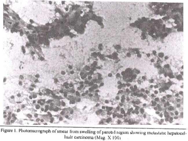
The clinical impression was parotid adenoma with a suspicion of malignancy. The smear showed clusters of large cells with abundant eosinophuilic granular cytoplasm and central round vesicular nuclei. Considering the swelling arising from parotid gland itself, a possibility of oncocytoma was raised. Surgical removal and histological evaluation was recommended.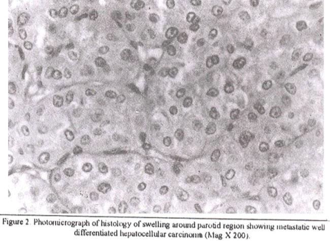
The patient underwent surgical removal of the jaw swelling in June, 1997. Frozen section showed a large cell carcinoma with features suggesting metastatic hepatocellular carcinoma. The clinicians were informed and past mdical history revealed that in October, 1996 the patient presented with liver mass and got anFNAB done on that mass by another surgeon. The clinical impression at that time was cavernous haeinangioma. Cytology slides and aspirated tissue fragments were sent to our department. A differential diagnosis of well differentiated hepatocellular carcinoma and hepatic adenoma was given favouring the former. After that, the patient was lost to follow- up. Four months. later he developed a parotid swelling and reported to E.N.T. Department in May, 1997. After frozen section diagnosis of the parotid mass, a radical parotidectomy was performed with removal of left superficial and deep parotid gland and ramus of left mandible. The permanent sections revealed the typical histology of a well differentiated hepatocellular carcinoma which had metastasized into the soft tissue around the parotid gland. The adjacent bony tissue of minus of mandible and surrounding skeletal muscle was invaded by the tumour. In December, 1997 the patient again developed a mass in oral cavity with a single cervical lymph node enlargement. The oral mass proved tobe a metastatic hepatocellualr carcinoma on histology while the lymph node was benign.
Discussion
Metastatic tumours involving head and neck region are unusual. Hepatocellular carcinoma metastasis tojaw and soft tissue of the oral cavity is a rare occurrence1. A review of literature revealed occasional case reports. Hepatocellular carcinoma metastasizes by direct extension, lymphatic route orhematogenously. Distant metastasis occur in 50% of cases. Bony metastasis are relatively infrequent and are involved in 11% of cases with the spine being the most important site of localization2. One study3 reported seven cases of metastatic tumours in head and neck region over a period of eighteen years. Out of these three had arisen from prirnaiy hepatocellular carcinoma with metastasis to paranasal sinuses. In another study4 fine needle aspiration findings of fifteen hepatocellular carcinomas were described, four in musculoskeletal, four in adrenal, four in regional lymph node, two in pancreas and one in pelvic region. Eleven of these cases had a biopsy proven hepatocellular carcinoma. In the remaining patients, the FNA diagnosis of hepatocellular carcinoma in metastatic sites was the initial diagnosis. In our case the nietastatic tumour had involved the left jaw including the mandibular ramus, skeletal muscle and soft tissue around parotid gland. Involvement of the major salivary gland by malignant neoplasm whose origin is outside the salivary gland occurs by direct extension, hematogenous mute or lymphatic route. The intimate relationship of the paraparotid and intraparotid lymphnodes with the parotid parenchyma frequently results in lymphatic migration of extrasalivary neoplasm to parotid region. Direct extension of non salivary gland tumours into the parotid regions occurs primarily from squamous cell carcinoma and basal cell carcinoma of overlying skin. Eighty percent of metastasis to this site have been from primary tumour elsewhere in the head-neck region and 20% have been from the infraclavicular site5. Hematogenous pathway is the route of metastasis from infraclavicular areas. In descending order of frequency the most common infraclavicular sites of pnmaiy tumour that metastasized to major salivary glands are lung, kidney and breast. Other infrequent sites include colon, rectum, prostate. ovary, pancreas, stomach, uterus and unnary bladder5. For most patients the primary tumour is known at the time of diagnosis of their salivaiy gland metastasis. In this case the FNA diagnosis of parotid swelling was suggestive of oncocytoma. Oncocytoma is a neoplasm made up of large polygonal cells with granular eosinophilic cytoplasm and comparatively small round nuclei. These cells can have cytologic resemblance to well differentiated hepatocytes. The FNA cytology slides were reviewed. On re- examination the
cell clusters showed striking similarity to well differentiated hepatocytes but the characteristic FNA features of hepatocellular carcinoma like presence of naked nuclei, endothelial cells derived from capillary vessels surrounding clusters of hepatocytes and intracytoplasmic bile pigment were not seen in this case. Initial diagnostic dilemma was due to lack of communication to the cytopathologist about the previous diagnosis of malignancy. The most important and helpful factor in resolving this dilemma was previous history. The importance of clinical history and radiographic and biochemical findings in the diagnosis of metastatic hepatocellular carcinoma cannot be overstated4. Patient may occasionally develop more than one primary malignant neoplasm, however in this case histologic similarity between the parotid tumour and a previously diagnosed neoplasm was a strong evidence in favour of metastasis. Farnilarity with the FNA cytology of hepatocellular carcinoma and proper medical history should allow its diagnosis in rare metastatic sites. Degree of differentiation in a hepatocellular carcinoma varies and may predict more or less aggressive behaviour. However, metastasis to head and neck area of this very well differentiated hepatocellular carcinoma within six months of initial diagnosis is another question mark about the reliability of tumour grading in predicting its histological behaviour.
References
1. Marker P. Clausen PP Metastasis to the mouth and jaws from hepatocellular carcinoma Acascrepofl Int. J. Oral.Maxillofae, Surg, 1991;20.371-4
2. Welhk K, Uayduk K, Ptitzer P Fine needle puncture cytology in metastatic hepatocellular carcinoma. Z. Gastroenterol., 1989;27:225-7.
3. Mochimatsu J, Tsukuda M. Furukawa Set al. Tumours metastatsizing to the head and neck: A report ofsevcn cases J.Laryngoi Otol., 1993;107:1171-3.
4. Dusenbery D. Carr Bl Fine needle aspiration diagnosis of hepatocellular carcinoma in metastatic sites. Ada. Cytol., 1996;40:443-9
5. Gary L, Ellis DDS, Paul L. Auselair. Turnouts of salivary gland. 1996. Third series, Fascicle-17 Washington DC. Armed Forces Institute of Pathology. 1996;pp:403-410.
Journal of the Pakistan Medical Association has agreed to receive and publish manuscripts in accordance with the principles of the following committees:


