

Itrat Mehdi ( Pakistan Medical Research Council, Research Centre, Jinnah Postgraduate Medical Centre, Karachi. )
Huma Qureshi ( Pakistan Medical Research Council, Research Centre, Jinnah Postgraduate Medical Centre, Karachi. )
Waquaruddin Ahmed ( Pakistan Medical Research Council, Research Centre, Jinnah Postgraduate Medical Centre, Karachi. )
Ghazala Mohyuddin ( Pakistan Medical Research Council, Research Centre, Jinnah Postgraduate Medical Centre, Karachi. )
Syed Ejaz Alam ( Pakistan Medical Research Council, Research Centre, Jinnah Postgraduate Medical Centre, Karachi. )
July 1998, Volume 48, Issue 7
Short Reports

Introduction
Helicobacter pylori a gram negative micro-acrophilic spiral gastric bacterium, was first isolated and cultured in 19821. It’s role is now well established in the pathogenesis of gastritis, gastric ulcer, duodenal ulcer, non-ulcer dyspepsia (NUD), gastric carcinoma and ly mphoproliferativc disorders of stomach2,9.
A variety of invasive and non-invasive diagnostic tests are currently available for the identification and coufirmation of H. pylori2,3,10,11. They all are invaluable ma given setting or pre-condition7.
Rapid Urcase CLO test is done on biopsy material obtained by gastroduodenoscope. It is relatively cheap, quick to perform. reasonably specific and sensitive i.e., upto 75-90%2,3,10-13. It however, becomes rather unreliable after antimicrobial therapy and is dependent on gastroduodenoscopy14.
A microbial culture can be done on biopsy material obtained through gastroduodenoscope. This is most specific, gold standard and confirmatory but it is not all that easy and also depends on an invasive procedure. It is expensive, time consuming, not universally available and vet provides additional valuable information on antibiotic sensitivity2,3,7,8,10. It has a low sensitivity and a low yield7. The organism is fastidious and it’s culture laborious2.
Histology and special stains for H. pylori are carried out on biopsy material retrieved by gastroduodenoscope. It depends on an invasive procedure, is rather expensive, requires technical expertise and takes lime to provide results2,6,10,15.
Urea Breath test (UBT) is carried out on subjects using 14C radioactive or 13C non-radioactive material, It is rapid, specific, sensitive and can be helpful in identifying a recent infection. It is expensive and has a significant false negativity3,16,17. Urea Breath Test has a specificity and sensitivity of 90—1 00%16. it is not universally available and treatment with proton pump inhibitors (PPI) or antibacterial can give false negative results17. It is dependent on radioactive material with its associated biohazard and require expensive equipment for analysis/interpretation.
Qualitative and quantitative detection of antibodies in patient’s serum against H. pylori, is also increasingly being used. This CLO antibody (IgG) test is simple. quick.
non-invasive, cheap and highly specific (sensitivity upto 95%)3,12 The reliability of this test is questionable in patients over 65 years of age18, it lacks validation and is subjected to inter-observer variation19. The antibody titre does not fall for 4-6 months, even after a complete and successful H. pylon eradication. Saliva can also be used for this test, but is relatively less scsnsitive20.
This study was carried out to determine the sensitivity. Specificity and predictive values of qualitative CLO antibody test in patients with upper gastrointestinal symptoms so that the test may be used as an effective bedside/outpatient screening test in patients of upper gastrointestinal symptoms to decide who might need an endoscopic evaluation.
Patients, Methods and Results
Thirty seven consecutive patients. in the age group of 15-65 years, presenting with upper gastrointestinal symptoms (like dyspepsia, pam, noctural pam, voiniting. hemateinesis) at the PMRC Research Centre. JPMC.. Karachi during 1996-1997 were included in the study.
Patients with history of peptic ulcer, surgery for peptic ulcer, recent endoscopy, associated varices. antinulcer therapy, antibacterial therapy, steroid therapy and Immunosuppression were excluded.
Patients were subjected to gastroduodenoscopuc evaluation (Olympus GIF 2T20/Fujinon Scope) under topical anaesthesia. Biopsies (one each) were taken from antrum and fundus and CLO urease test carried out by immersing the biopsy material into the prepared gel at 37°C for 30 minutes incubation as described elsewhere21 Blood samples (5 till) were taken from each patient and by a qualitative CLO antibody test was done with patients serum using Abbott Flex Pack TM-HP kit, as per manufacturers instructions. Two drops of buffered saline were used as control on the control pad, while one drop of patient’s serum was used on test pad and reading was taken at three minutes. Positive test was seen as a distinct red line below the control red line in the test area of the test pad window within three minutes, Eighteen of 37 patients (30 male, 7 female) were less than or equal to 40 years of age and 19 were over 40 years.
Thirty cases had an ulcer (19 anterior svall duodenal ulcer, 5 posterior wall duodenal ulcer, 3 multiple duodenal ulcers, I duodenal plus gastric ulcer and 1 pre-pyloric gastric ulcer, ulcer). Three ulcers were bleeding, three had associated duodenitis and one had associated esophagitis. Seven cases were non-ulcers: two antral gastritis, one gastritis, one erosive duodenitis, one duodenitis plus gastritis and two cases had normal endoscopy (Tables I and II).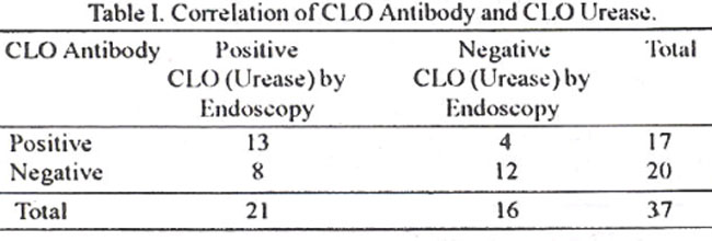
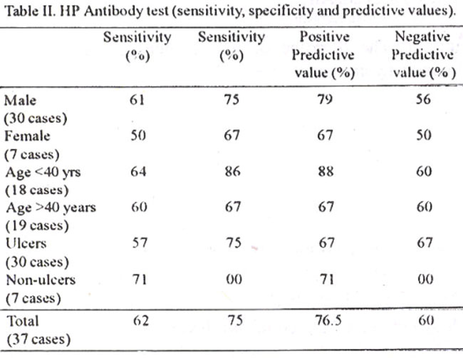
Seventeen of 37 were CLO antibody positive, while 13 out of these 17 were urease positive as well leaving only 4 behind as negative. Twenty cases were CLO antibody negative and 12 out of these 20 were urease negative as well. There were 8 cases who were CLO urease positive yet antibody negative. They probably represent a recent infection or less likely may be having ati inefficient or defective/suppressed immunological response. The overall sensitivity of CLO antibody assay was 62%, specificity 75%, a positive predictive value 76.5% and a negative predictive val ue of 60% (Table II).
Comments
Identification and isolation of helicobacter pylori and elucidation of its role in upper gastrointestinal disease process was a real breakthrough in a relatively short time1-8 In depth analysis of its microbiology, immunology, epidemiology and pathophysiology has revolutionised our understanding of upper gastrointestinal pathology. It has thus led to innovative advances in their managemeni. and prevention2-11.
A variety of commercial kits are increasingly made available to test for CLO antibody from serum; although laboratory based ELISA is more reliable, more specific and more sensitive3,12,18. ELISA assay, however, is expensive and time consuming requiring technical expertise and equipment. CLO antibody assay on the other hand is simple, quick. popular and cheaper. The test is however subject to inter observer variance and its correlation with endoscopic evaluation and compared to other available tests, is not veiy well documented/validated. As IgG remain elevated for upto 6-8 months after eradication, a pm and post treatment quantitative evaluation is highly desirable3. The other factors which can influence the CLO antibody assays are multiple strains infection, drug resistance, antigenic modulation, recurrent and repeated infection and appearance of mutant strains.
The screening for CLO antibodies in young patients is highly effective ill reducing the workload on gastroenterologists, screening out the patients who should/should not be subjected to endoscopic examination in busy outpatients/clinics in our setting22. This is particularly important in H. Pylon associated ulcer, gastritis. non-ulcer dyspepsia (NU D) and GI sy mptotus due to non-organic etiology. It will reduce workload, cufo down the expenditure, save valuable time and energy, avoid each and every one to be subjected to endoscopy. A quantitative CLO antibody screening test will probably become a routine clinical protocol. before an endoscopy, due to cost effectiveness and reducing the clinical workload. Lastly it is highly useful in sero- epidemiological studies of upper gastrointestinal diseases.
References
1. Warren JR. Marshall BJ Unidentified curved bacilli in the stomach of patients with gastritis and peptic ulceration. Lancet, 1984;131:311-314.
2. Penman ID, Palmer KR Helicobacter pylori The story so far Proe k Cull Physicians (Edinburgh) 1997;27:37-45.
3. Goodwin CS, Mendall MM, Northfield TC I Helicobacter pylori infection Lancet, 1997;349:265-269.
4. MsCarthy C, t’atchett S. Collins RM et al Long-tern prospeetive study of Helicobacter pylori tin nonulcer dyspepsia Dig I)is Ser . 1994;40:114-119.
5. Qureshi ll, Ahmed W, Syed Set al. Helicobacter py lori clearance and eradication with triple therapy in duodenal ulcer patients J Pak Med Assoc.. 1995:45;2-3.
6. Kaii J I, Jafarey NA. Alam SM et al Association of helicobacter py lori W ith acid peptic disease in Karachi J PaR Med Assoc. 1990;40:240-241.
7. Bourke 13, Jones N and Sherman P. Helicobacter pv ori infection and peptic ulcer disease in children Pediatr Infect. Dis. J , 1996;151-13.
8. Blaser MJ. Not all Helicobacter pylon strains are created equal. Should all be eliminated’? Lancet, 1997;349:1020-1022.
9. Louw JA. Lucke W, Jaskicwicz K, ct al Helicohacter pylori eradication in the African setting, with special reference to reinfection and duodenal ulcer recurrence Gut., 1995;36:544-547.
10. Sehnell GA, Sehubert TT. Barnes WG et al Comparison of urease, U & E and culture tests for campylohacter pylon. Gastroenterology, 1988;94:A410-A411.
11. Wesiblom lU, Madan E, Kept J, et al Evaluation of rapid unease test to detect campylobacterpylori infection J. CIin. Microbiol, 1988;26:1393-1394.
12. Marshall BJ. Helicobacter pylori Am J Gastroenterol. 1994,89 (Suppl)S116-S128.
13. Haq G, Ahmed W, Kazi JI, et al. Can Diagnosis of Helicobacter py lori be rapid and yet sensitive?J. Pak Sled. Assoc , 1991;41:139-140.
14. Peura t)A Helicobacter pylon: A diagnostic dilemma and a dilemma of diagnosis. Gastroenterology, 1995.109;313-315
15. Kolts BE, Joseph B, Achem SR, et el. Helicobacter pylon detection’ A quality and cost analysis Am J Gastroenterol., 1993;88:650-655.
16. Atharton JC and Spiller RC. The urea breath lest for Helicobacter pylori Gut, 1994;35:723-725.
17. Chey WD, Spybrook M, Carpenter S. et al Prolonged effect of omeprazole on the 14C-urea breath test, Am 3. Gastroenterol., 1996;91;89-92.
18. Newell DG, Hawtin PR, Stacey AR, et aI. Estimation of prevalence of Helocobacter pylon infection in an asymptomutic elderly population comparing (I4C) urea breath test and serology J. Clin pathol., 1991;44:385-387.
19. Vyas SK, Sharpstone I). Treasure J, et aI Pre-endoscopy screening using serodiagnosis of Helicobacter pylon infection Eur 3. Gastroenterol Hepatol. 1994;6:783-785.
20. Patel P, Mendall MA, Khulusi S, et al Salivary antibodies to Helicobacter pylon Screening dyspeptic patients before endoscopy Lancet, 1994;344:511-512.
21. Cliodna A, Me Nutty M, Dent JC et at. Detection of Compylobacter py or by the biopsy urease test: An assessment in 1445 patients Gut, 1989;30:1058-1062.
22.Lee A Therapeutie immunisation against Helicobaceter infection Gastroenterology, 1996;110:2003-2005.
Journal of the Pakistan Medical Association has agreed to receive and publish manuscripts in accordance with the principles of the following committees:


