

Naila Ehsan ( Departments of Gynaecology, Bolan Medical College, Quetta. )
Arshad Mehmood ( Departments of Obstetrics and Surgery, Bolan Medical College, Quetta. )
February 1998, Volume 48, Issue 2
Original Article

Abstract
Sixty-two cases of ectopic pregnancy admitted in the Department of Obstetrics and Cynaecology, Jinnah Postgraduate Medical Centre, Karachi from July, 1989 to December, 1990 were analysed to determine the incidence, risk factors, diagnostic features and management. During this period a total number of 10798 patients were delivered giving an incidence of 1:174 births or 5,7 per 1000 births. In 48,3% cases the cause was unknown, 16.1% followed by pelvic inflammatory disease, 16,1% had history of D & E for abortion, 6.4% had history of different type of pelvic surgery, 4.8% had IUCD in situ, 1,6% had recurrent ectopic pregnancy. Diagnosis was made clinically in 80.6% cases. Culdocentesis was performed in all cases except one and was diagnostic in 90.1% cases. Ultrasound was performed in 51 cases and showed positive results in 90.1% cases. Laparoscopy was performed in only 10 cases and had 100% accurate results. After thorough suscitation laparotomy was performed in all cases. Partial salpingectomy was done in 53.2%, salpingectomy and tubal ligation in 32.2%. (They had already completed their family) and salpingo- oophorectomy in 4.8% cases. Tubes were conserved in 6.2% cases (all of them were primigravidas). In single abdominal pregnancy, dead fetus and placenta was removed from the peritoneal cavity very carefully. The mortality rate in this study was 1.6% (JPMA 48:26, 1998).
Introduction
Ectopic pregnancy is a scrious disaster in the reproductive life of a woman and is a major gynecological emergency associated with increased mortality, decreased reproductive potential and recurrence1. It most commonly occurs between the ages of 20-35 years and is 2-3 times more common in non-white population2. Many centers have reported a progressively rising incidence of ectopic pregnancy in the last two or three decades3. History of pelvic inflammatory disease and pelvic surgery, present or past use of intrauterine contraceptive devices, therapeutic abortions, tuboplasties, ovulation induction drugs and previous ectopic pregnancy are likely risk factors for this changing pattern. Early and reliable diagnosis of ectopic pregnancy before tubal rupture is very important in preserving future fertility and allowing conservative treatment . Measurements of serum progesterone and serial beta human chorionic gonadotropin, direct vision by laparoscope, vaginal ultrasonography and uterine curettage are the tools used for early diagnosis of ectopic pregnancy4. Late diagnosis in these cases is due to lack of proper antenatal care, late arrival to tertiary are centres. lack of availability of diagnostic aids in teaching hospitals and high cost of these investigations which is beyond the reach of these patients. This study critically analyzed the epidemiology and aetiology of ectopic pregnancies in hospital deliveries and evaluated the clinical features, diagnosis and management.
Patients and Methods
This is a prospective study of 62 cases of Ectopic pregnancy. These cases were admitted and managed in the Department of Obstetrics and Gynaecology, Jinnah Postgraduate Medical Center, Karachi overa period of one and half year from July, 1989 to December, 1990. The patients mostly belonged to Karachi, interior Sindh and Baloch areas of Balochistan. After admission the detailed histories were recorded on a special proforma including the age, socioeconomic status, presenting complaints with special reference to amenorrhoea, severity and pattern of lower abdominal pain, duration and amount of vaginal bleeding, syncope, shoulder pain, urinary symptoms etc. Thorough menstrual history, complete obstetrical history with special reference to gravidity and parity was recorded. History of pelvic inflammatory disease, use of contraception and abdominal surgery was also noted. A thorough physical examination, including local, general and systemic was carried out with special consideration to signs of shock, abdominal tenderness, rigidity, abdominal distension, size of utems, adenexal mas and rocking of cervix. Investigations included complete blood picture, urinary HCG, abdominal ultrasonography, culdocentesis and laparoscopy. Diagnosis was based on clinical features, culdocentesis and ultrasound examination. In few cases laparoscopy was also performed. Laparotomy was done in all cases and other procedures as and when required.
Results
During one and a half year study period total number of deliveries was 10,798 and the incidence of ectopic pregnancy in 1:174 births or 5.7/1000 births. Two patients (3.2 percent) were below 20 years; 14 (22.5 percent) between 20-24 years; 30(48.3 percent) between 25-30 years and 16 (25.8 percent) were above 30 years of age. The mean age was 27.91 years. Twenty were nulliparas and among them 12 were pregnant for the first time. The maximum parity was 10 and the mean parity was 2.66. The majority of patients were of low parity and 14 were infertile for the last 3 years or more. In 30 (48.3%) patients the etiology was unknown. Evidence of pelvic inflammatory disease was assessed in 10(16.1%) patients and 10(16. 1%)hadD&E donefor incomplete abortionpreviously (Table I).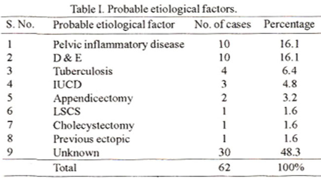
Abdominal pain was present in all followed by amenorrhea (80.6%) and syncopal attacks (6 1.2%). Fifteen patients were brought to hosiptal in the state of shock (Table II).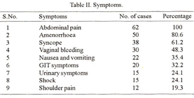
Painfollowing cervical excitation was found in 56(90.3%) cases, followed by abdominal tenderness and adnexal mass (Table III).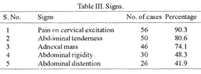
A clinical diagnosis was done in 50 cases. Culdocentesis was performed in all except one case and was positive in 90.1% of cases. Ultrasound was done in 51 and laparoscopy in 10 cases showed positive results in 46 (Table IV).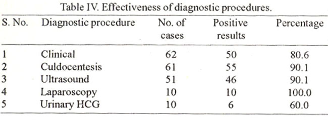
Fifty-six (90.3%) cases had acute variety of ectopic pregnancy and the remaining (9.6%) were of chronic type. There were 60 (96.7%) tubal pregnancies. One was abdominal which was post-term by 6 weeks and the other was in the accessory horn of the uterus. Out of 60 tubal pregnancies, 46 cases were of tubal rupture and 14 were of tubal abortion associated with intraperitoneal hemorrhage. After thorough preoperative resuscitation laparotomy was performed in all and partial salpingectorny in 34 (53.2%) cases. Tubes were conserved in 4 (6.4%) cases, all of them were primigravidas. In single abdominal pregnancy dead fetus and placenta was removed from the peritoneal cavity very carefully (Table V).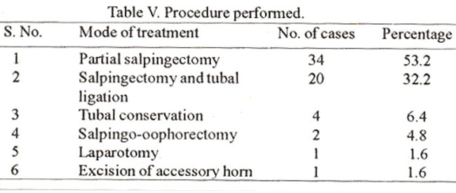
One patient died suddenly on 4th postoperative day. The exact cause of death was unknown but pulmonary embolism was suspected. The mortality rate in this study was 1.6%.
Discussion
The incidence of ectopic pregnancy in the present study in 1: 174, which is higher than otherstudies5,6. This is due to the fact that this center deals with the patients mainly belonging to low socio-econornic group and secondly that in this country a minority of women are delivered at hospitals whereas, majority of cases of ectopic pregnancy are managed in the hospitals. Incidence of ectopic pregnancy is 2-3 times more common in non-white women2 and all the cases in this study were of non-white race. Moreover, majority of the patients were in the age group of 25-30 years and of low parity. Several studies demonstrate that the risk increased consistently with maternal age and mostly in women of low parity7. Salpingitis damages the tubal mucosaand destroys its cilia. It further leads to fibrosis and scarring with narrowing of the lumen and blind pocket formation, thus favouring the tubal implantation. A case controlled retrospective study reported a 7-fold increase in the ectopic pregnancy rate in a group of women with laparoscopically verified salpingitis8. A study undertaken in Lund, Sweden from 1960 to 1975 provided the strongest aetiologic evidence of association of pelvic inflammatory diseas with ectopic pregnancy8. IUCD prevents implantation of the ovum in uterine cavity but leaves the other sites unprotected. There is a seven fold increased chance of having an ectopic pregnancy by its use. Previous operations on the pelvic or lower abdomen have increased risk of ectopic pregnancy9. In this study, in32 (51.7%) cases asignificantpast histoiy was available to think about a probable etiological factor like pelvic inflammatory disease, D&E, tuberculosis, IUCD use, abdominal surgery and recurrent ectopic pregnancy. In 30 (48.3%) cases, the cause remained unexplained. It might have been subclinical endosalpingitis, congenital tubal deformity, chromosomal and structural abnonnalities or genetic factors, Culdocentesis is less commonly performed, for the diagnosis of ectopic pregnancy, than in the past because transvaginal ultrasound and quantitative HCG measurements have better sensitivity and comparable specificity. But culdocentesis is likely to remain the most practicable and valuable diagnostic aid for ectopic pregnancy in Pakistan because of financial constraints and lack of provision of required expertise for the highly technological diagnostic facilities10. In this study, culdocentesis was found to be superior to abdominal ultrasonography as a diagnostic tool for ectopic pregnancy which is comparable to the findings of so many other studies10,11. Vaginal scanning when available is the most informative investigation in patients with suspected ectopic pregnancy. In circumstances here, it is neither possible nor practicable to have highly advanced sonographic facilities round the clock. Estimation of quantitative serum beta HCG was not carried out in the present study, as majority of the patients belonged to poor socioeconomic group and could not afford its expenses. Furthennore, all the patients came in the hospital at a late stage with ruptured ectopic pregnancy when clinical diagnosis and detection by culdocentesis and abdominal ultrasound was easy and the patient could not wait for the results of beta HCG.
Under all circumstances laparoscopy remains the final diagnostic test. short of laparotomy even in its earlier stages, but it is an invasive procedure and requires administration of general anaesthesia. Moreover, laparoscopic facilities cannot be made available for most of the cases in the local setup. In this study, laparoscopy was done only in ten cases, which had further strengthened the diagnosis. Similar to the observation of Dorfman et al12 , most of the ectopic pregnancies (96.7%) were present in the fallopian tubes. One was abdominal pregnancy and one was present in the accessory horn of the uterus, while other rare varieties like ovarian, cervical and interstitial pregnancies were not found. None of the ectopic pregnancies were diagnosed in unruptured stage, therefore, surgical treatment was only performed after stabilizing the general condition of the patient. Although the primary role of surgery in cases of ectopic pregnancy is to arrest hemorrhage, but improving the subsequent live birth rate and reducing the recurrent ectopic pregnancy rate, still remain the factors governing the choice of operation. Partial salpingectorny was performed in 34(53.2%), salpingectomy and tubal ligation in 20 (32.2%) and salpingo-oophorectomy in 2 (4.6%) of cases in this study. Ablative procedures i.e., salpingectomy has been the traditional method of treating both ruptured and unruptured pregnancies since Lawson Tail’s day and is still the method used in over 80% of cases in most countries9. Removal of the ipsilateral ovary has been advocated, but intrauterine pregnancy rate after salpingectomy and salpingooophorectomy were found to be similar, 27% versus 23%, but recurrent ectopic pregnancy was three times higher in patients after salpingectomy (external migration of 9vum) than salpingo- oophorectomy (16.7% versus 5,7%)1., However. most gynaecologists have healthy reluctance to remove noimal ovaries in women of reproductive age. Contralateml tubal ligation alongwith salpingectomy was attempted in patients of high parity. Tubal conservation was performed in4 (6.4%) cases all of them were primigravidas, but the fear of postoperative hemorrhage, recurrent pregnancies in the conserved tube have prevented their universal acceptance. There was a case of advanced abdominal pregnancy which was suspected clinically, but ultrasound on two occasions reported intra-uterine pregnancy. The case was six weeks post-mature and had a fetal death, oxytocin challenge test was negative. Laparotomy was performed on clinical suspicion and confirmed the diagnosis. Massive hemorrhage may occur in the course of operations for abdominal pregnancy due to lack of constriction of hypertrophied opened blood vessels after placental separation. To avoid this risk, one should be sure that blood vessels supplying the placenta can be effectively ligated before attempting removal of the organ. Inspite of this precaution patient had bled two liters during the location of exact site of placental attachment. Partial removal of placenta may lead to complications like infection, abscesses, adhesions, intestinal obstruction, wound dehesion and consumptive coagulopathy. Fortunately complete removal of placenta was possible in this case and patient did not develop any such complication. Maternal mortality in the present study was 1.6% which is comparable to other studies1.
References
1. Cuningham, F.G., Paul, C., Mac-Donald, N. et a!. Ectopic pregnancy: In Williams Obstetrics 18th ed. (Lin Paterson ed). Connecticut, Appleton and Lange, 1989, pp.511-32.
2. Zelenik, J.S. Ectopic pregnancy; An increasing social disease. Obstet. Gynecol., 1964;23:810-12.
3. Stock-Richard, J. The changing spectrum of ectopic pregnancy. Obatet. Gynecol., 1988;71 :885-88.
4. Caraon, A.S. and Buster, E.J. Ectopic pregnancy. N. Engi. J. Med., 1993;329:1174-81.
5. Weinatein, L., Morris, MB., Dottera, D. et al. Ectopic pregnancy - A new surgical epidemic. Obstet. Gynecol.. 1 983;6 1:698-701.
6. Bromham, David, R. Ectopic pregnancy update, I 9gg; 1:331.
7. Chung, C.S., Smith, R.C., Steinhoff, PG. et al. Induced abortion and ectopic pregnancy in subsequent pregnancies Am, 3. Epidemiol., 1982;1 15:879-87.
8. Westrom,L. Effect of acute pelvic inflammatory disease on fertility. Am. J. Obstet. Gynecol., 1975;121 :707-13.
9. Kadar,N. Ectopic pregnancy: A re-appraisal of aetiology diagnosis and treatment, in progress in obstetrics and gynaecology’, Vol. 3 (Stsd J. Ed). Edinburgh, Churchill Livingssone, 1983, p. 305.
10. Shaheen, N.R. Culdocentesis: An effective diagnostic tool. J.C.P.S.P., 1995;5 :272-4.
11. Tariq, TA. and Korejo, R. Culdoccntcsis in diagnosis of disturbed ectopic pregnancy. Stills useful procedure in developing countries. J. Pak. Med. Assoc., 1992;42:5-6.
12. Dorfman, S.F., Grimes, D.A., Cat, W. et sl. Ectopic pregnancy mortality United States 1979-1980 clinical aspect. Obstet. Gynecol., l984;64:386-388.
13. Sehenker, J.G., Eysl, F, and Polisnuk, W.Z. Fertility after tubal pregnancy. Surg. Gynaecol. Obatet., 1972;135:74-77.
Journal of the Pakistan Medical Association has agreed to receive and publish manuscripts in accordance with the principles of the following committees:


