

Ruqqiq Shafi Minhas ( Anatomy Department, Air University, Islamabad. )
Muhammad Yunus Khan ( Anatomy Department, CPSP Regional Centre, Islamabad. )
November 2016, Volume 66, Issue 11
Original Article

Abstract
Objective: To assess the effect of glucose on body length and body weight of chick embryos.
Methods: This experimental study was carried out at the College of Physicians and Surgeons Pakistan, Islamabad, from January 2013 to January 2014, and comprised chicken eggs. Fertilised eggs of Egyptian Fayyumi breed were injected with glucose (5% weight/volume solution) into egg albumen. The eggs were put in the incubator under standard conditions of temperature and humidity. Eggs were divided in two groups; control group A and experimental group B. Each group was subdivided in three subgroups. Eggs were opened on day 12 (A1, B1), day 15(A2, B2), and day 18(A3, B3) of incubation and the dissected-out embryos were compared with age-matched control subgroups. Effects of glucose were assessed by measuring the body weight and body length of embryos.
Results: Of the 180 eggs, there were 30(16.67%) in each of the 6 subgroups. The mean body length was 6.527±0.086cm in A1 and 5.287±0.035 in B1 (p=0.001); 9.560±0.095 in A2 and 9.237±0.114 in B2 (p=0.033); and 13.919±0.093 in A3 and 16.117±0.103 in B3 (p=0.000). Similarly, the mean body weight was 4.374±0.071 in A1 and 3.676±0.007 in B1 (p=0.001); 10.814±0.214 in A2 and 11.009±0.339 in B2 (p=0.619); and 18.142±0.123 in A3 and 22.87±0.067 in B3 (p=0.000).
Conclusion: Administration of glucose resulted in initial growth retardation of developing embryos but later on there was significant growth acceleration as the age advanced.
Keywords: Glucose, Growth retardation, Growth acceleration. (JPMA 66: 1444; 2016)
Introduction
Glucose, also known as dextrose, circulates in the blood as blood sugar. Raised level of glucose in the blood, hyperglycaemia, is a constant feature of diabetes mellitus (DM).1 This metabolic disorder is a known risk factor for many birth defects in infants born to diabetic mothers.2 Although mechanism of its teratogenicity is not understood completely, it is clear that hyperglycaemia plays a critical role. There is a positive correlation between hyperglycaemia during embryogenesis and congenital anomalies as evidenced by clinical3 and experimental data.4,5
Using animal models of streptozocin-induced diabetes, it is difficult to evaluate the effect of a single factor on the development. The administration of glucose provides an opportunity to access the direct effects of this individual metabolite.
Glucose acts as a fuel which powers the cellular machinery. It is essential for growth and metabolism, but there is accumulating evidence that an excess amount is detrimental to developing embryo as proved by animal-based research.4,5
Chicken (Gallus gallus domesticus) and their eggs have been used as research models because of easy availability and manipulation. Chicken genome has been sequenced and there are significant similarities between human and chicken genomes.6 This animal model system thus can be used to demonstrate the adverse effects of glucose on development and help to promote better understanding and prevention of unfavourable outcomes.
Macrosomia, an excess growth and fat storage, has been a risk factor for infants born to diabetic mothers.3 Reverse of macrosomia, i.e. growth retardation, has also been reported in pre-implantation mouse embryos exposed to elevated concentration of glucose due to alterations of intra-embryonic metabolites.7 Growth can be observed by measuring the body length and weight of the embryo.
Since diabetes is a rapidly growing public health problem in both developed and developing countries8 and it has increasing prevalence among the women in their reproductive years,9 this study was planned to see the effects of this metabolic disorder with excess glucose on the body growth of developing chick embryo.
Materials and Methods
This experimental study was carried out at the Anatomy Department, Regional Centre, College of Physicians and Surgeons Pakistan (CPSP), Islamabad, from January 2013 to January 2014, and comprised chicken eggs. Fertilised chicken eggs were injected with glucose and dissected out embryos were compared with controls.
Eggs belonging to Egyptian Fayoumi breed of gallus domesticus, were obtained from Poultry Research Institute, Rawalpindi, Punjab. They were divided into control group A and experimental group B. Each group was further divided into three subgroups, depending upon the day on which the eggs were opened to get the embryos. The day on which eggs were put in the incubator was taken as day 1. Eggs from subgroupsA1 and B1 were opened on day 12 of incubation, those from subgroupsB1 and B2 opened on day 15 of incubation, and those from subgroups A3 and B3 on day 18 of incubation. The cracked eggs and those stored in the refrigerator were excluded.
The experimental group was injected 0.3ml of 5% weight/ volume solution of glucose into egg albumen while the control group was injected with same volume of normal saline before putting into incubator. The dose of glucose was selected after giving preliminary doses and then choosing the one which was found to be teratogenic. The 5% solution of glucose was isotonic and this was to prevent any teratogenic effects that may result from change in osmolarity. The incubator (manufactured by Memmert Electric Company Germany) was thoroughly cleaned before putting eggs into it. Incubation was done under standard monitoring with temperature maintained at 38°Celsius and relative humidity was kept between 60-70%. Sufficient measures were taken to maintain a continuous electric supply.
The embryos were dissected out of the eggs on their respective days. After breaking the shell from the broader end in a bowl of water, embryos were cleanly extracted without any tractions and thus avoiding trauma. Embryos were weighed using a precision digital balance with 0.001g readability. The length was taken from the vertex, the highest point between the eyeballs, to the tip of coccyx along the curvature of spine by stretching a thread over the contours of the embryos between the above mentioned two points and length of thread was measured by the scale (Figure-1). SPSS 10 was used for data analysis. Student\\\'s t-test was applied to detect any significant difference in the means of body weight and body length of the chick embryos. P<0.05 was considered statistically significant.
Results
Of the 180 eggs, there were 30(16.67%) in each subgroup. After excluding the dead embryos, there were 28 in subgroup A1, 25 in B1, 28 in A2, 24 in B2, 30 in A3 and 28 in B3. The mean body length was 6.527±0.086cm in subgroup A1 and 5.287±0.035 in B1 (p=0.001); 9.560±0.095 in A2 and 9.237±0.114 in B2 (p=0.033); and 13.919±0.093 in A3 and 16.117 ±0.103 in B3 (p=0.000). Similarly, the mean body weight was 4.374±0.071 in A1 and 3.676±0.007 in B1, Figure-2 and 3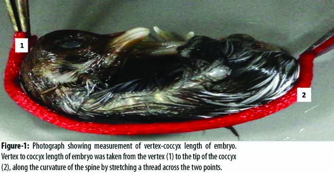
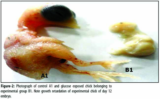
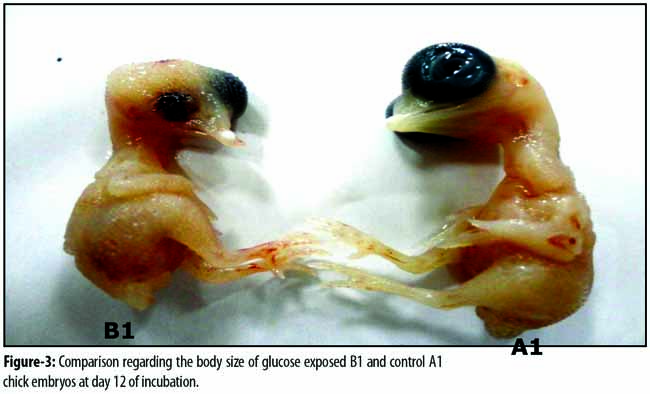
(p=0.001); 10.814±0.214 in A2 and 11.009±0.339 in B2 (p=0.619); and 18.142±0.123 in A3 and 22.87±0.067 in B3 (p=0.000) (Table)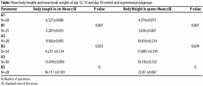
Discussion
The growth retardation is indicated by decrease in body weight and body length of glucose-exposed chick embryos. In our day 12 glucose-exposed embryos, the body length and body weight were significantly less as compared to those of the age-matched subgroup. This is in accordance with previous research work in which hyperglycaemia suppresses the cell cycle leading to slowing of the growth in chick embryos.10 Alteration of metabolites could be the basis of this growth inhibition as shown by work done by Moley and fellows by exposure of pre-implantation mouse embryos to elevated glucose.9 Excess glucose in mammalian post-implantation embryos resulted in ultra-structural changes in visceral yolk sac, such as reduced microvillus surface, fewer mitochondria and a sparser distribution of endoplasmic reticulum. Since visceral yolk sac is the prime route for uptake of nutrients during organogenesis, excess glucose likely to have inhibited the development in this way.11
Experimental embryos from our day 15 subgroup showed more body weight as compared to that of controls, but difference was not significant. As the age advanced, a significant weight gain was observed in 18-day-old embryos belonging to glucose-exposed subgroups. Increased birth weight was noted in offspring born to diabetic mothers. The reason for this could be that excess glucose delivered to foetus resulted in increased production of insulin by foetal pancreas. The combination of hyperinsulinaemia, an anabolic hormone, and hyperglycaemia, a major anabolic fuel, resulted in increased body fat and protein stores. It was proposed that foetal macrosomia resulted from not only due to an overabundance of glucose, but also of amino acids and lipids in the presence of maternal diabetes as all three classes of nutrients had elevated concentration in the circulation of pregnant diabetic women. Maternal diabetes also resulted in increased foetal adipose tissue mass as shown by assessing neonatal adiposity by measuring the skinfold thickness.12 Studies have been conducted in humans to assess the timings of foetal growth spurt in diabetic women. Foetal growth parameters like abdominal circumference and femur length, measured by ultrasound, were significantly higher from 18 weeks of gestation and thereafter. The difference increased progressively as the pregnancy advanced and accelerated growth rate persisted until 38 weeks of gestation.13 It can be postulated that an elevated glucose level at initial stages of development may result in early programming of embryos and subsequent growth acceleration. Previous studies have proposed that glucose level at the start of conception has affected the size of growing embryos.14,15 Moreover the insulin-like growth factor (IGF-1) might have played a role in increased weight gain in glucose exposed embryos since it is a regulator of foetal growth and increased levels of IGF-1 have been demonstrated in cord blood samples of foetuses born to diabetic mothers.16 Although our research manifested that administered glucose altered the normal growth of developing chick embryos, certain environmental factors and methodological limitations might have influenced the results. Therefore, further research is needed to ensure better understanding of the deleterious effects of excess glucose.
Conclusion
Glucose exposure caused the growth retardation in chick embryos till day 10 of incubation. However, a growth accelerating effect was observed in day 18 embryos. The study supports the need for better control of blood glucose levels in diabetic women who want to plan for pregnancy.
Disclaimer: None.
Conflict of Interest: None.
Source of Funding: None.
References
1.American diabetes association. Diagnosis and classification of diabetes mellitus. Diabetic Care 2005; 28: 37-42
2.Correa A, Gilbosa SM, Besser LM, Botto LD, Moore CA, Hobbs CA, et al. Diabetes mellitus and birth defects.Am J Obstet Gynecol 2008; 199: 237.
3.Yang J, Cummings EA, O\\\'connell C, Jangaard K. Fetal and neonatal outcomes of diabetic pegnancies. Obstet Gynecol 2006; 108: 644-50.
4.Liang J, Gui Y, Goas S, Li J, Song H. Elevated glucose induces heart defects by altering the expression of tbx 5 and tbx 20 in the developing zebrafish embryo. Birth Defects Res A Clin Mol Teratol 2010; 88: 480-6
5.Printer E, Haigh A, Nagy A, Madri JA. Hyperglycemia induced vasculopathy in murine conceptus is mediated via reduction of VEGF-A and VEGF receptor activation. Am J Pathol 2001; 158: 1199-206
6.Castelo R , Reymond A , Wyss C , Camara F, Parra G, Antonarakis SE, et al. Comparative gene finding in chicken indicates that we are closing in on the set of multi-exonic widely expressed human genes. Nucleic Acids Res 2005; 33: 1935-9
7.Moley KH, Chi MM, Manchester JK, McDougal DB, Lowry OH. Alterations of intraembryonic metabolites in preimplantation mouse embryos exposed to elevated concentration of glucose; a metabolic explanation for the developmental retardation seen in preimplantation embryos from diabetic animals. Biol Reprod1996; 54: 1209-16.
8.Guariguata L,Whiting DR, Hambleton I,Beagley J, Linnenkamp U, Shaw JE. Global estimates of diabetes prevalence for 2013 and projections for 2035. Diabetes Res Clin Pract 2014; 103: 137-49.
9.Lawence JM, Conteras R, Chen W, Sacks DA. Trends in prevalence of preexisting diabetes and gestational diabetes mellitus among a racially/ethnically diverse population of pregnant women, 1999-2005.Diabetes Care 2008; 31: 899-904.
10.Scott-Drechsel DE, Rugonyi S, Marks DL, Thornburg KL, Hinds MT. Hyperglycemia slows embryonic growth and supresses the cell cycle via cyclin D1 and p21. Diabetes 2013; 62: 234-42
11.Ellington SKL. Effects of excess glucose on mammalian post implantation embryo. Int J Dev Biol 1997; 41: 299-306
12.Nold JL, Georgieff MK. Infants of diabetic mothers. Pediatr Clin N Am 2004; 51:619-37.
13.Persson M, Pasupathy D, Hanson U, Norman M. Birth size distributation in 3,705 infants born to mothers with type 1 diabetes: a population based study. Diabetes Care 2011; 34: 1145-9.
14.Langer O. Fetalmacrosomia: etiologicfactors. ClinObstet Gynecol. 2000; 43: 283-97
15.Wong SF, Chan FY, Cincotta R, Oats JJ, McIntyre HD. Sonographic estimation of fetal weight in macrosomicfetuses: Diabetic versus non diabetic pregnancies. Aust N Z Obstet Gynaecol 2001; 41: 429-32.
16.Lindsay RS, Westgate JA, Beattie J, Pattison NS, Gamble G, Mildenhall LF, et al. Inverse changes in fetal insulin like growth factor (IGF)-1 and IGF binding protein-1 in association with higher birth weight in maternal diabetes. Clin Endocrinol 2007; 66: 322-8.
Journal of the Pakistan Medical Association has agreed to receive and publish manuscripts in accordance with the principles of the following committees:


