

Nadia Ghani ( Environmental Science Department, Lahore College for Women University Lahore, Pakistan. )
Anum Khalid ( Environmental Science Department, Lahore College for Women University Lahore, Pakistan. )
Arifa Tahir ( Environmental Science Department, Lahore College for Women University Lahore, Pakistan. )
July 2016, Volume 66, Issue 7
Original Article

Abstract
Objective: To examine the effects of airborne endotoxin on lung function impairment in exposure-response relationships among the workers of textile industry.
Methods: The cross-sectional study was conducted at Lahore College for Women University, Lahore, Pakistan, from January to August 2014, and comprised textile mill workers. The participants were divided into exposed and control groups. A questionnaire was used to ask workers about the potential adverse health effects of their occupation. The pulmonary function test was carried out by spirometer. Endotoxin levels in the samples were determined using the key quality characteristics limulus amebocyte lysate. The data was analysed to determine the correlation between the endotoxin exposure duration and pulmonary function test parameters.
Results: There were 200 subjects subdivided into 100 each inexposed and control groups. Overall, 160(80%) were not aware of safety measures and the remaining 40(20%) were partially practising. Changes in pulmonary function due to endotoxin exposure showed decreased force vital capacity, flow rate and peak expiratory flow parameters significantly different (p<0.05, p<0.001). The endotoxin concentration was between 12EU/m3 and 300EU/m3. Airborne endotoxin concentrations in textile plants exceeded the Dutch health-based guidance limit of 90EU/m3 and was associated with respiratory health effects.
Conclusion: Prolonged exposure to airborne endotoxin caused constant lung impairment. Proper safety measures should be adopted to avoid the inhalation of cotton dust.
Keywords: Endotoxin, Ameaobocyte Lysate assay, Pulmonary function, Force vital capacity. (JPMA 66: 803; 2016)
Introduction
Endotoxin (ET) is an outer membrane component of gram-negative bacteria comprising strong inflammatory agent. It is a key factor of interest for studies of lung ailments in occupational environment with organic dust (OD). A wide variety of occupational health hazards are present in the textile industry, the most obvious being OD, particularly cotton dust (CD). Workers are constantly exposed to it which impairs lung functions. Textile workers are exposed to airborne particulate from natural and synthetic fibrous materials in their work environment.1 Airborne ET has strong chronic health effects.2
ETs are complex lipopolysaccharides (LPS) which form an inherent fraction of the outer cell wall of all gram-negative bacteria and are responsible for the organisation and stability of the cell wall.3 ETs are responsible for toxic effects causing fever,4 multi-organ failure.5 septic shock,6 sepsis7 meningococcemia and severe morbidities like neurological disability (ND), hearing loss and loss of a limb.8
Prolonged exposure to such occupational environment has been associated with abnormal respiratory symptoms e.g., chest tightness. Bacteria growing in the CD themselves scan produce ET during storage and are responsible for producing the broncho-spastic response associated with byssinosis.9
Workers exposed to OD have been observed to exhibit byssinosis.10 Symptoms include coughing, shortness of breath and difficulty in breathing. These become more prominent when the affected person returns to work after the weekend, leading to the moniker "Monday Asthma".11
Limulus Amebocyte Lysate (LAL) analysis is widely used for ET measurement for environmental samples. This process uses an ET-triggered enzyme cascade from the horseshoe crab (Limulus polyphemus) to cleave a colorimetric substrate. As the LAL analysis is very sensitive, variability can arise due to differences in laboratory methods for sample collection, handling, storage, extraction, and analysis.12
LAL is used to measure biological activity of LPS that may be important in eliciting responses in the human lung and respiratory disease.13 Recently, in the Netherlands, Dutch Expert Committee on Occupational Standards (DECOS) has revised the earlier health-based occupational exposure limit and set a limit at 90 EU m-3. This exposure limit was based on a study which showed a "no-effect" for selected sensitive healthy subjects in an experimental setting of 6-h work exposure.14 The current study was planned to analyse the adverse toxic effects of ET concentrations in different units of textile industry, its effects on lungs and adequate measures that should be adopted to minimise these effects.
Subjects and Methods
The cross-sectional study was conducted at Lahore College for Women University, Lahore, Pakistan from January to August 2014, and comprised textile mill workers. After approval from the institutional review board, participants were divided into exposed and control groups, and all of them were categorised in three age brackets: <30; 30-40; and >40. The control group included those who had not been exposed to CD.
Samples were selected from two main textile industries of Lahore having all sub-units in working. Full-time employees having minimum experience of five years at the same place were included. Part-time workers, smokers and those who did not have medical fitness certificate were excluded. A modified, structured questionnaire of the American Thoracic Society (ATS) was developed by taking into consideration different working conditions and processes involved in the industry. Participants were asked about their exposure and potential adverse health effects of their occupation.
To establish the status of respiratory system, pulmonary function test (PFT) was conducted using aspirometer, according to the ATS procedure.15 The variables that were collected for each subject were the force vital capacity (FVC), force expiratory volume in one second (FEV1), FEV1 / FVC, vital capacity(VC), FEV1 / VC, and peak expiratory flow(PEF). The parameters was categorised as: <40=severe; 40-54=moderate; 55-70=mild; and >70=normal. Airborne ET concentrations were measured by using the LAL process through chromogenic method. A microbial air sampler (MAS) was used to collect air samples from two industries each on glass fibre filter to recognise probable causes of ET exposure. For this purpose five runs of 10 samples from each location were performed around 1.5 metres above the floor and measurements were taken. To evaluate the quantity of dust on the filters gravimetrically, the filters were pre- and post-weighed on an analytical balance. Inhalable dust concentration lower than the limit of detection (LOD) was allocated a significance of two-thirds of the LOD of the weighing scale. Extraction of ET was done under pyrogen-free conditions. The blank/sterile filters were extracted and assayed, and results were compared with spiked/blank concentration.
Quantitative kinetic chromogenic (KCA) LAL method (Cambrex, Verviers, Belgium; Lysate lot no. 3L433E, standard lot no. 3L2950) was used. ET concentration in units per cubic metre (EU/m3) were calculated by using the calculated volume of air sampled and ET concentrations[EU/ml X sample volume (ml)] / [time (min) X (rate (L/min) X 1 m3/1000 L].16
To ensure reproducibility and repeatability, the detection method was validated in the laboratory. By spike recovery of ET in samples that were diluted 10-fold the interference of the samples was assessed.
PFT data of exposed and control groups were presented as mean and standard deviation. The statistical differences among the groups were determined by one-way analysis of variance (ANOVA) in order to differentiate the variations. Data was considered statistically significant at p<0.05. Pearson\\\'s correlation coefficient was used to assess the relation between exposure airborne ET concentration and absorbance (nm).
Results
Of the 200 subjects, subdivided by 100 each in exposed and control groups. Overall, 160(80%) were not aware of safety measures and the remaining 40(20%) were partially practising. The airborne concentration of ET in the working area of several sections was found greater than the permitted threshold limit value (TLV) and workers were more apparent to have byssinosis and similar respiratory ailments. In production, maintenance, yarn sizing, warping and knotting sections ET concentrations were observed at high levels. Especially production and maintenance sections were more effected. Range of mean airborne ET concentration in work area was 40-300 EU/m3 and in the work area of above-mentioned sections was 40-150 EU/m3.
The spike recovery for LAL was lesser in weaving and dyeing section, at 45%. Reproducibility for all sub-sections was relatively alike. Standard curve for the test was projected by polynomial regression where R2wasless than 0.984 (0.22) (Figure-1)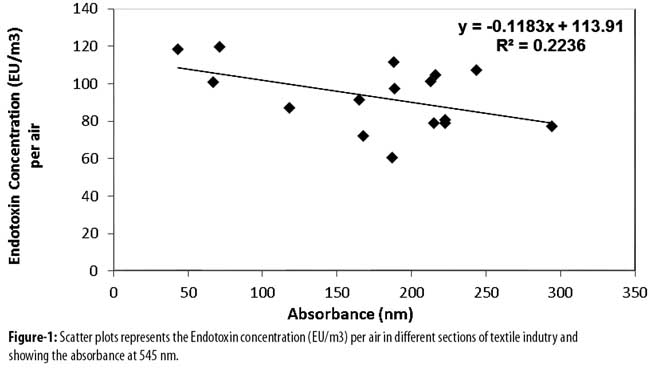
All functional parameters illustrated a decline with age in the exposed group as compared to controls(Table)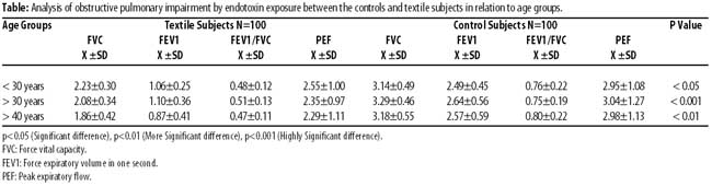
The mean values of FVC, FEV1, FEV1/FVC and PEF were usually lesser in the exposed group and the difference was larger and statistically significant (p<0.05), especially among the subjects ranging from 30 to 40 years. The mean values in the age group of > 40 years were smaller, and demonstrated a significantly (p<0.001) greater fall in lung function compared to controls and showed significantly higher prevalence of bronchial obstruction. Age, escalating time worked and increased duration of ET exposure were associated with a lower FEV1 and FVC. The mean values of FVC, FEV1, FEV1/FVC and PEF were usually lesser in the exposed group (Figure-2)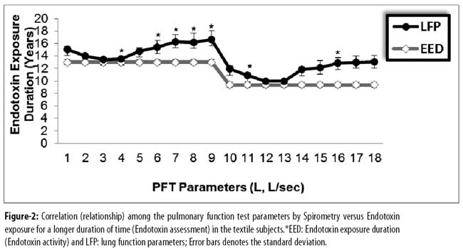
On the basis of World Health Organisation (WHO)\\\'s classifications, subjects were considered to have no constant ventilatory impairment, with FEV1 larger than 80% of the predicted value, those with FEV1 60-80% to have slight to moderate lung impairment, and those with FEV1<60% to have moderate to severe impairment.17 Ages of 40-49 were one-and a-half times and ages greater than or equal to 50 were almost two times probable to have byssinosis (Figure-3)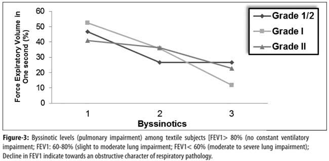
There was increased occurrence of equally obstructive as well as restrictive lung function abnormalities amongst workers when compared with control subjects, with the most common pattern being obstructive.
Discussion
Epidemiological studies coalesced through the exploit of the LAL assay on samples of airborne OD encompass revealed a relationship between the ET contact, e.g., component of the inhalable fraction of OD, and pulmonary signs or functional alteration.18 These observations have been verified by means of human exposure experiments.19
The LAL assay is used to detect enumerating airborne ET concentration from CD samples.20 A high association was observed in anticipated airborne ET concentration (log EU/m3) among two laboratories performing an assessment of duplicate samples of airborne CD, according to the study performed in cotton textile industry for the measurements of ET at work area.21 ET is an intoxicating, nonspecific stimulant of the immune system and results in unfavourable effects. The ET inhalation contact studies on humans reveal acute effects including decline in lung function as well as airway along with alveolar irritation.22
A study in Shanghai, China, explains that CD extract causes physiological effects in reducing airway hyper reactivity. In the pathogenesis of byssinosis as well as obstructive airway ailment, the ET is the chief moderator. Another Shanghai study illustrates that constant loss in lung function was more sturdily linked by exposure to ET than to CD.23 A study performed on pulmonary function (PF) in north-west Ethiopia found that among the CD-exposed personnel, byssinotics behaved conspicuously and demonstrated utmost acute as well as chronic alteration in PFs. It was also showed that FEV1 and FVC significantly decrease in the exposed group as compared with controls.24 In this study the symptoms were observed associated with lung function impairment. Textile workers are at risk for occupational lung disease, including byssinosis and chronic bronchitis. The initial phase of byssinosis is characterised by acute reversible symptoms, such as wheezing, chest tightness, shortness of breath or cough. These early symptoms are generally accompanied by reversible changes in PF (across-shift drops in FEV1). With continued exposure, the disease may progress to a stage in which symptoms are present throughout the work week and may eventually result in severe pulmonary disability.
Basic inherent limitation faced in this study was to convince the participants to contribute their input. Therefore, the size of sample could not be increased due to restrictions from industry owners and lack of willingness from workers to fillin the questionnaire.
Conclusion
Due to lack of safety measures, poor knowledge of workers and lack of law application, a large number of textile workers are exposed to airborne ET concentration which results in lung function impairment. Regular monitoring, proper rotation of workers and adoption of work-related hygiene programmes are highly recommended to reduce the adverse levels of intensity.
Acknowledgment
We are grateful to the Chairperson, Department of Environmental Science, for administrative support, and to the staff of the department lab for their cooperation.
Disclosures: None.
Conflict of Interest: None.
Funding: None.
References
1. Oldenburg M, Latza U, Baur X. Exposure-response relationships between endotoxin exposure and lung function impairment in cotton textile workers. Int Arch Occup Environ Health 2007; 80: 388-95.
2. Wang XR, Zhang HX, Sun BX, Dai HL, Hang JQ, Eisen EA. A 20-year follow-up study on chronic respiratory effects of exposure to cotton dust. Eur Respir J 2005; 26: 881-6.
3. Kim SE, Su W, Cho M, Lee Y, Choe WS. Harnessing aptamers for electrochemical detection of endotoxin. Anal Biochem 2012; 424: 12-20.
4. Su W, Kim SE, Cho M, Nam JD, Choe WS, Lee Y. Selective detection of endotoxin using an impedance aptasensor with electrochemically deposited gold nanoparticles. Innate Immun 2013; 19; 388-97.
5. Lawrence K. Pediatric Sepsis and Multiorgan Dysfunction Syndrome: Progress and Continued Challenges. Crit Care Nurs Clin North Am 2011; 23; 323-37.
6. Ding SJ, Chang BW, Wu CC, Chen CJ, Chang HC. A new method for detection of endotoxin on polymyxin B-immobilized gold electrodes. Electrochemistry Communications 2007; 9: 1206-11.
7. Gutsmann T, Howe J, Zahringer U, Garidel P, Schromm AB, Koch MH, et al. Structural prerequisites for endotoxic activity in the Limulus test as compared to cytokine production in mononuclear cells. Innate Immun 2010; 16: 39-47.
8. Kofyman A, Takayesu JK. Meningococcal disease. Afr J Em Med 2011; 1: 174-8.
9. Wang Y, Knoll W, Dostalek J. Bacterial pathogen surface plasmon resonance biosensor advanced by long range surface plasmons and magnetic nanoparticle assays. Analytical Chemistry 2012; 84: 8345-50.
10. Mberikunashe J, Banda S, Chadambuka A, Gombe NT, Shambira G, Tshimanga M, et al. Prevalence and risk factors for obstructive respiratory conditions among textile industry workers in Zimbabwe, 2006. Pan Afr J Med 2010; 17; 6:1
11. Liu AH. Hygiene theory and allergy and asthma prevention. Paediatr Perinat Epidemiol 2007; 3: 2-7.
12. Spaan S, Doekes G, Heederik D, Throne PS, Wouters IM. Effect of extraction and assay media on analysis of airborne endotoxin. Appl Environ Microbio 2008; 75: 3804-11.
13. Alwis KU, Milton DK. Recombinant Factor C assay for measuring endotoxin in house dust: Comparison with LAL, and (1-3)-beta-D-Glucans. Am J Ind Med 2006; 49: 296-300.
14. DECOS. Endotoxins: health based recommended exposure limit. A report of the Health Council of the Netherlands. 2010. The Netherlands: The Hague: Health Council of the Netherlands. Publication no 2010/040SH.
15. American Thoracic Society. Standardization of spirometry: 1987 update. Am Rev Respir Dis 1987; 136: 1285-98.
16. Lonzo I. Limulus amebocyte lysate QCL-1000 test kit manual. Catalogue number 50-648-U. 2001
17. World Health Organization. International classification of functioning and diability.Beta-2.Geneva: WHO, 1999.
18. Douwes J, Heederik D. Epidemiologic investigations of endotoxins. Int J Occup Environ Health 1997; 3: 26-31
19. Rylander R, Bake B, Fischer JJ, Helander I M. Pulmonary function and symptoms after inhalation of endotoxin. Am Rev Respir Dis 1989; 140: 981-6.
20. Dungan RS, Leytem AB. Airborne endotoxin concentrations at a large open-lot dairy in southern Idaho. J Environ Qual 2009; 38: 1919-23.
21. Mehta AJ, Wang XR, EisenEA, Dai HL, Astrakianakis G, Seixas N. Work area measurements as predictors of personal exposure to endotoxin and cotton dust in the cotton textile industry. Ann Occup Hyg 2008; 52: 45-54.
22. Reed CE, Milton DK. Endotoxin-stimulated innate immunity: a contributing factor for asthma. J Allergy Clin Immunol 2012; 108: 157-66.
23. Khan AJ, Nanchal R. Cotton Dust Lung Disease. Curr Opin Pul Med 2007; 13: 137-41.
24. Abebe Y, Seboxa T. Respiratory problems among textile mill workers in Bahir Dar, Ethiopia.Br J Ind Med 2007; 48:110-5.
Related Articles
Journal of the Pakistan Medical Association has agreed to receive and publish manuscripts in accordance with the principles of the following committees:


