

Huzaifa Ali Jaliawala ( Research Officer, Aga Khan University Hospital, Karachi. )
Fatima Mannan ( Research Officer, Aga Khan University Hospital, Karachi. )
Roger Christopher Gill ( Department of General Surgery, Aga Khan University Hospital, Karachi. )
Abdul Rehman Alvi ( Department of General Surgery, Aga Khan University Hospital, Karachi. )
June 2016, Volume 66, Issue 6
Case Reports

Abstract
An elderly gentleman presented to our emergency with a 10-day history of right upper quadrant pain and nausea with associated tenderness on examination. His white cell counts were raised with predominant neutrophils with ultrasound evidence of a heterogeneous sub-hepatic collection, not associated with fever, diarrhoea or vomiting. He had a similar episode 3 weeks ago, which resolved with antibiotics. Initially thought to be a perforated acute appendicitis or a liver abscess a CT scan was done to further substantiate our finding. This to the contrary revealed a perforated sub hepatic appendix. Patient was treated conservatively with IV fluids and antibiotics and the sub hepatic collection was aspirated under ultrasound guidance. He responded well to treatment and made an uneventful recovery.
Keywords: Sub-hepatic collection, Acute appendicitis, Perforation.
Introduction
Appendicitis is a common presentation in the Emergency Department worldwide. Prompt diagnosis and early intervention is crucial to avoid morbidity related to delayed diagnosis.1,2 This becomes a challenge when the presentation is deviant from what we see classically, making it important for surgeons to be familiar with these aberrant presentations to be able to diagnose appendicitis in time.
Case Presentation
An elderly male, presented to the Emergency Department with a 10-day history of gradually worsening right upper quadrant pain during the month of August, 2015 to the Aga Khan University Hospital, Karachi. It was constant, sharp, non-radiating, 8/10 in severity, aggravated on movement and mild exertion. It was associated with nausea and anorexia. There was no fever, vomiting, diarrhoea or constipation. The patient was haemodynamically stable. He had similar symptoms three weeks ago associated with fever, which resolved in outpatient setting with antibiotics.
On abdominal examination he had tenderness in the right upper quadrant. Bowel sounds were audible in all four quadrants. No visceromegaly was appreciated. His medical history revealed benign prostalic hypertrophy, Diabetes and Hypertension. Surgical history showed removal of kidney stones in the past. Family history was non-contributory.
Initially it was thought that he has either acute cholecystitis or a liver abscess, therefore an ultrasound was advised. This revealed a normal gall bladder with normal wall thickness with no pericholecystic fluid and normal biliary anatomy. However there was an heterogeneous sub hepatic collection adjacent to the gall bladder (Figure-1)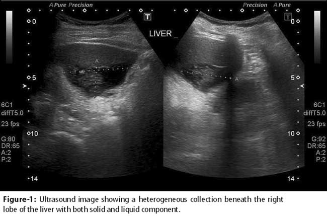 .
.
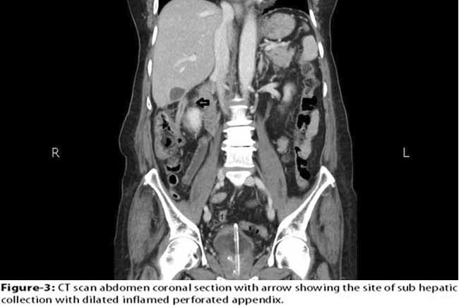
This raised the possibility of either a micro perforation from the gall bladder or a ruptured liver abscess. However this was not very convincing therefore a CT scan was performed. CT scan revealed appendix in the sub hepatic position which had perforated and was responsible for the sub hepatic collection evidenced on the ultrasonogram (Figure-2,3)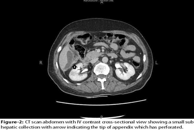
.
Investigations
Blood picture showed a white cell count of 15.1x10⁹, predominantly neutrophils. Liver Function Tests, Prothrombin Time, Activated Partial Thromboplastin Time, Electrolytes, Urea and Creatinine were within the normal ranges. Ultrasound of the abdomen showed evidence of a well-defined heterogeneous area along the inferior margin of right lobe of liver with solid and fluid component raising the possibility of a sub-capsular or ruptured liver abscess. CT scan revealed a perforated appendix located in the sub-hepatic region.
The differential diagnosis included:
1. Hepatic Abscess, 2. Acute cholecystitis, 3. Para-Colic Abscess.
Patient was admitted and broad spectrum antibiotics started along with Intravenous fluids. The abscess was aspirated under Ultrasound guidance. Fever and abdominal pain settled. Oral feeding was resumed gradually, which was well tolerated.
Outcome and Follow-Up
The patient responded well to the conservative approach to treatment. He was discharged on the third day of admission. At the time of discharge his white cell count was 8.9x10⁹, with normal differential. On follow up, a week later, in the outpatient clinic he had no complaints. He had resumed his normal diet and was tolerating it well.
Discussion
Clinical presentation of appendicitis varies on its anatomical location in the abdomen. The more commonly reported positions such as retro-caecal (74%), pelvic (21%), sub-caecal (1.5%), pre-ileal (1.5%), and post-ileal(0.5%) determine the classic presentation.3 However, there are rare variants present that distort the typical presentation making diagnosis a challenge.
Sub-hepatic location of the appendix is one such variant where the appendix is located in the right upper quadrant. King4 introduced us to the first case in 1955, whereas Palanivelu et al5 tabulated the incidence to be 0.08% from a study performed in India. Patients are usually misdiagnosed as acute cholecystitis, liver abscess, ureteric colic or acute pyelonephritis.5
CT scan is proven to be more beneficial to ascertain diagnosis in patients with atypical presentations of right upper quadrant pain with non-specific findings.6,7 As Sub-hepatic appendicitis is more common in the elderly; CT scan is indispensable in establishing a diagnosis.8,9
Conclusion
Sub-hepatic appendicitis complicated by perforation may lead to abscess formation. In our experience, it was adequately managed with conservative treatment with IV fluids, antibiotics and aspiration, saving the patient from unnecessary surgery.
Disclosure: The article and no part of it has been published in any other journal.
Conflict of Interest: There are no conflicts of interest to declare.
Financial Support: No financial support was required.
References
1. Omundsen M, Dennett E. Delay to appendicectomy and associated morbidity: a retrospective review. ANZ J Surg. 2006; 76:153-5.
2. Bickell NA, Aufses AH, Rojas M, Bodian C. How time affects the risk of rupture in appendicitis. J Am Coll Surg. 2006; 202:401-6.
3. Williams NS, Bailey H, Bulstrode CJK, Love RJM, O\\\'Connell PR. Bailey & Love\\\'s short practice of surgery. J Laryngology & Otology .2009; 123:258-9.
4. King A. Subhepatic appendicitis. AMA Arch Surg. 1955; 71:265-7.
5. Palanivelu C, Rangarajan M, John SJ, Senthilkumar R, Madhankumar MV. Laparoscopic appendectomy for appendicitis in uncommon situations: the advantages of a tailored approach. Singapore Med J. 2007; 48:737-40.
6. Bugliosi TF, Meloy TD, Vukov LF. Acute abdominal pain in the elderly.Ann Emerg Med. 1990; 19:1383-6.
7. Kraemer M, Franke C, Ohmann C, Yang Q, Acute Abdominal Pain Study Group. Acute appendicitis in late adulthood: incidence, presentation, and outcome. Results of a prospective multicenter acute abdominal pain study and a review of the literature. Langenbeck s Arch Surg. 2000; 385:470-81.
8. Ting JY, Farley R. Subhepatically located appendicitis due to adhesions: a case report. J Med Case Rep. 2008;2:339.
9. Ong EM, Venkatesh SK. Ascending retrocecal appendicitis presenting with right upper abdominal pain: utility of computed tomography. World journal of gastroenterology: World J Gastroenterol. 2009; 15:3576-9.
Journal of the Pakistan Medical Association has agreed to receive and publish manuscripts in accordance with the principles of the following committees:


