

Seyfettin Ciftci ( Department of Urology, University of Kocaeli, Kocaeli, Turkey. )
Ersan Ozbudak ( Department of Cardiovascular Surgery, University of Kocaeli, Kocaeli, Turkey. )
Hasan Yilmaz ( Department of Urology, University of Kocaeli, Kocaeli, Turkey. )
Murat Ustuner ( Department of Urology, University of Kocaeli, Kocaeli, Turkey. )
Levend Ozkan ( Department of Urology, University of Kocaeli, Kocaeli, Turkey. )
Mustafa Melih Culha ( Department of Urology, University of Kocaeli, Kocaeli, Turkey. )
April 2016, Volume 66, Issue 4
Case Reports

Abstract
Persistent lymphatic drainage is uncommon after most of the surgical operations. It is related with mechanical, nutritional and immunological problems as well as electrolyte imbalance and protein deficiency. It is most commonly seen in retroperitoneal surgeries including abdominal aortic surgery and retroperitoneal lymph node dissection. Conservative management is the first treatment choice and resolves the problem in most cases. However persistent high output drainage may not be resolved with conservative approach and surgical or invasive treatment may become necessary. Additionally, surgical management of persistent lymphatic drainage has not been sufficiently discussed in the literature. In this study, we present a case of persistent very high output lymphatic drainage after right radical nephrectomy which failed with conservative approach and was successfully treated with surgical management.
Keywords: persistent, high output, lymphatic drainage, surgical management
Introduction
Lymphatic drainage is uncommon after most of the surgical procedures.1 The incidence is not clearly known and any well-defined risk factor has not been established yet. It was reported in 12% of lower extremity vascular surgeries and found to be related with prolonged hospital stay and high wound infection.2 Persistent lymphatic drainage is also related with mechanical, nutritional and immunological dysfunction because of excessive loss of protein and lymphocytes.3 Most probable pathogenesisis is the obstruction or disruption of lymphatic drainage during the surgery.3 So, it is very common after abdominal aortic surgery which causes 81% of all surgical retroperitoneal lymphatic injuries.4 Persistent lymphatic drainage was also described in the urological surgeries including retroperitoneal lymph node dissection and radical nephrectomy.3 Conservative approaches including bed rest, salt restriction and medium chain triglyceride diet with high protein and low fat have been defined to overcome this postsurgical persistent problem.5 However, to the best of our knowledge, surgical management of persistent and very high output lymphatic drainage after radical nephrectomy and lymphadenectomy has not been discussed in the literature yet. In this study, we present a case of very high output lymphatic drainage after radical nephrectomy which failed with conservative management and was successfully treated with surgical ligation of lymphatic vessels.
Case Report
A 65-year-old male patient was admitted to our hospital with a complaint of haematuria in October 2014. A computed tomography (CT) scan revealed a 3.5x4x5 cm in diameter right solid renal mass (Figure-1A).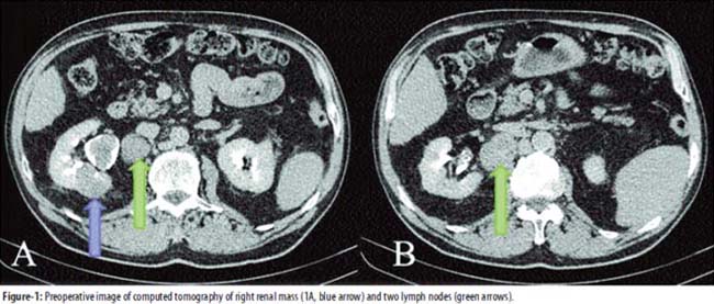
CT also revealed two lymph nodes (LN)in right renal hilum adjacent to the inferior vena cava (Figure-1). The largest diameter of biggest LN was 3.5 cm and the other 3 cm. A complete blood count and basic metabolic panel were unremarkable. In his past medical history, there was diabetes mellitus for 6 years. The patient underwent right retroperitoneal radical nephrectomy and excision of lymph nodes described in CT. A drainage catheter was left and operation was finished without any complication. Beginning on postoperative day 4, there was very high output lymphatic drainage through the catheter. Initially, the patient was managed with conservative approach including bed rest, medium chain triglyceride parenteral diet and total parenteral nutrition. However, the drainage reached 6000 ml per day at postoperative 10th day. Furthermore, electrolyte imbalance and severe protein deficiency (3.2 g/dL) (Normal range: 6.4-8.3 g/dL) occurred thereafter and was treated with electrolyte and protein supplement daily. Additionally, severe life-threatening hyponatraemia (110 mEq/L) (Normal range 136-145 mEq/L) occurred and it was treated with sodium supplementation. To follow-up the hyponatremia, electrolyte parameters were assessed twice daily. On postoperative day 20, high output drainage still continued with an amount of 5000-6000 ml per day despite the conservative management and intermittent blockage of the retro-peritoneal drainage catheter. So, we decided to re-operate the patient to ligate the lymphatic vessels. The patient again underwent retroperitoneal exploration through the previous incision. There were clearly visible two lymphatic vessels very close to the IVC which drained excessively during the exploration (Figure-2A).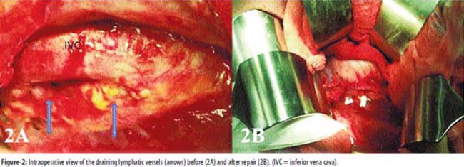
The lymphatic vessels were ligated with 5/0 non-absorbable suture in combination with a pair of previously prepared Teflon felt strips (Figure-2B), and fibrin glue (Tisseel® Baxter, Deerfield, IL, USA) was applied to cover the lymphadenectomy area. A drainage catheter was left and operation was completed without any complication. Drainage was 300 ml per day for the following day and steadily decreased almost to the amount of 50 ml per day. All biochemical parameters returned to the normal level within 4 days. Drainage catheter was removed 6 days after the second operation and patient was discharged on the following day. After one week and one month of discharge, ultrasonography scans were performed and no re-accumulation of fluid was detected in the operation site. Additionally, the patient was free of any symptom.
Pathology revealed a 7.5 cm type 2 papillary renal cell carcinoma with morphology of Fuhrman grade 2. One of the excised lymph node was diagnosed as metastatic and the other as benign.
Discussion
Although postsurgical lymphatic drainage is a rare and benign condition, it can be life-threatening if not treated correctly. Excessive and persistent lymphatic drainage causes electrolyte imbalance, protein and lymphocyte loss and therefore causes lymphopenia and infections.3 It was most commonly described after lower extremities vascular surgeries, abdominal aortic surgeries and retroperitoneal lymph node dissection.1,3,4 However, an accepted and clearly defined treatment has not been established due to its rarity. Conservative managements including bed rest, salt restriction and medium chain triglyceride diet with high vena cava thrombectomy was considered a successful conservative managements.3,5,6 Although salt restriction was recommended in those studies, severe life-threatening hyponatraemia occurred and sodium supplementation was required in our case. So, we recommend defining the electrolyte levels before starting salt restriction as conservative treatment. Except salt restriction, other conservative treatment options were applied to our case. Somatostatin was also described as an alternative treatment of choice besides closure of refractory lymphatic fistulae,5 whereas, we did not use this alternative.
Beside the above stated conservative treatment modalities, applicable materials were also discussed for the same purpose. Giberson et al described persistent lymphatic drainage in a paediatric age patient after the resection of abdominal wall lymphangiomatous tumour.7 The authors successfully treated the patient with application of fibrin glue through the drainage catheter. Albala et al described a case of persistent lymphatic drainage after pelvic lymphadenectomy and radical prostatectomy.8 They successfully managed the patient with instillation of diatrizoatemeglumine through a Jackson-Prattdrain.In our case, we also used fibrin glue in the second operation after ligation of lymphatic vessels to cover the lymphatic area. However, discussing surgical treatment of this postoperative persistent problem is limited. As surgical treatment, See etal and Selli et al reported the use of peritoneovenous shunting in persistent lymphatic leakage.9,10 Chylous ascites was accumulation of lymphatic drainage and seen in retroperitoneal lymph node dissection surgeries. Paracentesis and abdominal drain insertion are other surgical options that was used successfully to resolve the chylous ascites and persistent drainage. Baniel et al described peritoneovenous shunt and surgical approaches as choices to treat the chylous ascites after retroperitoneal lymph node dissection.6 Browne et al also described a 21-year-old male patient who underwent retroperitoneal lymph node dissection.3 The patient had persistent lymphatic drainage and the authors treated him with the combination of conservative approach and surgical management including paracentesis and insertion of a drain. As series, Steele et al. defined 15 patients who had prolonged lymphatic drainage after vascular surgery.11 The authors treated 5 patients with non-operative approach, whereas 10 patients with surgical re-exploration and ligation of lymphatic vessels. Moreover they used isosulfan blue to ensure correct and definitive ligation of lymphatic vessels. However these authors did not describe any high output drainage such in our case. Additionally, to the best of our knowledge, open surgical approach with ligation of lymphatic vessels which cause persistent high output leakage after radical nephrectomy has not been described in the literature.
Conclusion
Postsurgical persistent lymphatic drainage can be life-threatening and should be treated correctly. We added to literature a new case of persistent high output lymphatic drainage after radical nephrectomy and successfully treated with surgical approach. We recommend that, surgical treatment should be taken into consideration in this persistent condition if conservative management fails.
References
1. Skudder PA Jr, Geary J Lymphatic drainage from the groin following surgery of the femoral artery. J Cardiovasc Surg (Torino). 1987; 28:460-3.
2. Dunlop MG, Fox JN, Stonebridge PA, Clason AE, Ruckley CV. Vacuum drainage of groin wounds after vascular surgery: a controlled trial. Br J Surg. 1990;77:562-3.
3. Browne KM, Casey RG, Thornhill JA. Prolonged lymphatic leak after retroperitoneal lymph node dissection: a case report. J Med Case Rep. 2009; 3:8704.
4. Combe J, Buniet JM, Douge C, Bernard Y, Camelot G. Chylothorax and chylous ascites following surgery of an inflammatory aortic aneurysm. Case report with review of the literature]. J Mal Vasc. 1992; 17:151-6.
5. Leibovitch I, Mor Y, Golomb J, Ramon J. Chylous ascites after radical nephrectomy and inferior vena cava thrombectomy. Successful conservative management with somatostatin analogue. Eur Urol. 2002; 41:220-2.
6. Baniel J, Foster RS, Rowland RG, Bihrle R, Donohue JP. Management of chylous ascites after retroperitoneal lymph node dissection for testicular cancer. J Urol.1993; 150:1422-4.
7. Giberson WG, McCarthy PM, Kaufman BH. Fibrin glue for the treatment of persistent lymphatic drainage. J Pediatr Surg. 1988;23:1188-9.
8. Albala DM, Kevwitch MK, Waters WB. Treatment of persistent lymphatic drainage after laparoscopic pelvic lymph node dissection and radical retropubic prostatectomy. J Endourol. 1993; 7:337-40.
9. See WA, Kresowik TF, Jochimsen PR. Peritoneal venous shunting for the treatment of lymphatic ascites following retroperitoneal lymph node dissection. Urology. 1996; 48:783-5.
10. Selli C, Carini M, Mottola A, Barbagli G. Chylous ascites after retroperitoneal lymphadenectomy: successful management with peritoneovenous shunt. Urol Int. 1984; 39:58-60.
11. Steele SR, Martin MJ, Mullenix PS, Olsen SB, Andersen CA. Intraoperative use of isosulfan blue in the treatment of persistent lymphatic leaks. Am J Surg. 2003; 186:9-12.
Journal of the Pakistan Medical Association has agreed to receive and publish manuscripts in accordance with the principles of the following committees:


