

Shazib Sagheer ( Medical College, Cardiothoracic Surgery, Aga Khan University, Karachi. )
Abdul Malik Amir Humza Sohail ( Medical College, Cardiothoracic Surgery, Aga Khan University, Karachi. )
Yousaf Bashir Hadi ( Medical College, Cardiothoracic Surgery, Aga Khan University, Karachi. )
Saulat Fatimi ( Department of Surgery, Cardiothroacic Sugery, Aga Khan University, Karachi. )
March 2016, Volume 66, Issue 3
Student's Corner

Abstract
Mediastinal enteric cysts are a rare occurrence among adults and are usually asymptomatic. In most cases they are an incidental finding in the right hemi-mediastinum and are associated with vertebral anomalies. We report the unusual case of a 23 year old male who had a mediastinal mass on chest X-ray as an incidental finding. Chest Computed Tomography (CT) scan revealed no vertebral anomalies and a cystic mass in the left posterior mediastinum with features similar to those of a hydatid cyst. Posterolateral thoracotomy was done and the cyst was excised. Histopathology report revealed it to be an enteric cyst.
Keywords: Enteric cysts, Mediastinal mass, Thoracotomy, Bronchogenic cyst.
Introduction
Congenital foregut cysts arise as a consequence of malformations of the primitive intestine during embryogenesis and constitute 18% of primary mediastinal masses.1 They can be classified into one of the three categories: bronchogenic, intramural oesophageal or enteric.
Enteric cysts in the mediastinum are a rare occurrence; only 1-2% of all mediastinal cysts are enteric cysts.2 Histologically, their walls have a layer of smooth muscle and are lined by mucosal epithelium. 60% of them are diagnosed in infants before the age of 1 year.3 They are more common in children and usually present with respiratory symptoms, such as dyspnoea, stridor and cough; in adults they are mostly asymptomatic. They are usually found in the right posterior mediastinum and are associated with vertebral anomalies.4
Herein we present the case of a 23 year old male with a cyst in the left posterior mediastinum but with no identified vertebral anomalies. Upon histopathology, the cyst was identified to be an enteric cyst.
Case History
A 23 year old male was found to have a homogenous area of opacification with clear margins involving the left upper and mid lung zone, as an incidental finding on chest X-ray (Figure-1).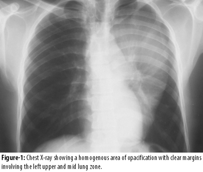
There was no evidence of lung collapse or pleural effusion. The patient was asymptomatic and all systemic examinations were unremarkable. Past medical and surgical history was non-contributory. The patient was a non-smoker, did not take alcohol and had no other addictions. There were no known allergies.
Computed Tomography (CT) scan report described a lobulated well-defined multiseptate lesion, measuring 18.9 ×13.3 ×8.5 cm, in the left posterior mediastinum in paravertebral location (Figure-2 and 3)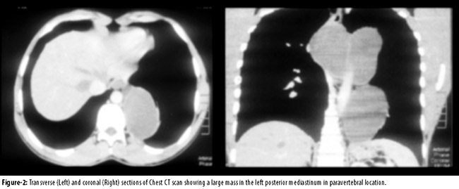
The features of the lesion were highly compatible with hydatid cyst Serological work-up for hydatid cyst was negative.
Posterolateral thoracotomy and excision of the cyst were performed. Intra-operatively, the cyst was not connected to the bronchi, vertebrae or esophagus and was successfully removed, preserving all adjacent structures. Post-operative recovery was uneventful.
Histopathology report of the excised mass described a multiloculated cyst, weighing 750 grams, filled with dark brown, thick material. Microscopically, the cyst wall was ulcerated and lined by tall columnar epithelium showing goblet cells. The underlying stroma showed sheets of haemosiderin laden macrophages and clusters of mucus glands. The glands were surrounded by mild to moderate lymphocytic infiltrate with their lumen being filled with pink amorphous material. The cyst was covered by smooth muscle, arranged in two layers, and did not contain any cartilage. Based on these findings the final diagnosis of an enteric cyst was made.
One other differential diagnosis was mediastinal lymphangioma. The radiological findings and the histological report did not identify any features suggestive of this and thus it was ruled out.
The patient was advised to undergo MRI scan of the spine to evaluate any associated vertebral anomalies. However, this could not be performed because of patient refusal.
Discussion
Enteric cysts, first described by Blasius in 1711,7 can be found anywhere in the alimentary canal. About 7-20% of enteric cysts are found in the thorax3 while only 1-2% of all mediastinal cysts are enteric cysts.2
They can present with a variety of symptoms depending upon the location of the cyst, the involved adjacent structures and the histological features. In children they are symptomatic and thus are diagnosed early. About 60% of enteric cysts are diagnosed in patients before the age of one year, usually due to respiratory symptoms.3 In adults they are symptomless and usually present as an incidental finding, as in our patient.
Mediastinal enteric cysts may also cause severe compressive symptoms like respiratory distress, tracheobronchial obstruction and dysphagia due to oesophageal compression. Compression of mediastinal vasculature and cardiac tamponade effect by compression of the heart can also occur.
The presence of gastric or pancreatic tissue in the cyst can result in wall ulceration, perforation and haemorrhage, leading to haemoptysis and haematemesis.3 Rupture into the pericardium resulting in cardiac tamponade has also been reported.3 Adenocarcinoma, squamous cell carcinoma and other malignancies can also develop in enteric cysts;1 there was no histological evidence of malignancy in our patient. In order to prevent the above-mentioned complications, the treatment of choice for such lesions is excision.6
Theories regarding the origin of enteric cysts point towards embryonic primitive foregut development anomalies.3 One of the most widely accepted theories about the origin of enteric cysts is split notochord syndrome which involves the persistence of neuroenteric band.6 Veenklaas, in 1952, theorized that incomplete separation of ectoderm and notochord could eventually result in spinal anomalies.6 Perhaps this is why most enteric cysts are associated with vertebral anomalies.
Also, enteric cysts usually lie in the posterior mediastinum and in most instances have no connection with the esophagus.8
Our case is unique in a number of ways. Firstly, enteric cysts are usually found in the right posterior mediastinum while in our case it was present in the left posterior mediastinum. Secondly, no vertebral anomalies were identified in our patient. In Ahmed, et al\\\'s series all patients but one had vertebral anomalies.4 In 2006, Zhang KR, et al reported vertebral anomalies in 12 out of 16 patients of enteric cyst.9 A right-sided posterior mediastinal mass with a vertebral defect is diagnostic of enteric cyst in more than 70% of cases.3 Because the strong association of enteric cysts with vertebral anomalies provides support to the notochord theory, the absence of any anomalies in this patient and other previously reported similar cases may point towards lacunae in the split notochord theory. Thirdly, features of the cyst on CT scan were consistent with hydatid cyst, and did not point towards the possibility of an enteric cyst. This raises questions about the accuracy of CT scan as a diagnostic tool for mediastinal masses. For mediastinal masses\\\' diagnostic purposes, it has already been pointed out that CT scan is relatively inaccurate to distinguish between cystic and solid masses.7
Conclusion
It is important to note that although enteric cysts are usually found in the right posterior mediastinum, they can be present in the left posterior mediastinum. Also, although most enteric cysts are associated with vertebral anomalies, the absence of any vertebral anomalies should not rule out the diagnosis of enteric cyst. In cases where on radiology a hydatid cyst is found in the posterior mediastinum, the possibility of an enteric cyst must be kept in mind as a differential diagnosis; CT scan, as seen in this case, may not be a very accurate tool for the diagnosis of mediastinal masses.
Disclosure: All the authors have declared no competing interest.
References
1. Suzuki K, Koyama S, Yamada S, Kawabata Y. Adenocarcinoma arising in a mediastinal enteric cyst. Intern Med 2006; 46: 781-4.
2. Davis Jr R, Oldham Jr H, SabistonJr D. Primary cysts and neoplasms of the mediastinum: recent changes in clinical presentation, methods of diagnosis, management, and results. Ann Thorac Surg 1987; 44: 229-7.
3. Iglesias Sentís M, Belda Sanchís J, Gimferrer Garolera JM, Catalán Biela M, Rubio Garay M, Ramírez Ruz J. [Mediastinal enteric cyst: unusual clinical presentation and histopathology]. Arch Bronconeumol 2004; 40: 185-7
4. Ahmed S, Jolleys A, Dark J. Thoracic enteric cysts and diverticulae. Br J Surg 1972; 59: 963-8
5. Le Pimpec-Barthes F, Cazes A, Bagan P, Badia A, Vlas C. [Mediastinal cysts: clinical approach and treatment]. Revue de Pneumologie Clinique 2010; 66: 52-2.
6. Birmole B, Kulkarni B, Vaidya A, Borwankar SS. Intrathoracic enteric foregut duplication cyst. J Postgrad Med 1994; 40: 228-30
7. Pisello F, Geraci G, Arnone E, Sciutto A, Modica G, Sciumè C. Acute onset of esophageal duplication cyst in adult. Case Report. G Chir 2009; 30: 17-20.
8. Carachi R, Azmy A. Foregut duplications. Pediatric Surgery International 2002; 18: 371-4.
9. Zhang KR, Jia HM, Pan EY, Wang LY. Diagnosis and treatment of mediastinal enterogenous cysts in children. Chin Med Sci J 2006; 21: 201-3.
Journal of the Pakistan Medical Association has agreed to receive and publish manuscripts in accordance with the principles of the following committees:


