

Yasemin Celik ( Department of Emergency Medicine, Bagcilar Education and Research Hospital, Istanbul )
Turker Yardan ( Department of Emergency Medicine, Ondokuz Mayis University Medical Faculty, Samsun, Turkey.. )
Ahmet Baydin ( Department of Emergency Medicine, Ondokuz Mayis University Medical Faculty, Samsun, Turkey )
Sabri Demircan ( Department of Cardiology, Ondokuz Mayis University Medical Faculty, Samsun, Turkey. )
March 2016, Volume 66, Issue 3
Original Article

Abstract
Objective: To investigate the role of N-terminal pro-brain natriuretic peptide and apelin in the assessment of right ventricular dysfunction in acute pulmonary embolism.
Methods: The prospective case-control study was conducted at Ondokuz Mayis University, Samsun, Turkey, from January 2008 to June 2009, and comprised adult patients with acute pulmonary embolism. A smaller group of healthy adults served as the control. Blood N-terminal pro-brain natriuretic peptide and apelin levels were measured on admission to the Emergency Department. SPSS 15 was used for data analysis.
Results: There were 56 cases and 20 controls in the study. Blood N-terminal pro-brain natriuretic peptide levels were higher in cases than the controls (p<0.05). Apelin levels were not different between the groups (p>0.05). Patients with right ventricular dysfunction had significantly higher peptide levels than those without the dysfunction (p<0.05). The cut-off value of peptide for the prediction of right ventricular dysfunction was 1000 pg/ml, with a sensitivity of 92.1% and specificity of 77.8%.
Conclusions: There was no significant change in plasma apelin levels in acute pulmonary embolism. The blood N-terminal pro-brain natriuretic peptide maybe a useful parameter in the assessment of right ventricular dysfunction in acute pulmonary embolism.
Keywords: Pulmonary embolism, Right ventricular dysfunction, NT-proBNP, Apelin. (JPMA 66: 306; 2016)
Introduction
Pulmonary embolism (PE) is a relatively common cardiovascular emergency. By occluding the pulmonary arterial bed, it may lead to acute life-threatening but potentially reversible right ventricular (RV) dysfunction.1 RV dysfunction has an important prognostic value in patients with PE. Patients with RV dysfunction are known to be at risk of subsequent clinical worsening and PE-related death.2
N-terminal pro-brain natriuretic peptide (NT-proBNP) is a member of natriuretic peptides and a useful marker in the evaluation of cardiac functions. NT-proBNP, also called BNPT, is known to be useful not only in diagnosis, but also in the risk stratification in various cardiac diseases, such as heart failure,3 pulmonary hypertension4 and acute PE.5 A few studies have revealed that BNPT has a potential to be a predictor of echocardiographic RV dysfunction in patients with acute PE.6,7
In 1998, Tatemoto et al.8 isolated a 36-amino acid peptide from an extract of the bovine stomach and named it apelin. Apelin is located in the endothelium of the myocardium, large vessels, and small veins and arteries.9 However, the most important source of apelin in the bloodstream is the lungs.10 Apelin shows positive inotropic as well as vasodilator effects. Changes in serum apelin levels were detected in different cardiovascular diseases. Although serum apelin value increases in the early stage of left ventricle (LV) failure, it decreases in the endstages.11 In addition, apelin values decrease in chronic pulmonary diseases.10 It was reported that the effect of exogenous apelin on vasomotor tone in a canine model of acute PE was complicated.12 However, to our best knowledge, no study has investigated the blood concentrations of apelin in patients with acute PE.
The current study was planned to investigate the role of serum BNPT and plasma apelin levels in the assessment of RV dysfunction in patients with acute PE.
Patients and Methods
The prospective case-control study was conducted at Ondokuz Mayis University, Faculty of Medicine, Samsun, Turkey, between January 2008 and June 2009, and comprised adult patients with acute PE who were admitted to the Emergency Department (ED). In addition, 20 age- and gender-matched healthy adults served as controls. The study protocol was approved by the institutional ethics committee.
After initial evaluation and physical examination, routine laboratory tests, chest X-ray, electrocardiography (ECG), echocardiography, spiral computed tomography (CT) of the thorax, and/or ventilation/perfusion (V/Q) lung scintigraphy were performed in all patients. PE was diagnosed with the presence of refilling defect in pulmonary arteries in spiral CT angiography and/or presence of perfusion defects in V/Q scintigraphy.1
Patients included presence of acute PE within the first 2 days, confirmation of PE diagnosis with spiral CT angiography and/or V/Q scintigraphy, normal LV systolic function on echocardiography, and serum creatinine <1.5mg/dl. Those with no echocardiography at the time of admission and those with history of chronic pulmonary disease were excluded.
Patients were divided into two groups based on the presence or not of RV dysfunction on echocardiography. Moreover, according to the presence of RV dysfunction and systolic blood pressure (SBP), patients were divided into three groups as:13 a) Massive PE: presence of RV dysfunction finding in echocardiography and SBP<90 mmHg, b) Submassive PE: presence of RV dysfunction finding in echocardiography and SBP>90 mmHg, and c) Lower-risk PE: presence of normal RV function in echocardiography and a normal systolic arterial pressure.
Two-dimensional echocardiography was performed as soon as possible after admission using a standard commercial ultrasound machine (Vivid 7, GE Vingmed, Horten, Norway) with a 2.5-MHz transducer. RV end-diastolic diameter (EDD) was measured from either the apical or subcostal four-chamber view. An experienced cardiologist blinded to the BNPT and apelin levels and the clinical data performed these examinations. According to the patients\\\' echocardiography findings, RV dysfunction was confirmed by at least one of the following findings:14 i) RV hypokinesis (asymmetric or delayed contraction, usually in the base of the RV), ii) paradoxical septal systolic motion, iii) RV dilatation (EDD>30mm or RVEDD/LVEDD >1 in apical four-chamber view).
Peripheral venous blood samples (8 ml) were collected from the patients on admission. One-half of each sample was placed into a tube with aprotinin+ethylenediaminetetraacetic acid (EDTA) for obtaining plasma, while the other half was placed into a tube without anticoagulant for obtaining serum. Samples were centrifuged at 2500 g for 15min. The same procedure was also applied to blood samples of the control group. Samples were stored at -70°C in a deep freezer until analysis. Serum BNPT levels were measured by using electrochemiluminescence immunoassay (ECLIA) with Roche Diagnostics Elecsyspro BNP E-170 test kit in an Elecsys 2010 device (Roche Diagnostics, Mannheim, Germany). Plasma apelin levels were measured using the competitive enzyme immunoassay method with Apelin-36 (human) enzyme-linked immunosorbent assay (ELISA)/Phoenix test kit in a Biotech Synergy 4 device.9,15,
All statistical calculations were done using SPSS15. Values were reported as median (min-max) for data that was not distributed normally and non-parametric tests were used. Comparisons between groups were done using the Mann-Whitney U test for two groups, and Kruskal-Wallis analysis of variance (ANOVA) followed by Bonferroni-corrected Mann-Whitney U test for more than two groups. Correlations between the variables were evaluated using Spearman correlation analysis. P<0.05 was accepted as a statistically significant difference for Mann-Whitney U test, Kruskal-Wallis analysis of variance and Spearman correlation analysis, whereas a value of p<0.017 was accepted for the Bonferroni-corrected Mann-Whitney U test. Receiver operating characteristic (ROC) curves for predicting RV dysfunction determined by echocardiography were generated from the data. Sensitivity and specificity were also calculated for BNPT levels.
Results
There were 56 patients with acute PE with a median age of 62 years (inter-quartile range [IQR]: 24-94 years); and 31(55%) of the patients were females. There were 20 healthy controls with a median age of 55 years (IQR: 27-78 years); and 11(55%) of the controls were females (p>0.05 each) (Table-1).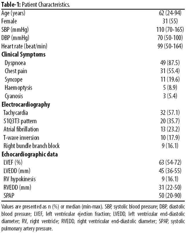
Serum BNPT levels were significantly higher in patients than in the control group (Table-2).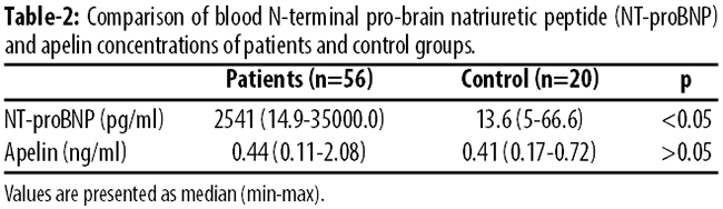
There was a positive correlation between BNPT and systolic pulmonary artery pressure (SPAP) (r=0.57; p<0.05) and between BNPT and RV diameter (r=0.44; p<0.05),as well as between SPAP and RV diameter(r=0.61;p<0.05).There was no statistically significant difference between the patient and control groups with respect to plasma apelin levels (p>0.05). There was a weak negative correlation between apelin and RV diameter (r=-0.27; p<0.05). However, there was no correlation between apelin and BNPT or between apelin and SPAP (p>0.05).
There were 38(67.8%) patients with RV dysfunction and they had significantly higher BNPT levels than those without RV dysfunction. In addition, patients with and without RV dysfunction had higher BNPT levels than the control group (Table-3).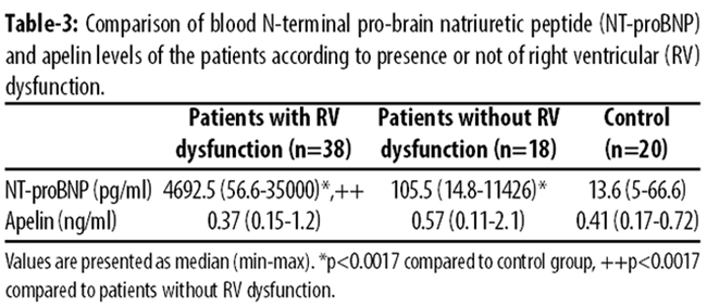
The cut-off value for prediction of RV dysfunction was 1000pg/ml for BNPT, which was identified by ROC analysis. The area under the curve (AUROC) was 0.882 (95% confidence interval [CI], 0.781; 0.982), which indicated good discriminative power. A BNPT value >1000 pg/ml had a sensitivity rate of 92.1%, specificity rate of 77.8%, positive predictive value (PPV) of 89.7%, and negative predictive value (NPV) of 82.4%. A BNPT level >1000 pg/ml was associated with a risk ratio of 40.83 (95% CI, 6.68-290.22) for the diagnosis of RV dysfunction. There was no significant difference between patients with and without RV dysfunction with respect to apelin levels (p>0.05).
Besides, 18(32.1%) patients were diagnosed as having Low-risk PE, 14(24.9%) with Submassive PE and 24(43%) with Massive PE. All of these groups had higher BNPT levels than the control group (p<0.05). Moreover, the Low-risk PE group had lower BNPT levels than the Massive PE and Submassive PE groups (p<0.05). There was no significant difference between these groups and the control group with respect to apelin levels (p>0.05) (Table-4).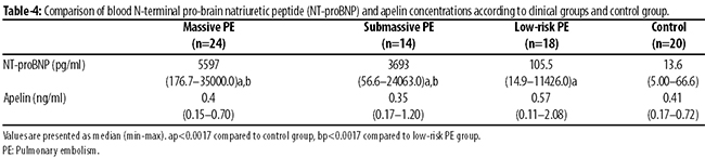
Discussion
RV functions take an important place in acute PE for the evaluation of the clinical condition and determination of the treatment strategy. As hypoxia causes pulmonary vasoconstriction, it raises Pulmonary alveolar proteinosis (PAP) immediately. This condition leads to increased RV after load, RV dilatation and RV dysfunction.1 Presence of RV dysfunction in PE is related with poor prognosis. Haemodynamic malfunction after RV dysfunction is pointed out as indicating the need for aggressive treatment in these patients.16 It was suggested that early thrombolytic treatment in haemodynamically stable patients with RV dysfunction reduces mortality, PE recurrence and pulmonary hypertension in the late period of PE.17,18
For more than a decade, there has been increased interest in the assessment of the clinical course of PE with cardiac biomarkers. It is known that plasma BNP and BNPT levels increase after ventricle wall tension. These indicators are used for diagnosis of LV failure related to ventricle dysfunction.19 In addition, increased BNP was detected especially in acute PE and the other conditions related to increased RV volume and pressure.4,20 BNP, which is a C-terminal peptide-active biological part of proBNP, and NT-proBNP, which is an inactive N-terminal part, are related to acute PE.6,7
In this study, the role of BNPT and apelin in the assessment of RV dysfunction in acute PE was evaluated. We found that BNPT levels were significantly higher in patients compared with controls. There was a positive correlation between BNPT and SPAP and between BNPT and RV diameter, as well as between SPAP and RV diameter. Kruger et al.21 reported that there was a positive relevance between RV end-diastolic pressure and BNP levels. Similar to our results, Choi et al.6 reported that there was a positive correlation between BNPT and SPAP in acute PE. Our results showed that serum BNPT levels are increased in acute PE and that this is related to RV functions and SPAP in acute PE.
In our study, patients with RV dysfunction had significantly higher BNPT levels than patients without RV dysfunction. In addition, patients with and without RV dysfunction had higher BNPT levels than the control group. It was detected that the cut-off value of BNPT for diagnosis of RV dysfunction was 1000 pg/ml. This cut-off value for RV dysfunction diagnosis had 92.1% sensitivity and77.8% specificity. Similar to our results, it was reported that patients with RV overload had higher BNPT levels than patients without RV overload.5 Abul et al.22 reported that the optimal cut-off value to predict RV dysfunction was 530pg/ml for BNPT (sensitivity: 80% and specificity: 47%) in haemodynamically stable patients with acute PE. The cut-off value for RV dysfunction in the present study was higher than in that study, and can be explained by the fact that both haemodynamically stable and unstable patients with acute PE were included in the present study. The cut-off of 1000pg/ml of BNPT should be validated by further studies. Our study revealed that BNPT levels are associated with RV dysfunction and that BNPT can be used as a marker for the presence of RV dysfunction in patients with acute PE and normal LV function.
In our study, patients were classified into Massive, Submassive or Low-risk groups according to clinical severity. BNPT levels of the Massive and Submassive PE groups, which were more severe clinically, were significantly higher than those in the low-risk PE group. Similar to our results, it was reported that BNPT may be helpful in the assessment of clinical severity in acute PE.5We suggest that echocardiography need not be ordinarily ordered in patients with normal BNPT levels, because RV function will be almost normal and patients can be considered to be in the Low-risk group. If BNPT levels are elevated, echocardiography should be performed for further risk stratification and assessment of RV function.
Measurement of BNPT concentrations can also be helpful for determination of the treatment strategy as well as for prediction of clinical outcome in acute PE. Vuilleumier et al.23 suggested that the BNPT level could be an efficient tool for the identification of very low-risk non-massive PE patients, who could be treated in an outpatient setting. It was reported that out-of-hospital treatment is safe in haemodynamically stable patients with PE with low (<500 pg/ml) BNPTlevels.24 Binder et al.25 reported that patients with BNPT levels <1000 pg/ml could reliably be identified as a low-risk group, as indicated by the high NPV for in-hospital death and the combined primary end-point "complicated clinical course". They suggested a simple risk stratification algorithm for patients with PE, with the use of BNPT or troponin testing as an initial step that should be followed by echocardiography in the case of elevated levels of the biomarker. Andreassen et al.26 reported that BNPT levels may be significant to determine clinical severity and long-term mortality in patients with idiopathic hypertension, chronic pre-capillary hypertension and PE. In our study, we did not evaluate the patients with respect to clinical outcome.
Apelin was identified as a novel adipokine and became popular in cardiovascular disease in recent decades.27 It has an inotropic as well as vasodilator effect. Moreover, apelinplays a role in body fluid balance.28 Although these effects show that apelin has important endocrine regulator function for the heart, but the clinical practice and benefits of apelin are unclear. Apelin contributes to the pathophysiology of diseases such as heart failure, parenchymal pulmonary disease, pulmonary hypertension, and atherosclerosis related to the apelin-APJ (Apelin receptor) signal pathway.9,29 It was suggested that the most important source of apelin in blood is the lungs.10
Different results were obtained from studies that evaluated apelin levels in cardiac diseases. Chen et al.11 reported that in patients with different stages of LV failure, it was detected that although plasma apelin levels were increased in the early stage of heart failure, they decreased when the heart failure progressed. In a recent study, it was reported that while BNP levels were increased in heart failure, apelin levels were decreased.28 In another study, apelin and BNPT levels were compared in patients with chronic parenchymal lung disease, idiopathic pulmonary hypertension and heart failure. In that study, apelin levels were significantly decreased in these patient groups. However, while BNPT levels were increased in heart failure and pulmonary hypertension, there was no change in chronic parenchymal lung disease. In that study, it was interpreted that the decrease in apelin levels in both heart failure and parenchymal lung disease maybe related with the secretion of apelin dominantly from the pulmonary vasculature.10
In our study, the relation between plasma apelin level and PE was also evaluated. There was no significance difference between the patient and control groups with respect to apelin levels. Although there was a weak negative correlation between apelin and RV diameter, but there was no correlation between apelin and BNPT or between apelin and SPAP. There was no significant difference between patients with and without RV dysfunction and the control group with respect to apelin levels. In addition, there was no significant difference between the clinical and control groups with respect to apelin levels. According to these results, it can be concluded that apelin levels did not change in acute PE on admission.
Only one study, conducted by Feng et al.12 has investigated apelin levels in PE. In that study, they induced acute PE in dogs with autologous blood clots to assess the effect of apelin on pulmonary and systemic circulation in the acute phase of acute PE. They found that the content of apelin messenger ribonucleic acid (mRNA) in the lung tissue increased during the first several hours but decreased at 24 hours (h) after the induction of acute PE. However, the expression of apelin and APJ proteins in the pulmonary arteries did not change significantly within 24h after acute PE induction. The expression of apelin in the bronchial epithelial cells had significantly increased at 1h and decreased at 24h after acute PE induction. These results revealed that release of apelin from pulmonary tissue showed periodic variations in acute PE. In that study, it was concluded that the effect of apelin on vasomotor tone is complicated, and it can cause variable changes in pulmonary and systemic arterial pressures under different physiological and pathological conditions.
Our study has several limitations. The study is monocentric and limited by the small sample size, which precludes the possibility of any strong conclusions. Prospective and controlled studies involving larger numbers of acute PE patients are needed. In addition, only one blood sample was collected from each patient on admission for measuring BNPT and apelin levels. When the periodic variations of apelin release are considered, serial measurements of BNPT and apelin are needed for further comments. In this study, patients with renal failure and LV dysfunction were excluded. The results of this study cannot be generalised for all acute PE cases.
Conclusion
Serum NT-proBNP, or BNPT, levels are elevated in acute PE. Serum BNPT may serve as an indicator of clinical severity. Measurement of BNPT levels may be a useful approach in the diagnosis of RV dysfunction in patients with acute PE. The possibility of RV dysfunction in patients with plasma BNPT levels >1000 pg/ml should be strongly considered. There was no significant change in plasma apelin levels in patients with acute PE on admission.
Acknowledgements
We are grateful to Dr. Ahmet Tevfik Sunter for statistical assistance, and to the Scientific Research Foundation of Ondokuz Mayis University, Samsun, Turkey, for financial assistance.
*Presented at the 2nd EurAsian Congress on Emergency Medicine, October 28-31, 2010 Antalya-Turkey.
References
1. Torbicki A, Perrier A, Konstantinides S, Agnelli G, Galiè N, Pruszczyk P, et al. Guidelines on the diagnosis and management of acute pulmonary embolism: the Task Force for the Diagnosis and Management of Acute Pulmonary Embolism of the European Society of Cardiology. Eur Heart J 2008; 29: 2276-315.
2. Yardan T, Altintop L, Baydin A, Yilmaz O, Guven H. B-type natriuretic peptide as an indicator of right ventricular dysfunction in acute pulmonary embolism. Int J Clin Pract 2008; 62: 1177-82.
3. Waldo SW, Beede J, Isakson S, Villard-Saussine S, Fareh J, Clopton P, et al. Pro-B-type natriuretic peptide levels in acute decompensated heart failure. J Am Coll Cardiol 2008; 51: 1874-82.
4. Fijalkowska A, Kurzyna M, Torbicki A, Szewczyk G, Florczyk M, Pruszczyk P, et al. Serum N-terminal brain natriuretic peptide as a prognostic parameter in patients with pulmonary hypertension. Chest 2006; 129: 1313-21.
5. Pruszczyk P, Kostrubiec M, Bochowicz A, Styczy?ski G, Szulc M, Kurzyna M, et al. N-terminal pro-brain natriuretic peptide in patients with acute pulmonary embolism. Eur Respir J 2003; 22: 649-53.
6. Choi HS, Kim KH, Yoon HJ, Hong YJ, Kim JH, Ahn Y, et al. Usefulness of cardiac biomarkers in the prediction of right ventricular dysfunction before echocardiography in acute pulmonary embolism. J Cardiol 2012; 60: 508-13.
7. Abul Y, Karakurt S, Ozben B, Toprak A, Celikel T. C-reactive protein in acute pulmonary embolism. J Investig Med 2011; 59: 8-14.
8. Tatemoto K, Hosoya M, Habata Y, Fujii R, Kakegawa T, Zou MX, et al. Isolation and characterization of a novel endogenous peptide ligand for the human APJ receptor. Biochem Biophys Res Commun 1998; 251: 471-6
9. Japp AG, Newby DE. The apelin-APJ system in heart failure: pathophysiologic relevance and therapeutic potential. Biochem Pharmacol 2008; 75: 1882-92.
10. Goetze JP, Rehfeld JF, Carlsen J, Videbaek R, Andersen CB, Boesgaard S, et al. Apelin: a new plasma marker of cardiopulmonary disease. Regul Pept 2006; 133: 134-8.
11. Chen MM, Ashley EA, Deng DX, Tsalenko A, Deng A, Tabibiazar R, et al. Novel role for the potent endogenous inotrope apelin in human cardiac dysfunction. Circulation 2003; 108: 1432-9.
12. Feng JH, Li WM, Wu XP, Tan XY, Gao YH, Han CL, et al. Hemodynamic effect of apelin in a canine model of acute pulmonary thromboembolism. Peptides 2010; 31: 1772-8.
13. Kucher N, Goldhaber SZ. Cardiac biomarkers for risk stratification of patients with acute pulmonary embolism. Circulation 2003; 108: 2191-4.
14. Golpe R, Pérez-de-Llano LA, Castro-Añón O, Vázquez-Caruncho M, González-Juanatey C, Veres-Racamonde A, et al. Right ventricle dysfunction and pulmonary hypertension in hemodynamically stable pulmonary embolism. Respir Med 2010; 104: 1370-6.
15. Francia P, Salvati A, Balla C, De Paolis P, Pagannone E, Borro M, et al. Cardiac resynchronization therapy increases plasma levels of the endogenous inotrope apelin. Eur J Heart Fail 2007; 9: 306-9.
16. Lualdi JC, Goldhaber SZ. Right ventricular dysfunction after acute pulmonary embolism: pathophysiologic factors, detection and therapeutic implications. Am Heart J 1995; 130: 1276-82.
17. Goldhaber SZ. Thrombolytic therapy in venous thromboembolism. Clinical trials and current indications. Clin Chest Med 1995; 16: 307-20.
18. Goldhaber SZ, Elliott CG. Acute pulmonary embolism: part II: risk stratification, treatment, and prevention. Circulation 2003; 108: 2834-8.
19. Ruskoaho H. Cardiac hormones as diagnostic tools in heart failure. Endocr Rev 2003; 24: 341-56.
20. Bal L, Thierry S, Brocas E, Van de Louw A, Pottecher J, Hours S, et al. B-type natriuretic peptide (BNP) and N-terminal-proBNP for heart failure diagnosis in shock or acute respiratory distress. Acta Anaesthesiol Scand 2006; 50: 340-7.
21. Krüger S, Graf J, Merx MW, Koch KC, Kunz D, Hanrath P, et al. Brain natriuretic peptide predicts right heart failure in patients with acute pulmonary embolism. Am Heart J 2004; 147: 60-5.
22. Abul Y, Ozsu S, Mentese A, Durmus I, Bektas H, Pehlivanlar M, et al. Carbonic anhydrase IX in the prediction of right ventricular dysfunction in patients with hemodynamically stable acute pulmonary embolism. Clin Appl Thromb Hemost 2014; 20: 838-43
23. Vuilleumier N, Le Gal G, Cornily JC, Hochstrasser D, Bounameaux H, Aujesky D, et al. Is NT-proBNP superior to clinical scores for risk stratification in non-massive pulmonary embolism? J Thromb Haemost 2010; 8: 1433-5.
24. Agterof MJ, Schutgens RE, Snijder RJ, Epping G, Peltenburg HG, PosthumaEF,et al. Out of hospital treatment of acute pulmonary embolism in patients with a low NT-proBNP level. J Thromb Haemost 2010; 8: 1235-41.
25. Binder L, Pieske B, Olschewski M, Geibel A, Klostermann B, Reiner C, et al. N-terminalpro-brain natriuretic peptide or troponin testing followed by echocardiography for risk stratification of acutepulmonary embolism. Circulation 2005; 112: 1573-9.
26. Andreassen AK, Wergeland R, Simonsen S, Geiran O, Guevara C, Ueland T. N-terminal pro-B-type natriuretic peptide as an indicator of disease severity in a heterogeneous group of patients with chronic precapillary pulmonary hypertension. Am J Cardiol 2006; 98: 525-9.
27. Lv D, Li H, Chen L. Apelin and APJ, a novel critical factor and therapeutic target for atherosclerosis. Acta Biochim Biophys Sin (Shanghai) 2013; 45: 527-33.
28. Chandrasekaran B, Kalra PR, Donovan J, Hooper J, Clague JR, McDonagh TA. Myocardialapelin production is reduced in humans with left ventricular systolic dysfunction. J Card Fail 2010; 16: 556-61.
29. Quazi R, Palaniswamy C, Frishman WH. The emerging role of apelin in cardiovascular disease and health. Cardiol Rev 2009; 17: 283-6.
Journal of the Pakistan Medical Association has agreed to receive and publish manuscripts in accordance with the principles of the following committees:


