

Xu Ziqiang ( Transplantation Center, the First Affiliated Hospital of Wenzhou Medical University, Wenzhou, China. )
He Yunqiang ( Transplantation Center, the First Affiliated Hospital of Wenzhou Medical University, Wenzhou, China. )
Fu Hongxing ( Transplantation Center, the First Affiliated Hospital of Wenzhou Medical University, Wenzhou, China. )
Wang Jinjun ( Transplantation Center, the First Affiliated Hospital of Wenzhou Medical University, Wenzhou, China. )
Cai Yong ( Transplantation Center, the First Affiliated Hospital of Wenzhou Medical University, Wenzhou, China. )
March 2016, Volume 66, Issue 3
Original Article

Abstract
Objective: To evaluate the effects on filtration membrane of glomerulus after islet transplantation in a rat model of streptozotocin-induced diabetic nephropathy.
Methods: The experimental case-control study was conducted at Wenzhou Medical University, Wenzhou, China from January to May 2015, and comprised male Sprague Dawley rats obtained from the Laboratory Animal Centre of Wenzhou Medical University. The rats were intraperitoneally injected with streptozotocin to induce diabetic nephropathy. Diabetic rats were divided into two groups; the islets group received islets transplantation under the kidney capsule; and the diabetic nephropathy (DN) group consisted of untreated diabetic nephropathy rats. The control group consisted of non-diabetic rats. Islets were surgically transplanted under the kidney capsule. Kidney function and blood glucose were measured and pathological changes in the kidney were observed by electron microscope, while the expressions of Wilms\\\' tumour-1, caspase-3 and transforming growth factor-beta 1 were tested by immunohistochemical method and Western blot analysis.
Results: Each of the three groups had 6 rats each with body weights ranging from 180g to 220g. Reduced urinary protein excretion and alleviated damage of podocytes and glomerular basement membrane were seen in the islet-transplanted rats. The alleviation of podocyte damage was related to alteration in the synthesis of caspase-3, transforming growth factor-beta 1, and Wilms\\\' tumour-1 protein in the glomerulus.
Conclusion: Diabetic nephropathy rats after islet transplantation can ameliorate the damage of podocytes and basement membrane by inhibiting the pathway of transforming growth factor-beta 1.
Keywords: Diabetic nephropathy; Islet transplantation; Podocytes; Glomerular basement membrane, TGF-b1.
Introduction
Diabetic nephropathy (DN) is the most common cause of end-stage renal disease (ESRD).1 Reportedly, 40% diabetic patients develop nephropathy irrespective of glycaemic control.2 Glucose-dependent pathways, such as advanced glycation, play an important role in the development of diabetic renal disease. In early-stage, hyperglycaemia-induced impairment of the glomerulus filtration membrane, including podocytes and the glomeruli basement membrane (GBM), could lead to the occurrence of proteinuria.3,4 Furthermore, persistent proteinuria would lead to the damage of kidney structure and promote the fibrosis of kidney.
Islet transplantation is the most effective measure for type 1 diabetes. It is reported that islet transplantation can ameliorate albuminuria and alleviate the damage of kidney.5 But few researches have addressed the effect of islet transplantation on glomerulus filtration membrane and its precise mechanism. The current study was planned to evaluate the beneficial effects of islet transplantation on glomerular filtration structure in DN rats.
Subjects and Methods
The experimental case-control study was conducted at Wenzhou Medical University, Wenzhou, China from January to May 2015, and comprised male Sprague Dawley rats obtained from the Laboratory Animal Centre of Wenzhou Medical University. All rats were freely fed with water and rodent chow. All animal procedures were based on international guidelines and were approved by the Wenzhou Medical University Animal Policy and Welfare Committee.
Twelve rats received a single dose of streptozotocin injection (STZ; Sigma Aldrich,USA) 55mg/kg intraperitoneally to induce the DN model. One week later, blood samples from tail vein were collected to measure the blood glucose. Rats were considered diabetic if the blood glucose level was between 288mg/dl and 540mg/dl for more than two consecutive days without fasting. At week 8 after the modelling, diabetic rats were divided into two groups. The first group (Islets group) received islets transplantation under the kidney capsule; and the second group (DN group) consisted of untreated DN rats. The control group consisted of six non-diabetic rats.
Twelve male Sprague Dawley rats (body weight: 200-250g) were used as donors. Islets from two donor rats were supplied for each recipient. Islet isolation was performed according to the method described in literature.6 The islets were harvested by reversely perfusion of collagenase V into common bile duct and purification by Histopaque (Sigma-Aldrich, USA) density gradient followed by manual picking. A small incision was performed on the right flank of the recipient rats and the right kidney was exposed. About 800-1000 islet equivalent (IEQ) islets were transplanted under the kidney capsule of the diabetic rats.
After transplantation, blood glucose levels of all groups were measured once a week until the rats recovered euglycaemic state (defined as blood-glucose <10 mmol/L). Also, 24-h urine specimen was collected by metabolic cages and measured by Fully Automatic Biochemistry Analyser (HITACHI 7600, Japan).
Four weeks after islet transplantation, a midline laparotomy was performed on experimental animals. The kidney was in situ perfused with saline by aortic cannulation. A part of right kidney, including transplanted islets, was immersed into 10% neutral formalin solution and 2.5% glutaraldehyde for histopathological analysis. The other part of right kidney and the left kidney was divided into pieces and immediately frozen in liquid nitrogen for Western blot assays.
Portions of renal cortical tissue from four rats per group were prepared according to the method described by other investigators7 and detected by electron microscope (Philips 301).
The kidney tissues were embedded in paraffin and cut into 4-µm for next measure. Haematoxylin and eosin (H&E) staining and immunofluorescence analysis with rabbit monoclonal antibody against insulin (Santa Cruz, USA) were used to observe the survival and secretion function of transplant islets under the kidney capsule.
For immunohistochemical (IHC) staining, the expression of Wilms\\\' tumour-1 (WT-1), caspase-3 and transforming growth factor-beta 1 (TGF-b1) was detected by immunoperoxidase staining with antibodies, including rabbit monoclonal WT-1 (Santa Cruz, USA), rabbit polyclonal active-caspase-3 (Santa Cruz, USA) and rabbit polyclonal TGF-b1 (Santa Cruz, USA). Briefly, renal tissues were sliced into 4mm sections. After removal of the paraffin by xylene and dehydration by graded alcohol, slides were transferred into a 10mmol/L citrate buffer solution and then heated at 80°C for 5 minutes for antigen retrieval. After washing, 3% peroxide was applied for 20 minutes to block the activity of endogenous peroxidase. To avoid non-specific staining, slides were incubated with normal goat serum at room temperature for 20 minutes. The primary anti-rat WT-1, caspase-3 or TGF-b1 monoclonal antibody (MoAb) was added respectively at 1:150 dilution and kept at 4°C overnight. Negative control sections were stained under identical conditions by substituting the primary antibody with equivalent concentrations of normal rabbit immunoglobulin G (IgG). Slides were detected using horseradish peroxidase-conjugated streptavidin, and peroxidase activity was identified by reaction with 3, 3\\\'-diaminobenzidine tetrahydrochloride substrate. Sections were then counterstained with Mayer\\\'s haematoxylin, dehydrated and mounted. To evaluate WT-1, caspase-3 and TGF-b1 staining, glomerular grid field was scored semi-quantitatively by image-ProPlus system (Media Cybernernetics, USA). The results of cell immune-staining were scored (no staining -, weak staining +, moderate staining ++ and strong staining +++).
The expression of TGF-b1 proteins was examined by Western blot using total lysate as previously described.8 Kidney cortex tissues were homogenised in lysis buffer and protein concentrations were determined using a Micro bicinchoninic acid (BCA) Protein Assay kit. Equal amount of protein was separated on a sodium dodecyl sulfate (SDS)-polyacrylamide gel, blotted onto nitrocellulose membranes, and probed with specific antibodies against TGF-b1 (Santa Cruz, USA), and b-Actin (Santa Cruz Biotechnology) was used as control. The antigen-antibody complex was developed by enhanced chemiluminescence (Amersham, Piscataway, NJ, USA), exposed in the dark room and analysed for integral absorbance of the protein bands using Quantity One software Version 4.6.2 (Bio-Rad, USA).
Data was presented as means ± standard error of the mean (SEM). Statistical analyses were performed using one-way analysis of variance (ANOVA), and p<0.05 was considered significant. All statistical analyses were performed using SPSS 13.
Results
Each of the three groups had 6 rats each with body weights ranging from 180gto 220g. The levels of blood glucose (BG) in DN rats decreased obviously significantly at 24h after islet transplantation (p<0.05). At week 4 after transplantation, the levels of BG and 24-hour urine protein from all groups were tested. All rats in the Islets group were normoglycaemic. The level of BG in DN group was higher than that in Islets group (28.6±0.58 versus 9.48±0.31mmol/L; p<0.01). Islets were adjacent to the kidney capsule, and some red blood cells were buried into transplant islets. Also, the transplant islets produced insulin by immunofluorescence image (Figure-1A).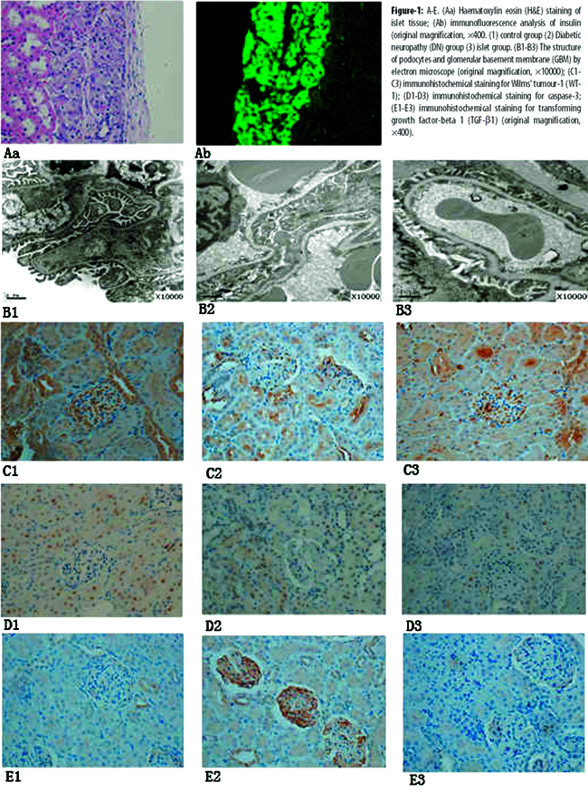
This suggested that the blood glucose level in DN rats returned to normal level after islet transplantation.
There was a marked decrease in 24-hour urine protein in the Islets group compared to the DN group (54.07±2.00 versus 9.45±1.38 mg;p<0.01). Irregular thickening of glomerular basement membrane (GBM)and the fusion of podocytes was observed in the DN group (Figure-1-B). However, the fusion of podocytes\\\' foot process was rare and the damage of GBM was modified in the Islets group (Figure-1. B3). WT-1 protein is an optimal marker of podocytes.9 WT-1 immunostaining was observed in all groups (Figure-1-C). The scores of WT-1-positive podocytes were the lowest in DN group compared to the Islets group and control groups (17.17±0.51 versus 33.28±0.57;p<0.01 and 17.17±0.51 versus 26.33±0.95; p<0.01). It suggested that islet transplantation could attenuate the level of proteinuria and ameliorate GBM damage and podocytes in DN rats.
Compared with DN group, the expression of caspase-3 protein was significantly decreased in the islets group. Immunostaining of caspase-3 was markedly increased in glomeruli of DN rats (Figure-1-D), but very limited numbers of positive cells were found in the control and Islets groups (Table).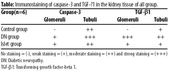
The expression of TGF-b1 in kidney tissues were examined in all groups. An increased expression of TGF-b1 in the DN group was observed by IHC analysis and Western blot assays compared with the control group (p<0.01). The expression of TGF-b1 in the Islets group decreased significantly after islet transplantation (Figure-2).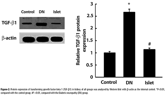
Discussion
The present study found that the damage in kidney from early-stage DN rats was ameliorated after islet transplantation. The possible mechanism was related to the alleviation of the damages of podocyte and GBM, and the reduction in urine protein by inhibiting the pathway of TGF-b1.
Recently, some clinical studies discovered that islet transplantation could reverse the early kidney lesion for diabetic patients, whereas the specific mechanism is still unclear.5,10 Clinically, diabetic nephropathy is mainly characterised by increased proteinuria resulting from irregular thickening of basement membrane and damages to podocytes in the early stage.11 Thus, a rat model in our study was established for early-stage DN with a single dose of STZ. At 12 weeks after modelling, the amount of proteinuria increased significantly. Electron microscopy showed that foot processes of podocytes were partially effaced and basement membranes were irregularly thickened. Immunohistochemically, there was a decrease of WT-1 (podocytes markers). These changes in rats used in our study were consistent with the clinical changes of early-stage diabetic nephropathy. The rats were used as recipients of islet transplantation.
Impairment of glomerular filtration barrier is a major cause of proteinuria in the procedure of early-stage diabetic nephropathy. The filtration barrier is mainly composed of endothelial cells, basement membrane and epithelial cells (podocytes), among which podocytes were the most critical for the composition of the filtration membrane and their impairment would lead to the formation of proteinuria.12 It was reported that the podocytes were discovered to have a higher level of apoptosis,9 decrease, detach from GBM13 and be partially effaced at foot processes14 in the pathogenesis of DN. However, the remaining podocytes failed to fill up the impaired areas. As a consequence, the filtration membranes became more permeable, where proteinuria was produced. As another important structure of the filtration barrier, the GBM was selectively permeable. As an extracellular matrix component of the glomerular filtration barrier, the GBM acts as a size-selective and charge-selective physical filter barrier.15 In the early stage of diabetic nephropathy, it is irregularly thickened, and the structure is thus impaired through which macromolecular substances pass.16 In our study, the damages to of podocyte and GBM in DN rats were ameliorated after islet transplantation.
As a pleiotropic cytokine, TGF-b1 plays an important role in the progression of DN. The secretion of TGF-b1 may be enhanced in the kidney in case of sustained hyperglycaemia. TGF-b1 was found to induce the podocytes to experience the epithelial-to-mesenchymal transition (EMT) and cause glomerularsclerosis.17 Besides, TGF-b1 was discovered to activate the Notch signal pathway to facilitate damage and apoptosis of podocytes.18 Following the application of anti-TGF-b1 antibody, the apoptosis of podocytes was apparently inhibited.19 Krag et al20 discovered that the high filtration could not promote the thickening of GBM in the early stage of diabetic nephropathy. However, GBM got significantly thicker due to high expression of TGF-b1 and high filtration. Wang et al21 discovered that the thickening of GBM was apparently lessened by inhibiting the expression of downstream signals of the TGF-b1 pathway. All of these studies have suggested that it is possible to inhibit the impairment of glomerular filtration barrier and delay the progression of glomerular sclerosis by regulating the synthesis of TGF-b1.
In our study, it was demonstrated that sustained hyperglycaemia in early-stage DN rats activated the pathway of TGF-b1 and led to the damage of filtration membranes, which is in line with previous studies.22 As an effective way for treating diabetic nephropathy, islet transplantation can reverse the recipients\\\' sustained hyperglycaemia. The blood glucose level was found to drop significantly in rats after islet transplantation and can be kept in the normal range. So, the activation of TGF-b1 pathway was inhibited after islet transplantation.
Conclusion
The study demonstrated that a novel mechanism by which proteinuria in early stage DN rats after islet transplantation can be ameliorated is related to modifying the barrier of glomerular filtration by inhibiting the pathway of TGF-b1. Besides, islet transplantation for late-stage DN rats is worthy of further research from the perspective of treatment timing and mechanism.
Acknowledgments
We are grateful to the Science Technology Department of Zhejiang Province (2013C37006) for financial assistance. Thanks are also due to Fu Hongxing and other colleagues in the Wenzhou Medical University.
Trial Grant No 2013C37006.
Reference
1. Shera AS, Jawad F, Maqsood A, Jamal S, Azfar M, Ahmed U. Prevalence of chronic complications and associated factors in type 2 diabetes. J Pak Med Assoc 2004; 54: 54-9.
2. Ian H, Tessa C, Yoshio N, Patrick J, Noel S, Himmelfarb J. Temporal trends in the prevalence of diabetic kidney disease in the United States. JAMA 2011; 305: 2532-9.
3. Wolf G, Chen S, Ziyadeh FN. From the periphery of the glomerular capillary wall toward the center of disease: Podocyte injury comes of age in diabetic nephropathy. Diabetes 2005; 54: 1626-34.
4. Jung HS, Jeffrey H. The glomerular basement membrane as a barrier to albumin. Nat Rev Nephrol 2013; 9: 470-7.
5. Remuzzi A, Cornolti R, Bianchi R, Figliuzzi M, Porretta-Serapiglia C, Oggioni N, et al. Regression of diabetic complications by islet transplantation in the rat. Diabetologia 2009; 52: 2653-61.
6. He Z, Wang F, Kumagai-Braesch M, Permert J, Holgersson J. Longterm gene expression and metabolic control exerted by lentivirus transduced pancreatic islets. Xenotransplantation 2006; 13: 195-203.
7. Tian Y, Lv G, Yang Y, Zhang Y, Yu R, Zhu J, et al. Sodium valproate ameliorates diabetes-induced fibrosis and renal damage by the inhibition of histone deacetylases in diabetic rat. Int J Clin Exp Pathol 2014; 7: 3028-37.
8. Zheng JJ, Wu CZ, Lin Z, Guo Y, Shi L, Dong P, et al. Curcumin up-regulates phosphatase and tensin homologue deleted on chromosome 10 through microRNA-mediated control of DNA methylation - a novel mechanism suppressing liver fibrosis. FEBS J 2014; 281: 88-103.
9. Zhou H, Kajiyama H, Tsuji T, Hu X, Leelahavanichkul A, Vento S, et al. Urinary exosomal Wilms\\\' tumor-1 as a potential biomarker for podocyte injury. Am J Physiol Renal Physiol 2013; 305: 553-9.
10. Figliuzzi M, Bianchi R, Cavagnini C, Lombardi R, Porretta-Serapiglia C, et al. Islet transplantation and insulin administration relieve long-term complications and rescue the residual endogenous pancreatic ? cells. Am J Pathol 2013; 183: 1527-38.
11. Ziyadeh FN, WolfG. Pathogenesis of the podocytopathy and proteinuria in diabetic glomerulopathy. Curr Diabetes Rev 2008; 4: 39-45.
12. Lenoir O, Jasiek M, Hénique C, Guyonnet L, Hartleben B, Bork T, et al. Endothelial cell and podocyte autophagy synergistically protect from diabetes-induced glomerulosclerosis. Autophagy 2015; 11: 1130-45.
13. Hu GH, Jiao B. Mechanism of podocyte detachment: Targeting transmembrane molecules between podocytes and glomerular basement membrane. Biomed Aging Pathol 2013; 3: 36-42.
14. Thomas MC. Pathogenesis and progression of proteinuria. Contrib Nephrol 2011; 170: 48-56.
15. Miner JH. Organogenesis of the kidney glomerulus: focus on the glomerular basement membrane. Organogenesis 2011; 7: 75-82.
16. Jefferson JA, Shankland SJ, Pichler RH. Proteinuria in diabetic kidney disease: a mechanistic viewpoint. Kidney Int 2008; 74: 22-36.
17. Meeteren L, Dijke P. Regulation of endothelial cell plasticity by TGF-beta. Cell Tissue Res 2012; 347: 177-86.
18. Niranjan T, Bielesz B, Gruenwald A, Ponda MP, Kopp JB, Thomas DB, et al. The Notch pathway in podocytes plays a role in the development of glomerular disease. Nat Med 2008; 14: 290-8.
19. Nam BY, Paeng J, Kim SH, Lee SH, Kim do H, Kang HY, et al. The MCP-1/CCR2 axis in podocytes is involved in apoptosis induced by diabetic conditions. Apoptosis 2012; 17: 1-13.
20. Krag S, Nyengaard JR, Wogensen L. Combined effects of moderately elevated blood glucose and locally produced TGF-beta1 on glomerular morphology and renal collagen production. Nephrol Dial Transplant 2007; 22: 2485-96.
21. Wang A, Ziyadeh FN, Lee EY, Pyagay PE, Sung SH, Sheardown SA, et al..Interference with TGF-beta signaling by Smad3-knock out in mice limits diabetic glomerulosclerosis without affecting albuminuria. Am J Physiol Renal Physiol 2007; 293: 1657-65.
22. Satirapoj B. Diabetic kidney disease: important mechanisms and treatment. J Nephrol Soc Thai 2009; 15: 126-39.
Journal of the Pakistan Medical Association has agreed to receive and publish manuscripts in accordance with the principles of the following committees:


