

Raza Hussain ( Department of Surgical Oncology, Shaukat Khanum Memorial Cancer Hospital and Research Centre, Lahore, Pakistan. )
Arif Jamshed ( Department of Radiation Oncology, Shaukat Khanum Memorial Cancer Hospital and Research Centre, Lahore, Pakistan. )
Hassan Iqbal ( Department of Surgical Oncology, Shaukat Khanum Memorial Cancer Hospital and Research Centre, Lahore, Pakistan. )
Sadaf Usman ( Department of Radiation Oncology, Shaukat Khanum Memorial Cancer Hospital and Research Centre, Lahore, Pakistan. )
Muhammad Irfan ( Department of Radiation Oncology, Shaukat Khanum Memorial Cancer Hospital and Research Centre, Lahore, Pakistan. )
Abu Bakar Hafeez Bhatti ( Department of Surgical Oncology, Shaukat Khanum Memorial Cancer Hospital and Research Centre, Lahore, Pakistan. )
February 2016, Volume 66, Issue 2
Original Article
Long term survival and impact of various prognostic factors in T1, T2 oral tongue cancer in Pakistan

Abstract
Objective: To determine the outcome in patients with early squamous cell carcinoma of oral tongue, and the impact of various prognostic factors on survival.
Methods: The retrospective study was conducted at Shaukat Khanum Memorial Cancer Hospital and Research Centre, Lahore, Pakistan, and comprised records of patients treated for squamous cell carcinoma of early stage tongue between March 2003 and October 2009. Various factors, including demographics, risk factors, stage, and grade of the tumour were determined. Kaplan Meier curves were plotted to determine the 5-year overall survival, relapse-free survival, local control, regional control, and loco-regional control.
Results: A total of 137 patients with early oral tongue tumours were treated. With a median follow-up of 46 months, the overall survival of T1, T2 early tongue tumour was 73% and 64%. The incidence of occult metastasis was 30%. The overall survival for Stage I/II and III/IV was 78% and 50% (p=0.002). Patterns of failures included; local 19 (13%), regional 22 (16%), loco-regional 4 (3%) and distant 5 (4%) respectively. The 5-year local control, regional control, loco-regional control was 86%, 82% and 72%. The only significant predictor of overall survival was clinical and pathological N stage in T1 patients, and surgical procedure, grade, pathological N stage in T2 cases.
Conclusions: Treatment of early squamous cell carcinoma of oral tongue effectively achieved local control and disease-free survival. Nodal stage was the most important prognostic factor in terms of survival and recurrence.
Keywords: Squamous cell carcinoma, Early tongue, Survival, Prognostic factors. (JPMA 66: 187; 2016).
Introduction
Tongue remains the most common sub-site for tumours of the oral cavity. Almost half-a-million new cases are diagnosed each year with oral cancer, and tongue accounts for more than 50% of these tumours.1 Tumours of oral cavity present at a late stage in our country where factors like low socio-economic status, lack of awareness, inappropriate diagnosis and unlicensed options might delay the diagnosis and appropriate referral to a specialised institutions. The incidence of early stage tongue cancer is low and not much has been reported about the management of squamous cell carcinoma of the early stage oral tongue (SCCOT) from Pakistan.
In our institution, a total of 373 patients were treated for anterior tongue tumours between 2003 and 2009, and 137 (37%) of them were stage T1/T2. Majority of the literature on early tongue tumours is retrospective in nature and has been reported with other sub-sites of the oral cavity. Surgery is the primary management with adjuvant treatment dictated by the pathological report. Neck nodal metastases remain the most important prognostic factors in the management of oral tongue tumours. As far as management of neck in early oral tongue cancer is considered, the house remains divided between elective neck dissection (END) and wait-and-watch policy.2,3 Although there has been no significant improvement in terms of the survival in SCCOT in the past few decades and for stage 1 and 2, but it has been reported between 75% and 89%.3-6 We do not have any information on survival outcomes in Pakistani population with early tongue tumours.
The current study was planned to determine survival outcomes in patients with T1/T2 SCCOT in our population and analyse various prognostic factors.
Patients and Methods
The retrospective study was conducted at Shaukat Khanum Memorial Cancer Hospital and Research Centre, Lahore, Pakistan, and comprised records of patients treated for SCCOT between March 2003 and October 2009. Head and neck database the hospital was used to extract patient files. The study was granted exemption from formal ethic review by the hospital ethics committee. Inclusion criteria included all patients with T1 and T2 SCCOT. Those with metastatic disease, excision of the tongue tumour at an outside facility, previous history of radiation, and previous neck surgery were excluded.
After histological diagnosis all patients had undergone a complete staging work-up in the Head and Neck clinic. Staging work-up included baseline magnetic resonance imaging (MRI) of face and neck, chest X-ray, and routine blood tests. All cases were discussed by a multidisciplinary team to devise the treatment protocol. Ultrasound abdomen and bone scan was reserved for patients with suspicion of distant metastatic disease.
All patients had undergone partial glossectomy with a gross clear margin of 1cm. Patients with tumour size >1cm in greatest dimension or tumour thickness of >4mm underwent selective neck dissection (SND). SND I-III was performed in all T1 and less than 3cm T2 tumours. Greater than 3cm T2 tumours underwent SND I-IV. Bilateral neck dissection was performed in patients with radiological evidence of significant contralateral neck node or tumour crossing the midline. Primary tumour was oriented to determine margins from tumour. Sub-levels of the neck specimen were divided by the surgeon in the operating room and sent to the pathologist in separately labelled containers.
Adjuvant treatment (post-operative radiation or chemo-radiation) was given within 4-6 weeks of surgery. It was planned in accordance with the pathology report. Tumours with pathological T3 (pT3), pT4, tumour thickness >4mm, close margins, lymphovascular invasion (LVI), perineural invasion (PNI) and/or N2/N3 nodal disease were subjected to post-operative radiation (PORT). Patients with extracapsular spread, positive margins and >2 metastatic lymph nodes received chemo-radiation. Pathological node-negative patients received 60Gy in 30 fractions to primary operative site and ipsilateral neck, whereas in pathological node-positive patients same dose was delivered to primary site and bilateral neck. Dose was 66Gy in 33 fractions in case of positive margins. Radiation was delivered for five days in a week. Patients were treated using Co-60 or 6MV linear accelerator. Customised blocks or multileaf collimators were used. Cisplatin75mg/m2 was administered intravenously (IV) 3 weekly on day 1 and 22 in concurrent settings.
SPSS19 was used for statistical analysis. Local control (LC), regional control (RC) and loco-regional control (LRC) were determined by subtracting the date of event from the date of biopsy. Overall survival (OS) and relapse-free survival (RFS) were determined by Kaplan Meier curves. Univariate analysis was performed with Log rank test to determine significance of the various prognostic factors.
Results
There were 137 patients in the study with a median age of 55 years (range: 15-85 years). There were 74 (54%) males and 63(46%) females. Overall, 27(20%) were smokers, 22(16%) used betel nuts and 12(9%) were naswar-chewers. The N stage of the patients at the time of presentation was 112(82%) N0, 14(10%) N1 and 11(8%) N2b (Table-1).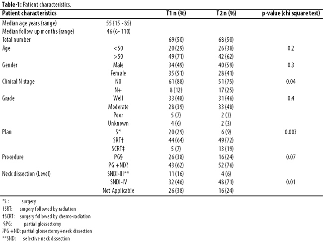
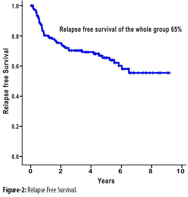
Of the total, 95(69%) patients underwent both partial glossectomy and END. Only 26(19%) patients underwent surgery only. The 5-year OS of stage I/II and III/ IV was 78% and 50% (p=0.002). The 5-year OS and RFS of the whole group was 68% and 65% (Figures 1 and 2).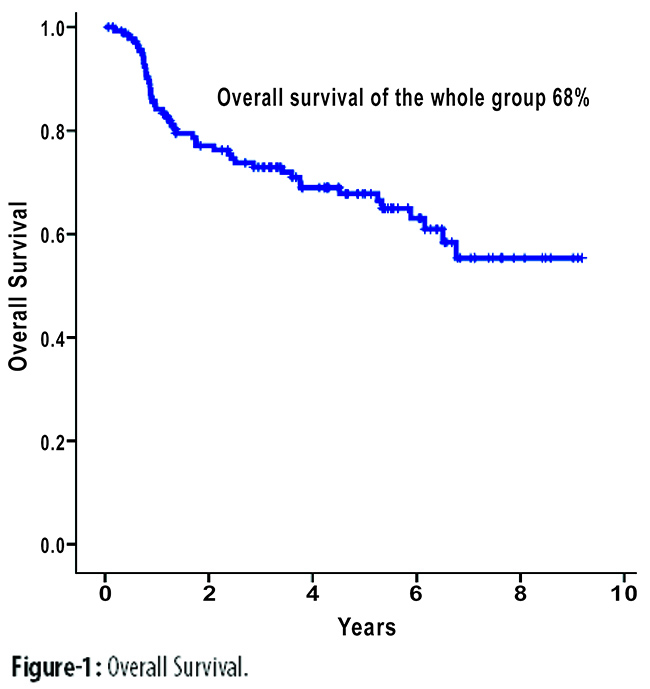
At a follow-up of 5 years, LC, RC, LRC, and distant control for the whole group were 86%, 82%, 72% and 96% respectively. The incidence of occult metastasis was 29 (39%) patients. The actual 5-year OS of T1 and T2 tumours was 73% and 62% with a median follow-up time of 46 months (range: 6-110 months) of the whole group (Figure 3).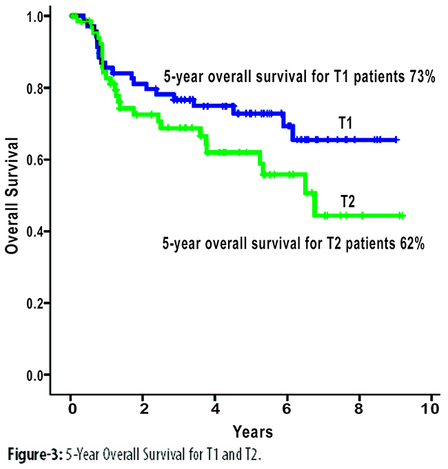
In respect to OS, both clinical and pathological N stage were significant prognostic factors in T1 patients and in respect to OS, surgical procedure, grade, pN stage were significant prognostic factors in T2 SCCOT. Four out of 45(9%) patients died of underlying medical conditions.
Patients having surgical margins closer to 5mm in both T1 and T2 groups had more incidence of local failure though surgical margins had no impact on OS and RFS (Table-2).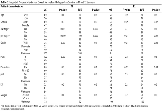
Patterns of failure included: LC19, RC 22, LRC 4 and distant 5 patients (Table-3).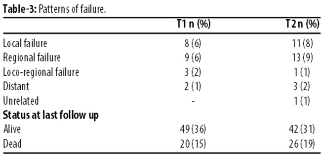
None of the patients undergoing surgery alone reported local failure and majority of the failures were reported in patients receiving surgery followed by radiation (SRT) in both the groups.
All clinical T1 (cT1) patients with regional recurrence had pathologically node-positive disease. Only 2(1.5%) patients with pT2N0 developed regional recurrence. Six (27%) patients were salvaged out of a total of 22 neck recurrences. A total of 4 patients failed loco-regionally and 5 developed distant metastatic disease who were clinically node-negative, and 3 were moderately differentiated SCCOT. Five patients developed distant metastatic disease within the first two years of the follow-up.
Tumour thickness was reported in 42(31%) patients and due to the small number it was difficult to extract its prognostic value. A total of 95 patients underwent neck dissection, of these 43 patients had pathologically node-positive disease. Nine out of these 43 patients showed skip metastasis. Eight patients underwent bilateral neck dissection and only one patient had pN2c disease.
All patients with T2 N+ disease received either post-operative radiotherapy or chemo-radiation (Table-4).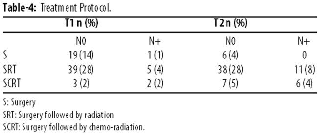
Discussion
Surgery remains the treatment of choice for the management of early tongue carcinoma. The RFS and OS for the whole group was 65% and 68%. The OS for stage I-II in our study was 78%. Various prognostic factors have been reported in the literature to determine the survival of patients with SCC of the tongue. For the past many decades, the single most important prognostic indicator of survival for this disease is the regional spread to the ipsilateral and contralateral neck nodes.4 In our group, pathological stage showed significant prognostic value in OS and RFS for T1, T2 SCCOT. A 20-year study reported OS and disease-specific survival to be 79% and 86%.1 In that study all the patients had clinically node-negative disease. Various other studies have reported the survival in the range of 75-89%.3,5-7 When comparing the prognosis, oral tongue has poor outcome than the rest of the sub-sites of the oral cavity.5
The debate continues in respect to the management of neck for early SCC of oral tongue. Various studies have advocated END. The rational to perform END is the high incidence of occult metastatic disease, to stage neck and to determine the various prognostic factors in the final pathological report that further determines the use of adjuvant treatment.7,8-10 A study reported the incidence of occult metastatic disease in T1-T2 tongue tumours to be as high as 47%.11 The incidence of occult metastatic disease in our study was 39% (29 patients), which is comparable to published data.1,7,12 In our setting, majority of the patients belong to low socioeconomic class and come from distant parts of the country, making close monitoring impractical. Furthermore, in our experience the patients who present with recurrent neck disease, do so late and most of the times they are unsalvageable, further supporting the notion of END. Only 6 (22%) out of a total of 22 regional failures in our study were salvaged. The other school of thought advocates wait-and-watch policy rather than performing END.2,3 Various prospective and retrospective studies have shown similar disease control in observing the neck instead of performing END in early SCC of oral tongue.2,3 A large retrospective study of 359 patients with T1-T2 N0 early tongue tumour reported disease-free survival for END and wait-and-watch policy was 74% and 68% respectively (p=0.53). The OS for both the groups was 60% at 5-year follow-up (p=0.24).2 The most significant aspect of the wait-and-watch policy is close meticulous surveillance of the patients both clinically and with ultrasound. A prospective study in 2009 reported having salvaged all the neck recurrences due to strict routine follow-up.3 A study published by our centre earlier reported that in patients with early T1 N0 well-differentiated carcinoma of the oral cavity, END can be safely avoided due to less than 10% incidence of occult metastatic disease.4 Another study also reported observing neck in a very selective group of patients with small T1 superficial tumours and reported a disease-specific survival rate of 92%.1
No single prognostic factor can predict the incidence of occult metastasis in early SCC of oral tongue. Various factors like tumour thickness or grade of histology have proven to have impact on the development of neck nodes.13,14 A recent meta-analysis published in 2009 has stratified that tumour thickness >4mm is the most reliable predictor of occult disease.14 The shortcoming in our study is the inadequate reporting of the tumour thickness. Tumour thickness was only reported in 35% patients and subgroup analysis in these patients did not carry statistical power to determine the prognostic value. With the advent of sophisticated technological advances, like frozen sections and intraoperative ultrasound, tumour thickness can be predicted in patients with early tongue SCC, thus helping in a selective group of patients in which END can be avoided.15,16 Ultrasound has shown to be highly accurate in predicting thickness of tongue tumours.
In the last 10 years, sentinel lymph node biopsy (SLNB) has been performed as an alternative to END, especially in early tongue SCC. SLNB has shown sensitivity of as high as 96%.17 Limitations to this procedure include procedure expertise and sophisticated equipment and is recommended at centres where it is performed at least on 10 patients in a year. Our centre is a high-volume centre for neck dissection, performing almost 10neck dissections a month. SLNB has been used by our breast surgeons for almost 10 years and it is possible that we can incorporate the technique in early tongue tumours, further selecting a subgroup of patients in whom END can be avoided.
Literature reveals that PORT has proven to improve LRC in head and neck tumours.18-21 Guidelines suggest that positive surgical margin and the presence of extra-capsular spread patients should be treated with concurrent chemo-radiation (CRT).22,23 In our study, 18 patients received CRT. In SCCOT the main reasons for failure are LC, RC, and LRC. Although no formal reporting of the RT-associated toxicity was documented in our study, but xerostomia remains the most worrisome long-term side effect after conventional radiotherapy. Recently with the advent of intensity-modulated radiation therapy (IMRT), many centres have started incorporating IMRT in adjuvant settings in the treatment of oral cancer and have shown acceptable LC.24 Patterns of failure in our study included: LC 19, RC 22, LRC 4 and distant 5. The patterns of local and regional failure in the current study are comparable withpublished studies.25-27
Elective neck radiation has shown advantage as it can be used as an alternative to neck dissections. Elective whole neck irradiation has shown better regional control than no neck treatment.28 Though role of PORT in multiple node-positive patients is well established, its role in early tongue tumour with N1 stage is under-reported.29-31 A study in 2010 reported that in patients with T1 T2- N1 SCC of oral cavity, adjuvant RT improved OS.32 In our study all the patients with pN1 stage received post-operative radiation. Previous reports have shown that majority of the recurrences with tongue tumours occur within the first 2 years of follow-up.33 Our recurrence patterns are no different from published studies.
Conclusion
Due to the high incidence of occult metastatic disease, END could not be avoided in our settings. We have already employed customised templates to report various prognostic variables, especially tumour thickness and depth of invasion in histopathology. Our centre already has the facility of IMRT, but since we are the only regional tertiary care cancer facility and due to immense tumour burden, we are struggling to do IMRT in all early tongue cancer patients. We are incorporating SLNB in early tongue tumours in our setting, but we can further select a subgroup of patients in whom END can be avoided.
References
1. Ganly I, Patel S, Shah J. Early stage squamous cell cancer of the oral tongue--clinicopathologic features affecting outcome.Cancer. 2012; 118:101-11.
2. D\\\'Cruz AK, Siddachari RC, Walvekar RR, Pantvaidya GH, Chaukar DA, Deshpande MS, et al. Elective neck dissection for the management of the N0 neck in early cancer of the oral tongue: need for a randomized controlled trial. Head Neck.2009 ; 31:618-25.
3. Yuen AP, Ho CM, Chow TL, Tang LC, Cheung WY, Ng RW,et al. Prospective randomized study of selective neck dissection versus observation for N0 neck of early tongue carcinoma. Head Neck. 2009; 31:765-72.
4. Shim SJ, Cha J, Koom WS, Kim JE, Lee CG, Choi EC, et al. Clinical outcomes for T1-2N0-1 oral tongue cancer patients underwent surgery with and without postoperative radiotherapy [serial online]. RadiatOncol. 2010; 27:5:43.
5. Al-Rajhi N, Khafaga Y, El-Husseiny J, Saleem M, Mourad W, Al-Otieschan A, et al. Early stage carcinoma of oral tongue: prognostic factors for control and survival. Oral Oncol. 2000; 36:508-14.
6. Kligerman J1, Lima RA, Soares JR, Prado L, Dias FL, Freitas EQ , et al. Supraomohyoid neck dissection in the treatment of T1/T2 squamous cell carcinoma of the oral cavity. Am J Surg. 1994; 168:391-4.
7. Iqbal H, Bhatti AB, Hussain R, Jamshed A. Regional failures after selective neck dissection in previously untreated squamous cell carcinoma of oral cavity .Int J SurgOncol. 2014; 2014: 205715.
8. Huang SF, Kang CJ, Lin CY, Fan KH, Yen TC, Wang HM, et al. Neck treatment of patients with early stage oral tongue cancer: comparison between observation, supraomohyoid dissection, and extended dissection. Cancer. 2008; 112:1066-75.
9. Haddadin KJ, Soutar DS, Webster MH, Robertson AG, Oliver RJ, MacDonald DG. Natural history and patterns of recurrence of tongue tumors. Br J Plast Surg. 2000; 53:279-85.
10. Lydiatt DD, Robbins KT, Byers RM, Wolf PF. Treatment of stage I and II oral tongue cancer. Head Neck. 1993; 15:308-12.
11. Fakih AR, Rao RS, Patel AR. Prophylactic neck dissection in squamous cell carcinoma of the oral tongue: a prospective randomized study. Semin Surg Oncol. 1989;5:327-30.
12. Pimenta ATM, Da Silva FAR, Carvalho AL, Pinto CA, Kowalski LP. Predictive factors of occult metastasis and prognosis of clinical stages I and II squamous cell carcinoma of the tongue and floor of the mouth. Oral Oncol. 2004; 40:780-6.
13. Fakih AR, Rao RS, Borges AM, Patel AR. Elective versus therapeutic neck dissection in early carcinoma of the oral tongue. Am J Surg. 1989; 158:309-13.
14. Huang SH, Hwang D, Lockwood G, Goldstein DP, O\\\'Sullivan B.Predictive value of tumor thickness for cervical lymph node involvement in squamous cell carcinoma of the oral cavity. Cancer. 2009; 115:1489-97.
15. Mark Taylor S, Drover C, Maceachern R, Bullock M, Hart R, Psooy B, et al. Is preoperative ultrasonography accurate in measuring tumor thickness and predicting the incidence of cervical metastasis in oral cancer? Oral Oncol. 2010; 46:38-41.
16. Yuen APW, Ng RWM, Lam PKY, Ho A. Preoperative measurement of tumor thickness of oral tongue carcinoma with intraoral ultrasonography. Head Neck. 2008; 30: 230-4.
17. Civantos FJ, Zitsch RP, Schuller DE, Agrawal A, Smith RB, Nason R, et al. Sentinel lymph node biopsy accurately stages the regional lymph nodes for T1-T2 oral squamous cell carcinomas: results of a prospective multi-institutional trial. J Clin Oncol. 2010; 28:1395-400.
18. MacComb WS, Fletcher GH.Planned combination of surgery and radiation in treatment of advanced primary head and neck cancer. Am J Roentgenol. 1957; 77:462-7.
19. Kramer S, Gelber RD, Snow JB, Marcial VA, Lowry LD, Davis LW, et al. Combined radiation therapy and surgery in the management of advanced head and neck cancer: final report of study 73-03 of the Radiation Therapy Oncology Group. Head Neck Surg. 1987; 10:19-30.
20. Tupchong L, Scott CB, Blitzer PH, Marcial VA, Lowry LD, Jacobs JR, et al. Randomized study of preoperative versus postoperative radiation therapy in advanced head and neck carcinoma: long-term follow-up of RTOG study 73-03. Int J Radiat Oncol Biol Phys. 1991; 20:21-8.
21. Peters LJ, Goepfert H, Ang KK, Byers RM, Maor MH, Guillamondegui O, et al. Evaluation of the dose for postoperative radiation therapy of head and neck cancer: first report of a prospective randomized trial. Int J Radiat Oncol Biol Phys. 1993; 26:3-11.
22. Bernier J, Domenge C, Ozsahin M, Matuszewska K, Lefèbvre JL, Greiner RH, et al. Postoperative irradiation with or without concomitant chemotherapy for locally advanced head and neck cancer. N Engl J Med. 2004; 350:1945-52.
23. Cooper JS, Pajak TF, Forastiere AA, Jacobs J, Campbell BH, Saxman SB, et al. Postoperative concurrent radiotherapy and chemotherapy for high-risk squamous-cell carcinoma of the head and neck. N Engl J Med. 2004; 350:1937-44.
24. Gomez DR, Zhung JE, Gomez J, Chan K, Wu AJ, Wolden SL, et al. Intensity-modulated radiotherapy in postoperative treatment of oral cavity cancers. Int J Radiat Oncol Biol Phys. 2009; 73:1096-103.
25. Hicks WL Jr, North JH Jr, Loree TR, Maamoun S, Mullins A, Orner JB, et al. Surgery as a single modality therapy for squamous cell carcinoma of the oral tongue. Am J Otolaryngol. 1998; 19:24-8.
26. Yuen AP, Lam KY, Chan AC, Wei WI, Lam LK, Ho WK, et al.Clinicopathological analysis of elective neck dissection for N0 neck of early oral tongue carcinoma. Am J Surg. 1999; 177:90-2.
27. Sessions DG, Spector GJ, Lenox J, Haughey B, Chao C, Marks J. Analysis of treatment results for oral tongue cancer.Laryngoscope. 2002; 112:616-25.
28. Spaulding CA, Korb LJ, Constable WC, Cantrell RW, Levine PA.The influence of extent of neck treatment upon control of cervical lymphadenopathy in cancers of the oral tongue.Int J Radiat Oncol Biol Phys. 1991; 21:577-81.
29. Day TA, Davis BK, Gillespie MB, Joe JK, Kibbey M, Martin-Harris B.Oral cancer treatment.Curr Treat Options Oncol. 2003; 4:27-41.
30. Grecula JC, Schuller DE, Rhoades CA, Au JL-S,Nag S, Bauer CJ, et al. Intensification regimen 2 for advanced head and neck squamous cell carcinomas. Arch Otolaryngol Head Neck Surg. 1999; 125:1313-8.
31. Bernier J, Cooper JS, Pajak TF, van Glabbeke M, Bourhis J, Forastiere A, et al. Defining risk levels in locally advanced head and neck cancers: a comparative analysis of concurrent postoperative radiation plus chemotherapy trials of the EORTC (#22931) and RTOG (#9501). Head Neck. 2005; 27: 843-50.
32. Shrime MG, Gullane PJ, Dawson L, Kim J, Gilbert RW, Irish JC, et al. The impact of adjuvant radiotherapy on survival in T1-2N1 squamous cell carcinoma of the oral cavity.ArchOtolaryngol Head Neck Surg. 2010; 136:225-8.
33. Meoz RT, Fletcher GH, Lindberg RD. Anatomical coverage in elective irradiation of the neck for squamous cell carcinoma of the oral tongue. Int J Radiat Oncol Biol Phys. 1982; 8:1881-5.
Related Articles
Journal of the Pakistan Medical Association has agreed to receive and publish manuscripts in accordance with the principles of the following committees:


