

Muhammed Mubarak ( Department of Histopathology, Sindh Institute of Urology and Transplantation (SIUT), Karachi, Pakistan. )
September 2015, Volume 65, Issue 9
Editorial

The term focal segmental glomerulosclerosis (FSGS) describes the morphological appearance of glomerular lesions under the light microscope (LM) resulting from a large number of both primary and secondary glomerular diseases.1,2 But a widespread use of the term has led to a misconception in the minds of many that it is a single disease, which is in fact, not the case. The histopathological diagnosis of FSGS should not be equated with an end diagnosis. In some instances, the renal pathologist may guide the clinician about the possibility of underlyng disease causing this particular morphological pattern. However, more often than not, the renal pathologists are only able to covey simply the diagnosis of FSGS, along with its classification into a particular type.3 It is the responsibility of the nephrologists to further explore the underlying diseases causing this particular pattern of glomerular injury and when none is found, FSGS is labeled as idiopathic or primary FSGS (Figure-1A).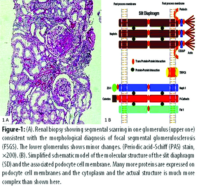
Even the idiopathic variety of FSGS is not a single entity. It is a highly heterogeneous disease with varied presentations, clinical courses, responses to treatment, prognoses, and recurrences following transplantation. To give some examples, some patients with FSGS respond to steroids, some do not; some patients present in childhood, some as adults; some present with nephrotic syndrome (NS), while others present with mild proteinuria. Some cases of FSGS, but not all, recur in transplanted kidneys. This degree of phenotypic heterogeneity does raise some pertinent questions. Do these phenotypic differences reflect differences in the underlying biological processes of the disease? Does the lesion of FSGS represent a "final common pathway" far downstream along the course of a number of distinct biological processes affecting the kidneys? How will genetics help in understanding the phenotypic differences among the individuals affected by this lesion? How much is the contribution of nature (genes) vs. nurture (environment) in individual cases of FSGS? And last but not the least is the question of how genetics will help in the ultimate management of the condition in patients?4
The above questions are of significant clinical importance for a number of reasons. FSGS has overtaken membranous nephropathy (MN) as the most common cause of NS in adults in many parts of the world and more importantly, its incidence is on the rise not only in adults but also in children.5 Secondly, it has emerged as the leading cause of end-stage renal disease (ESRD) in many countries of the world.6 This has stimulated marked interest in research on the pathogenesis of FSGS in recent past. The focus of research has shifted from morphology to molecular front and during the past few years, the molecular basis of FSGS has been extensively investigated in the familial as well as in the sporadic cases and mutations in a number of genes encoding podocyte proteins have been described. As a result of this activity, the number of novel genes and their protein products is increasing day by day.7
Most of these genes are mainly or almost exclusively expressed in the podocytes and are involved in the organization and functioning of the slit diaphragm (SD) and the actin cytoskeleton of the podocyte foot processes.7 Mutations in these genes lead to both structural and functional alterations in the podocytes and lead to a variety of clinical manifestations ranging from mild proteinuria to overt NS. The mutations in podocyte genes result in either autosomal recessive FSGS (NPHS1, NPHS2, LAMB2, PLCE1, ARGHDIA, MYO1E and other genes) or autosomal dominant FSGS (ACTN4, CD2AP, INF2, TRPC6, WT1, ARHGAP24 and other genes) or mitochondrial disorders (CoQ6, tRNALeu, tRNATyr, tRNAIle). Figure 1B shows the molecular organization of the SD and the associated common podocyte proteins. FSGS can also be caused by genes that are widely expressed in other tissues and cell types and cause syndromic FSGS like WT1, PAX2 and LMX1B. Mutations in any of the above genes result in the disruption of the complex network of these proteins, foot process effacement and loss of SD. As podocytes play an important role in the structural and functional integrity of glomerular filtration barrier (GFB), any injury to the podocyte not only affects the structure of the podocyte or SD, but also disrupts the adjacent structures and consequently alters glomerular perm selectivity to the macromolecules in the blood.4,7
Autosomal dominant forms of FSGS typically manifest in adulthood, and contain gain-of-function mutations; however, penetrance may be incomplete with variable expression of the disease phenotype. Many adult patients with familial FSGS present with non-nephrotic range proteinuria or they may be asymptomatic. Rate of progression to ESRD is often slow in autosomal dominant forms of the disease. Mutations are rarely found in adults with isolated sporadic FSGS. The most common cause of the autosomal dominant FSGS is the INF2 gene mutations (16%), followed by mutations in the TRPC6 (12%) and the ACTN4 (3.5%) genes. On the other hand, the autosomal recessive mutations are mostly loss-of-function mutations and tend to manifest early in life from first month to adolescent age and are usually severe in nature and lead to rapid progression to ESRD. Mutations in the NPHS1, NPHS2 and PLCE1 genes have been found in most of the severe cases of congenital and early onset NS, whereas, mutations in the LAMB2 and WT1 genes are found in syndromic NS. The prevalence rates of gene mutations have varied among the studies. In a large European study, 66% of the children presenting with NS in their first year of life were found to carry mutations in the NPHS1, NPHS2, WT1 and LAMB2 genes.8 We also investigated the prevalence of mutations in NPHS1 and NPHS2 genes in childhood NS and found a prevalence of 5.5% and 3.4%, respectively.9 However, our study also included older children and adolescents. Our mutation rates are low as compared with studies from Europe and United States, but more or less similar to other regional studies. Mutations in all the identified genes collectively account for the majority of cases presenting with the FSGS in the early years of life and also for adult FSGS. Nonetheless, there is a sizeable minority of cases whose genetic causes are still unknown.4,9
In addition to the above single gene disorders leading to proteinuric disorders, many common genetic polymorphisms have been found to predispose to disease, alter its course, response to treatment or rate of progression to ESRD. In this context, our group investigated the association of angiotensin converting enzyme (ACE)-II polymorphism with the risk of NS in children. It was found that certain genotypes of ACE-II predispose to the development of NS. However, no association of ACE-II polymorphism was found with response to steroid therapy.10
In conclusion, recent progress in the molecular investigation of FSGS has paved the way for understanding the heterogeneity of the disease and extricating the interplay between the genes and the environment. This will ultimately translate into targeted molecular therapies likely to halt the progression of the disease into ESRD.
References
1. D\'Agati VD, Kaskel FJ, Falk RJ. Focal segmental glomerulosclerosis. N Engl J Med. 2011; 365:2398-411.
2. Rood IM, Deegens JK, Wetzels JF. Genetic causes of focal segmental glomerulosclerosis: implications for clinical practice. Nephrol Dial Transplant. 2012; 27:882-90.
3. Shakeel S, Mubarak M, Kazi JI, Jafry N, Ahmed E. Frequency and clinicopathological characteristics of variants of primary focal segmental glomerulosclerosis in adults presenting with nephrotic syndrome. J Nephropathol. 2013; 2:28-35.
4. Pollak MR. Inherited podocytopathies: FSGS and nephrotic syndrome from a genetic viewpoint. J Am SocNephrol. 2002; 13:3016-23.
5. Shakeel S, Mubarak M, Kazi JI. Frequency and clinicopathological correlations of histopathological variants of pediatric idiopathic focal segmental glomerulosclerosis. Indian J Nephrol. 2014; 24:148-53.
6. Kitiyakara C, Eggers P, Kopp JB. Twenty-one-year trend in ESRD due to focal segmental glomerulosclerosis in the United States. Am J Kidney Dis. 2004; 44:815-25.
7. Zenker M, Machuca E, Antignac C. Genetics of nephrotic syndrome: new insights into molecules acting at the glomerular filtration barrier. J Mol Med (Berl). 2009; 87:849-57.
8. Hinkes BG, Mucha B, Vlangos CN, Gbadegesin R, Liu J, Hesselbacher K, et al. Nephrotic syndrome in the first year of life: two thirds of cases are caused by mutations in 4 genes (NPHS1, NPHS2, WT1 and LAMB2).Pediatrics. 2007; 119: e907-19.
9. Abid A, Khaliq S, Shahid S, Lanewala A, Mubarak M, Hashmi S, et al. A spectrum of novel NPHS1 and NPHS2 gene mutations in pediatric nephrotic syndrome patients from Pakistan. Gene. 2012; 502: 133-7.
10. Shahid S, Abid A, Mehdi QS, Firasat S, Lanewala A, Naqvi SAA, et al. Association of the ACE-II genotype with the risk of nephrotic syndrome in Pakistani children. Gene. 2012; 493:165-8.
Journal of the Pakistan Medical Association has agreed to receive and publish manuscripts in accordance with the principles of the following committees:


