

Fahad Pervaiz ( Faculty of Pharmacy and Alternative Medicines, The Islamia University of Bahawalpur, Bahawalpur, Pakistan. )
Sahrish Rehmani ( Faculty of Pharmacy and Alternative Medicines, The Islamia University of Bahawalpur, Bahawalpur, Pakistan. )
Sana Majid ( Faculty of Pharmacy and Alternative Medicines, The Islamia University of Bahawalpur, Bahawalpur, Pakistan. )
Hina Anwar ( Faculty of Pharmacy and Alternative Medicines, The Islamia University of Bahawalpur, Bahawalpur, Pakistan. )
July 2015, Volume 65, Issue 7
Original Article

Abstract
Objective: To evaluate the biological markers that are commonly assessed in breast cancer to estimate a patient\'s response to endocrine therapy and their prognosis for better clinical outcomes.
Methods: The retrospective study was conducted at Bahawalpur Institute of Nuclear Oncology and comprised record of early breast cancer patients who gave positive diagnostic tests for hormone receptors status i.e. immunohistochemical test and were treated during 2007-2013. Data of oestrogen, progesterone and human epidermal growth factor receptor 2 expression status was analysed. SPSS 12 was used for statistical analysis.
Results: Overall record of 345 patients was studied of whom 149(43%) were identified to have positive hormone receptor status.. The age of the patients ranged from 24 to 86 years with 97(65%) in 25-50 years, 46(30.8%) 51-75 years and 6(4.08%)in 76-100 years. Besides, 76(51%) patients had carcinoma of right breast; 86(58%) were diagnosed as Stage III, 55(37%) Stage II and 8(5.3%) Stage IV. Those diagnosed with oestrogen receptor (positive status) were 16(10.7%), human epidermal growth factor receptor 2 over-expression 13(8.7%), oestrogen/progesterone hormone receptor positivity (or luminal A) 76(51%) and 35(23.4%) patients were positive for all the three receptors.
Conclusion: About half of the patients were diagnosed with a positive hormone status and it was observed that in most of the cases disease was metastasised to distant organs.
Keywords: Oestrogen, HER2-neu, Chemotherapy, Breast cancer. (JPMA 65: 747; 2015)
Introduction
Cancer is any malignant growth or tumour which is caused by abnormal and uncontrolled cell proliferation, which invades and destroys adjacent tissues and may subsequently spread to other parts of the body through the lymphatic system or the blood stream. Breast cancer (malignant breast neoplasm) is known to originate from breast tissue, most commonly from the inner lining of milk ducts or the lobules that supply the ducts with milk. Breast cancer is considered the world\'s most common cancer and is known to be the second major cause of cancer deaths among women in the United States.1 In US and Western Europe the average age at diagnosis is 63 years, while it is around 51 years in Iran.2
It has been demonstrated that breast cancer in younger age groups tends to be more advanced and more aggressive than in older age groups. Breast neoplasms in younger age groups were of higher grade, with hormone receptor-negative status, poorly differentiated tumour grade, greater extent of lymphovascular invasion and amplification of human epidermal growth factor receptor 2 (HER2) than breast neoplasms in older age groups.3-6
For better prediction of a patient\'s response to endocrine therapy and their prognosis, biological markers i.e. oestrogen (ER) and progesterone receptor (PR) status are assessed. An important prognostic factor is metastasis to the lymph nodes which indicates an advanced disease status with the probability that cancer cells have spread to distant sites. At diagnosis, 30% to 50% breast cancers have spread to the sentinel lymph node.7-9
Patients with tumours that are diagnosed with positive ER and/or PR status have lower risks of mortality compared to women with negative ER and/or PR disease.10-14 Clinical trials have also demonstrated that the survival advantage for women with hormone receptor-positive tumours is increased by treatment with adjuvant hormonal and/or chemotherapeutic regimens.15
HER2-neu, which encodes a receptor tyrosine kinase, is amplified and over-expressed in 20-25% breast cancers and such tumours are often resistant to hormone therapy. Despite a general inverse relationship between HER-2/neu over-expression and ER and/or PR expression, a fraction of patients are both HER2-neu- and hormone receptor-positive.16
No special attention has been given on hormone receptor status in breast cancer in southern Punjab, Pakistan. To develop a better understanding, the current study was planned to evaluate the biological markers that are commonly assessed with breast cancer to estimate a patient\'s response to endocrine therapy and their prognosis for better clinical outcomes.
Patients and Methods
The retrospective study was conducted at Bahawalpur Institute of Nuclear Oncology (BINO) in Punjab province of Pakistan, and comprised record of early breast cancer patients who gave positive diagnostic tests for hormone receptor status like immunohistochemical (IHC) test for ER and PR status and fluorescence in situ hybridisation (FISH) test for HER-2/neu, and were treated during 2007-2013. The proforma was designed to collect information regarding various parameters associated with breast cancer directly by patient counselling. Records of patients having primary or recurrent ductal, lobular and other types of breast carcinoma were evaluated for hormone receptor status. The pathology reports were reviewed for patient\'s demographics, site of tumour, histological type of carcinoma, grade and stage of carcinoma at diagnosis, hormonal status, nodal status, treatment strategy, additional associated conditions and adverse drug reactions (ADRs) due to chemotherapy.
Records of patients with IHC data representing positive status of either ER, PR or HER2-neu. World Health Organisation (WHO) classification of breast tumours17 was used. Carcinomas were graded according to the modified Bloom and Richardson method.18 The hormone receptor status was determined using either IHC methods alone for ER and PR, and HER2-neu status was determined using IHC and FISH. Appropriate positive controls were included for each immune stain. Treatment strategy mostly prescribed for breast cancer included chemotherapy with Fluorouracil, Adriamycin Cyclophosphamide/Doxorubicin and Cyclophosphamide (FAC/CAF), Taxol, Adriamycin and Cyclophosphamide (TAC), Fluorouracil, Epirubicin and Cyclophosphamide (FEC) and patients having positive hormonal status were recommended treatment with anti-oestrogens i.e. Tamoxifen, Letrozole and Anastrozole. Patients with positive HER2-neu status were prescribed herceptin. In addition, radiotherapy was recommended for most patients and mastectomy along with axillary clearance was also done. Treatment provided to patients was according to the National Comprehensive Cancer Network (NCCN) guidelines based on patient\'s histopathological tumour type and positive hormonal and HER2-neu status (Figure).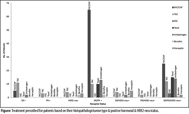
Using SPSS 12, Chi-square test and odds ratio (OR) were applied for statistical analysis Pvalues at 95% confidence interval (CI) were calculated to estimate magnitude and precision of association among the cases.
Results
Overall record of 345 patients was studied. Of them, 149(43%) were identified to have positive hormone receptor status. The age of these149 patients ranged from 24 to 86 years, with 97(65%) in 25-50 years, 46(30.8%) 51-75 years and 6(4.08%) in 76-100 years. Besides, 76(51%) patients had carcinoma of the right breast; 86(58%) were diagnosed as Stage III, 55(37%) Stage II and 8(5.3%) Stage IV. Those diagnosed with ER-positive status were 16(10.7%), HER2-neu over-expression 13(8.7%), ER/PR receptor-positivity (or luminal A) 76(51%) and 35(23.4%) patients were positive for all the three receptors.
Overall, 135(90.6%) cases were of invasive ductal carcinoma (IDC), 9(6.04%) invasive lobular and 5(3.5%) of other types (papilloma, Paget\'s disease, metastatic carcinoma breast, focal invasion comedo type, and ductal papilloma with mucinous carcinoma). The cases were diagnosed at different grades: 38(25.5%) of G1 (low grade), 50(33.5%) of G2 (intermediate grade), 42(28.1%) of G3 (high grade) and 19(12.7%) of G4 (undifferentiated) (Table-1).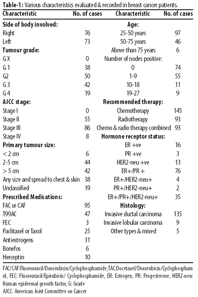
Of the 135 IDC cases, 73(54%) were luminal A (positive for both ER and PR), 32(23.7%) were luminal B, 16(11.8%) were ER+/PR- and 13(9.6%) were negative for both ER/PR receptors but positive for HER2-neu. In IDC patients having stage II (n=52), 4 were ER+, 5 were HER2-neu+, 29 were ER+/PR+ and 14 were triple positive (luminal B). In stage III IDC patients (n=79), 10 cases were positive for ER, 8 of them were positive for HER2-neu, 45 were positive for ER/PR (luminal A) and 16 were triple positive (luminal B). In stage IV cases (n=4), 2 were ER+, 2 were triple and none was ER/PR and HER2-neu positive. In the lobular carcinoma cases (n=9), 3 were PR-positive, 2 were ER/ HER2-neu positive, 1 was PR/HER2-neu positive, 2 were ER/PR/HER2-neu positive and 1 was ER/PR positive. Three of the lobular carcinoma cases were of stage II, 5 of stage III and 1 of stage IV. In the other types of breast tumour (n=5), 1 was ER/PR positive, 2 were ER/HER2-neu positive, 1 was PR/HER2-neu positive and 1 was triple positive while none of them showed positivity for HER2-neu.
Combined chemotherapy and radiotherapy was prescribed in 93(62%) cases, only chemotherapy in 145(97%)and only radiotherapy in 93(62%). Mastectomy and axillary clearance was done in 120(80.5%) patients.
Chemotherapy with Fluorouracil, Adriamycin and Cyclophosphamide (FAC) was prescribed to 95(63.7%) patients, while Taxol, Adriamycin and Cyclophosphamide (TAC) to 47(31.5%). Fluorouracil, Epirubicin, Cyclophosphamide (FEC) was prescribed in 3(0.02%), Tamoxifen to 31(20.8%) and herceptin to 8(5.3%).
OR was applied to compare IHC subtypes with premenopausal vs. postmenopausal, stage II vs. stage III and IV, lymph nodes positive status vs. negative status, histology IDC vs. other types and histologic grade and I and II vs. III and IV (Table-2).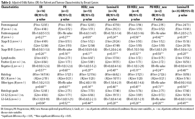
Characteristics of the 149 cases with IHC data were also evaluated (Table-3).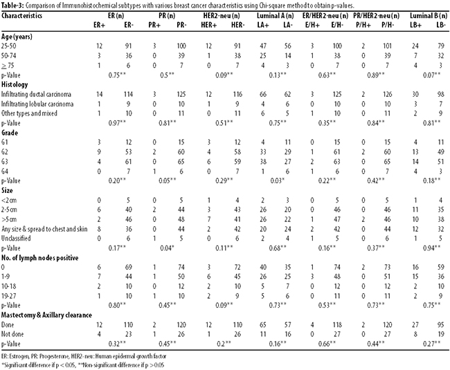
Discussion
One of the most important parameters in breast cancer management and patient survival is the hormone status and responsiveness of tumour to hormone. In developed countries many studies have been carried out to evaluate the hormone receptors and HER2-neu status. In US numerous studies have been used to demonstrate and evaluate differences in hormone receptor status and histology by race and ethnicity among women.19-22
The relationship between tumour size and lymph node involvement is clinically well known and is found to be the most powerful indicator for poor prognosis in breast cancer patients.23 Pathological data of patients with breast cancer suggested that at diagnosis, tumour-infiltrated lymph nodes\' presence is common with estimates ranging from 30% to 50% of cases depending mainly on breast tumour size.24 Young age at diagnosis as an independent predictor of poor survival, as has been revealed by numerous studies conducted in Europe and the Us.7,25,26 In young women, risk profile is worse than in older women. It was seen that young women with breast tumours had a tendency to have larger tumour sizes, more positive lymph nodal status, more negative hormone receptors status, higher tumour grades at diagnosis than the older women.3
Both sides of the body were equally involved in our study; 76(51%) cases of the right breast and 73(49%) of the left side. WHO classification of breast tumours was used to classify breast cancer histopathology among the 149 cases. IDC was found to be most prevalent compared to other histological types such as invasive lobular and other types such as papilloma, paget\'s disease, metastatic carcinoma, focal invasion comedo type, and ductal papilloma with mucinous carcinoma. Most of the patients were in the age range of 25-50 years (65%). Compared to other age groups, younger patients were more prone to the development of breast cancer than the older patients. A similar study in Jordan showed 240(89.9%) cases were ductal, 22(8.2%) were lobular and 5(1.9%) of mixed ductal and lobular carcinomas, while patients\' age ranged from 27 to 89 years.27
In this study, 16 (10.7%) cases were ER-positive, 3(2.01%) were PR-positive, 13(8.72%) were HER2-neu-positive, 76(51.0%) were luminal A (ER+/PR+) positive, 35(23.4%) were luminal B (ER+/PR+/HER2-neu+) positive, 4(2.68%) were ER+/HER2-neu positive and 2(1.34%) were PR+/HER2-neu+ positive.
Treatment also depends on the histopathological stage, grade and the breast cancer cells with ER, PR or HER2-neu receptors on their surface. Treatment provided to patients was according to the NCCN guidelines. Most of the patients were receiving FAC/CAF, TAC, and FEC with anti-oestrogens for treating their positive hormonal status, and patients with HER2-neu were recommended treatment with herceptin.
There was a significant difference between different parameters compared with IHC subtypes in our study, but only p-values for comparison between American Joint Committee on Cancer (AJCC) stage and IHC subtypes had significant relationship (p<0.05), for ER (p=0.01), PR (p=0.04) and HER2-neu (p=0.02). PR status showed statistically insignificant difference when compared with tumour size, while there was insignificant association with other variables when compared with receptor subtypes (p>0.05 each). Various similar studies conducted on this aspect showed marked differences in their results which may be due to differences in the race, ethnicity, geographical distribution, lifestyle, environmental factors, risk factors involved etc. One US study revealed that among women having age range of 30-89 years at diagnosis, compared to ductal carcinoma cases, the lobular and ductal/lobular carcinomas were more likely to be diagnosed with stage III/IV, tumour size 5.0cm, and nodal-positive status. More aggressive tumour phenotypes were associated with inflammatory carcinomas, and mucinous, tubular and papillary tumours were associated with less aggressive phenotypes. The studied histological types of breast tumour greatly differed in their clinical presentations, and the differences found in their hormone receptor status and grading point to their markedly different aetiologies. Using OR p-values showing significant statistical relation of HER2-neu with premenopausal vs postmenopausal status (p=0.01) and for ER with histological grade I-II vs. III-IV (p=0.04) were also found during this study. This reveals the following facts; about half of the patients had positive hormonal status, luminal A IHC subtype was more prevalent among patients compared to other subtypes. Advanced stage of disease was present in most of the patients. There was statistically significant difference between various parameters.
Conclusion
Further research should be carried out to understand the various trends and relationships between different variables associated with breast cancer for better prognosis of breast cancer patients in order to obtain better clinical outcomes with improved survival.
References
1. American Cancer Society. Cancer Facts and Figures 2004; 3: 10.
2. Kollias J, Elston CW, Ellis IO, Robertson JF, Blamey RW. Early-onset breast cancer-histopathological and prognostic considerations. Br J Cancer 1997; 75: 1318-23.
3. Shannon C, Smith IE. Breast cancer in adolescents and young women. Eur J Cancer 2003; 39: 2632-42.
4. Carey KA, David SH, Gloria B, Chaitanya RA, John AF, Yi Zhang, et al. Young age at diagnosis correlates with worse prognosis and defines a subset of breast cancers with shared patterns of gene expression. J Clin Oncol 2008; 26: 3324-30.
5. Gnerlich JL, Deshpande AD, Jeffe DB, Sweet A, White N, Margenthaler JA. Elevated breast cancer mortality in young women (<40 Years) compared with older women is attributed to poorer survival in early stage disease. J Am Coll Surg 2009; 208: 341-7.
6. Nagi S El, Muhieddine S, Mazen KK, Maya C, Ziad KS, Fady BG, et al. Effects of young age at presentation on survival in breast cancer. BMC Cancer 2006; 6: 194.
7. Jatoi I, Hilsenbeck SG, Clark GM, Osborne CK. Significance of Axillary Lymph Node Metastasis in Primary Breast Cancer. J Clin Oncol 1999; 17: 2334-40.
8. Brian EH, Leslie B, Ronald K. Hormones and the Etiology of Cancer. In: Robert CB, Donald WK, Raphael EP, Ralph RW, James FH, Emil F, eds. Holland-Frei Cancer Medicine. 5th ed. Hamilton, Ontario: B.C. Decker, 2000.
9. Henderson BE, Ross RK, Bernstein L. Estrogens as a cause of human cancer: the Richard and Hinda Rosenthal Foundation award lecture. Cancer Res 1988;48:246-53.
10. Henderson BE, Ross RK, Pike MC, Casagrande JT. Endogenous hormones as a major factor in human cancer. Cancer Res 1982; 42: 3232-9.
11. Key TJ, Pike MC. The role of oestrogens and progestogens in the epidemiology and prevention of breast cancer. Eur J Cancer Clin Oncol 1988; 24: 29-43.
12. Ferguson DJ, Anderson TJ. Morphological evaluation of cell turnover in relation to the menstrual cycle in the "resting" human breast. Br J Cancer 1981; 44: 177-81.
13. Henderson BE, Ross RK, Pike MC. Hormonal chemoprevention of cancer in women. Science 1993; 259: 633-8.
14. Fisher B, Redmond C, Fisher ER, Caplan R. Relative worth of estrogen or progesterone receptor and pathologic characteristics of differentiation as indicators of prognosis in node negative breast cancer patients: findings from National Surgical Adjuvant Breast and Bowel Project Protocol B-06. J Clin Oncol 1988;6:1076-87.
15. Aaltomaa S, Lipponen P, Eskelinen M, Kosma VM, Marin S, Alhava E, et al. Hormone receptors as prognostic factors in female breast cancer. Ann Med 1991; 23: 643-8.
16. Lethaby AE, Mason BH, Harvey VJ, Holdaway IM. Survival of women with node negative breast cancer in the Auckland region. N Z Med J 1996; 109: 330-3.
17. WHO. Histological typing of breast tumours, 2nd edn, international histological classification of tumours no. 2. WHO, Geneva; 1981.
18. Bloom HJG, Richardson WW. Histological Grading and prognosis in breast cancer. Br J Cancer 1957; 11: 359-77.
19. Pegoraro RJ, Karnan V, Nirmul D, Joubert SM. Estrogen and progesterone receptors in breast cancer among women of different racial groups. Cancer Res 1986; 46: 2117-20.
20. Avima MR, Flora L, Yochanan W, Abraham G, Esther ARN, Angela C. Estrogen and progesterone receptors in breast cancer patients: Epidemiologic characteristics and survival differences. Cancer 1989; 64: 196-202.
21. Susan MG, Josée D, Peter G, Susan C, David PW. Hormone receptor status of breast tumors in black, Hispanic, and non-Hispanic white women: An analysis of 13,239 cases. Cancer 1996;77:1465-71.
22. Joslyn SA. Hormone receptors in breast cancer: racial differences in distribution and survival. Breast Cancer Res Treat 2002;73:45-59.
23. Utah, Atlanta. Relation of tumor size, lymph node status, and survival in 24,740 breast cancer cases. Cancer 1989; 63: 181-7.
24. Foster RS. The biologic and clinical significance of lymphatic metastases in breast cancer. Surg Oncol Clin N Am 1996;5:79-104.
25. Rodríguez-Cuevas S, Macías CG, Franceschi D, Labastida S. Breast carcinoma presents a decade earlier in Mexican women than in women in the United States or European countries. Cancer 2001;91:863-8.
26. Swanson GM, Lin CS. Survival patterns among younger women with breast cancer: the effects of age, race, stage, and treatment. J Natl Cancer Inst Monogr 1994;16: 69-77.
27. Sughayer, MaherA, MahaM, Al-Khawaja, Massarweh S, Mahmoud Al-Masri. Prevalence of hormone receptors and HER2/neu in breast cancer cases in Jordan. Pathol Oncol Res 2006;12:83-6.
Journal of the Pakistan Medical Association has agreed to receive and publish manuscripts in accordance with the principles of the following committees:


