

Asadullah Makhdoom ( Department of Orthopaedic Surgery & Traumatology, Liaquat University of Medical & Health Sciences Jamshoro. )
Lachman Das Maheshwari ( Sindh Government Hospital Kohsar, Latifabad Hyderabad. )
Muhammad Ayoub Laghari ( Department of Orthopaedic Surgery & Traumatology, Liaquat University of Medical & Health Sciences Jamshoro. )
Syed Muhammad Tahir ( Department of Burn Surgery, Liaquat University of Medical & Health Sciences Jamshoro. )
Syed Muhammad Ali ( Department of Orthopaedic Surgery & Traumatology, Liaquat University of Medical & Health Sciences Jamshoro. )
Khaleeque Ahmed Siddiqui ( Department of Orthopaedics, Isra University Hyderabad. )
July 2015, Volume 65, Issue 7
Original Article

Abstract
Objective: To compare the outcome of the open proximal and distal fractures of tibia treated by Nasser Awais External Fixtator with T-clamp.
Methods: The descriptive case series was conducted from August 2009 to July 2012 at Department of Orthopaedic Surgery & Traumatology, Liaquat University of Medical & Health Sciences, Jamshoro, and comprised in-patients of open proximal and distal fractures of tibia who were divided into two equal groups: group A had distal and group B had proximal patients. All patients had extra-articular open fractures Gustilo I, II, IIIA and IIIB of proximal and distal end of tibia between 15 and 60 years of age who had arrived within 8 hours of the injury. Below-knee plaster cast was applied for 02 to 03 weeks after the removal of Nasser Awais External Fixtator and all patients were followed up for 12 months. SPSS 17 was used for statistical analysis.
Results: Overall, there were 30 patients; 15(50%) in each of the two groups. The mean age in group A was 28.9±9.43 years and 34.3±14.60 years in group B. There were 11(73.3%) males and 4(26.7%) females in group A, and 15(100%) males in-group B. Mean fracture union time in group A was 17.20±2.93 weeks (range: 11-23 weeks) and in group B it was 23.53±2.44 weeks (range: 19-28 weeks). Ankle joint stiffness occurred in 2(6.7%) cases that were in group A, and knee joint stiffness was seen in 2(6.7%) cases and they were in group B.
Conclusion: Nasser Awais External Fixator with T-Clamp was a safe technique and promoted the union with a low complication rate with less union time in distal tibial fracture compared to proximal fracture.
Keywords: T-clamp, NAEF, Open proximal and distal tibia fractures. (JPMA 65: 727; 2015).
Introduction
In open fractures external fixation is a percutaneous technique of immobilisation of bone. External fixation is easily applicable, frame-adjustable, minimises blood loss and is also a useful salvage procedure for the complications occurring from extremity trauma.1 Open proximal and distal tibial fractures are common presentations at orthopaedic emergency and trauma centres.2 Fractures of distal tibia, particularly metaphyseal region treatment choice, is influenced by the involvement of the fracture to the Plafond with comminution, displacement and soft-tissue envelope injury. For extensive soft tissue injury and wound contamination, the successful treatment is external fixation.3
In 1981 Professor Syed Muhammad Awais of the Department of Orthopaedic Surgery, King Edward Medical College and Mayo Hospital Lahore, introduced the first Naseer-Awais External Fixator4 (NAEFS-NA-1). From 1982 to 1984, five papers were presented on the development and use of these scientific instruments in national and international conferences within the country and abroad. For fracture and leg lengthening, mono-lateral and multi-axial NA-1 fixator is used.5 Several problems were encountered, mainly too many free joints, and were stabilised with this system (Figure-1).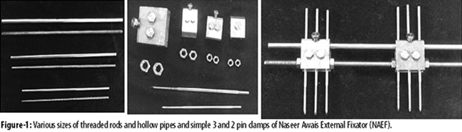
In 1998 more designs of NA-Fix were conceived and followed by Ilizarov\'s method.
Naseer-Awais External Fixation System (NA-5) was another advancement of the same department. NA-5 is in fact a Modular External Fixation System (MEFS) in which components of different sizes are assembled together and used in different situations. Present MEFS is unique in its simple design, dependable strength, easy and fast application, and its ability to be used for the management of a wide range of problems. Technically, both compression and distraction could be produced separately or simultaneously at two levels of same bone. T-shaped clamp was developed in 2004 as its advancement to allow Shanz Screw insertion for the small distal and proximal fracture fragment stabilisation. Rotating clamps for each Shanz Screw is a choice now available to prepare the construct like T-clamps with rotating pin clamps and static Pin Plates of different sizes (Figure-2).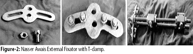
NAEF is cost-effective and well tolerated by patients compared to other sophisticated dynamic axial fixators because in our circumstances, most patients belong to poor socio-economic conditions and it is easy to apply on them. Application of compression and distraction can be achieved through this procedure and it also helps to enhance healing of the fracture in the open fractures of tibia.6,10 Compared to other unilateral external fixators are not available in our country. It is more stable because at least four Shanz Screw can be introduced through T-clamp.
Literature showed that distal tibial fracture because of it subcutaneous anatomy is common. Presently proximal tibial fracture is not uncommon. This study was designed to evaluate the union time, and to compare the complications like non-union and joint stiffness between two groups using NAEF, hypothesising that NAEF with T-clamp is equally effective in both types of fractures.
Material and Methods
The descriptive case series was conducted from August 2009 to July 2012 at Department of Orthopaedic Surgery & Traumatology, Liaquat University of Medical & Health Sciences, Jamshoro, and comprised in-patients of open proximal and distal fractures of tibia who were divided into two equal groups: group A had distal and group B had proximal patients. Extra-articular open fractures Gustilo I, II, IIIA and IIIB of proximal and distal ends of tibia in adults of either gender were included. Patients having multiple fractures, associated head injury, co-morbidities like hypertension and diabetes were excluded. Gustilo type IIIC intra-articular fractures of proximal and distal end of tibia were also excluded. Convenience sampling was used to manage the study population.
After admission, detailed examination was done, focussing on the fractured limb, soft tissue injury with stability of joints above and below the fracture site. NAEF was applied within 48 hours of the admission and repeated surgical toilet was done to deal with soft tissue injuries, which were finally managed as indicated, including delayed primary closure, partial thickness skin graft and local faciocutaneous flap.
Post-operative radiograph was taken on the same or next day of surgery. Clinical assessment and examination of wound was done twice a week till the wounds were fully healed and the patients were mobilised by non-weight-bearing movement with the help of either crutches or walking frames. The standard review regime was every week for 4 weeks, 3 weeks for 3 months, then every 6 weeks for 4 months, and finally 8 weeks for 6 months.
Compression and distraction alternatively was performed at weekly interval after the first 03 weeks of application of external fixators in both groups to enhance osteoblastic activity for the achievement of early callus formation. Dynamisation of external fixator was done when the patient achieved full weight-bearing.
Fractures were considered united when tenderness or movement at fracture site was clinically absent and fracture line was not visible on the radiograph. Below-knee plaster cast was applied for 02 to 03 weeks after the removal of NAEF and all patients were followed up for 12 months.
Records of every patient, from admission to discharge, and their assessment in the review clinics till the final check-up, were maintained through a designed proforma and data was analysed using SPSS 17.
Results
Overall, there were 30 patients; 15(50%) in each of the two groups.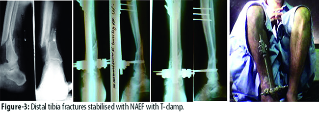
The mean age in group A (Figure-3) was 28.9±9.43 years and 34.3±14.60 years in group B (Figure-4). 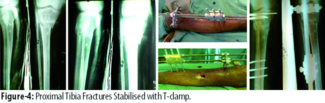
There were 11(73.3%) males and 4(26.7%) females in group A, and 15(100%) males in-group B. In terms of mode of injury, there were 17(56.7%) cases of road traffic accident (RTA); 8(53.3%) in group A and 9(60.0%) in group B. Besides, there were 5(16.7%) cases of firearm injury, 3(10.0%) each related to fall from height and assault, and 1(3.3%) each of blast and machine injury.
According to Gustilo classification, 11(36.7%) type IIIA were seen; 6(40.0%) in group A and 5(33.3%) in group B. There were 9(30.0%) cases of type IIIB; 3(20.0%) in group A and 6(40.0%) in group B (Table-1).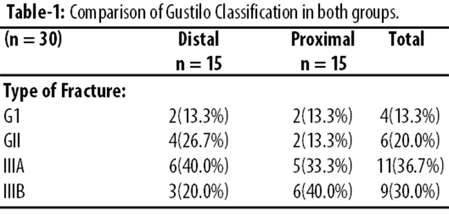
Mean partial weight-bearing walking was 11.800±3.88 weeks (range: 8-19 weeks) in group A and 15.13±2.19 (range: 12-20 weeks) in group B. Mean full weight-bearing was 16.60±3.04 weeks (range:11-23 weeks) in group A and 17.46±2.97 (range: 10-22 weeks) in group B. Mean fracture union time in group A was 17.20±2.93 weeks (range:11-23 weeks) and in group B it was 23.53±2.44 weeks (range: 19-28 weeks).
NAEF T-clamp was removed in all cases. Highly significant difference was seen in the two groups with mean NAEF T-clamp time of 20.06±3.22 weeks in group A (range: 14 to 25 weeks) and 25.93±2.40 in group B (range: 22-30 weeks). Complications were pain during walking and pain site osteolysis. Out of 9(30.0%) cases of pain during walking, 3(20.0%) cases were found in-group A and 6(40.0%) cases were in-group B. Pin site osteolysis was seen in 9(30.0%); 5(33.3%) in group A and 4(26.7%) in group B. Pin tract infection was observed in 10(33%) cases in group A and 11(36%) in group B. Ankle joint stiffness was seen in 2(6.7%) cases in group A and knee joint stiffness was seen in 2(6.7%) cases that were found in group B (Figure-5).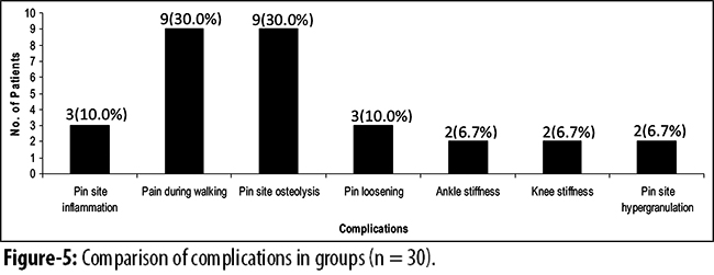
Natural healing and closure of the injury was observed in 5(17%) cases, delayed primary closure was done in 12(40%), partial thickness skin graft was done in 8(27%), while local fasciocutaneous flap was done in 5(17%) cases to deal with the skin defect. Clinical results were excellent in 13(86.6%) patients in group A and 12(80%) patients in group B, good in 1(6.6 %) patients in group A and 2(13.3%) in group B, and fair in 1(6.6 %) patient in group A and 1(6.6 %) in group B (Table-2).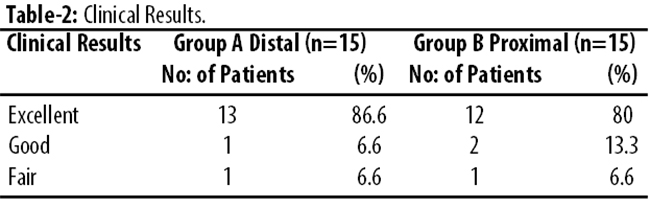
Discussion
Proximal and distal tibia fractures with intra-articular extension have become a challenge for orthopaedic surgeons because altered procedures of treatment have been recommended, including limited internal with external fixation, as well as open reduction and internal fixation.7
In the 20th century two studies reported satisfactory percentage of good to excellent short and long-term results with surgical methods.8
In this study, majority of patients were males. The male-to-female ratio seen in group A was 2.75:1 compared to group B which was 100% male. This can be attributed to Pakistani setup where the female population largely works indoor or in agricultural fields and does not travel much. The higher rate of fracture among males clearly correlated with the lifestyle of male, especially in our part of the world. The males are more involved in outdoor activities and the young male are more enthusiastic about life and are likely to be careless drivers. Females usually have a sedentary lifestyle and are less involved in driving which is a common cause of accidents. Earlier studies have reported 4:1 and 2.1:1 male-female ratio.9,10
The age of our study population ranged from 15 to 60 years. The fractures were most common in the 2nd and 3rd decades in our study. Earlier series also show higher incidence of fractures in younger age groups. One showed age range from 20 to 60 years with a mean of 32 years11 and another12 showed average age of 37 years.
RTA was the most common cause of fracture of tibia.13 Due to limited sources of income, motorbike is the main mode of transportation of the middle class and in accidents involving motorbikes, tibia is commonly affected. The farmers, housewives, retired people have a comparatively lesser fracture rate as they do not travel frequently, whereas workers and labourers tend to have serious injuries commonly due to industrial accidents, automobile accidents, housewives sustaining fractures through fall from height when they climb up some ladder or stool to pick up objects from the shelves.
The present study showed the most common mechanism of tibial fractures as RTAs with 17(56.7%) patients; 8(53.3%) in group A and 9(60.0%) in group B. They were followed by firearm injury 5(16.7%), fall from height 3(10.0%), assault 3(10.0%) and blast 1(3.3%). In one study,16 the leading cause of limb injury accounting for 76.8% (53 patients) was RTA, assault in 7(10.1%), fall 3(4.3%), gunshot 4(5.8%) and other causes in 2.9% patients.
In our study, majority of cases were seen in type IIIA 11(36.7%), while 9(30.0%) cases were seen in type IIIB. One series14 of 30 patients of tibia fractures, 5(16%) in type I, 6(20%) in type II, 12(40%) in type IIIA and 7(23%) were in type IIIB. Another study15 stated that 8, 12 and 13 fractures were grade II, III, and I respectively.
Longer the duration of hospital stay, greater the burden on the patient financially and psychologically. Ideal treatment should therefore minimise the duration of hospital stay. The mean hospital stay in this study in group A (distal) was 12.9±9.03 days and in group B (proximal) it was 22.0±12.17 days (range: 1-45 days). It is comparable to other studies where the mean hospitalisation period was 21.25 days.12
Weight-bearing is very important for callus formation during fracture healing. Weight-bearing increases the formation of bone in fracture healing and lack of weight-bearing decreases the amount of woven bone that is formed in healing of fractures. Weight-bearing is essential for articular cartilage regeneration. Weight-bearing is also helpful in preventing deep vein thrombosis (DVT). Movements of the ankle and weight-bearing improve venous peak velocity and assist in thromboprophylaxis. Factors that affect adversely fixation stability include the degree of comminuting and fracture, inadequate reduction or fixation of the fracture, and severe bone demineralisation.16 In our study, partial weight-bearing walking was 11.800±3.88 weeks with the range from 8 to 19 weeks in group A and partial weight bearing walking was 15.13±2.19 (range: 12-20 weeks) in group B. While full weight-bearing was 16.60±3.04 weeks (range: 11-23 weeks) in group A and 17.46±2.97 weeks (range: 10-22 weeks) in group B. One study17 on external fixation over the tibia presented average time to partial weight-bearing walking as 36 days and to full weight-bearing as 65 days.
Fracture healing depends upon systemic and local factors.18 Multiple patient factors have been shown to contribute in delayed union and non-union of fractures. One of them is malnutrition, which often goes unrecognised. Adequate protein is required for healing, and inadequate caloric intake has been shown to contribute to delay in union and non-union. Cigarette smoking has been shown in numerous clinical and experimental studies to have an adverse effect on fracture healing.19 Infection prevents union at fracture site. The soft tissue dissection and, disruption of the bone blood supply by the plate-screw-bone construct should be minimised to promote callus formation. Stable fixation is essential for fracture healing. The implant used must resist physiological loads to allow fracture union by limiting fracture gap stress, provide sufficient stability to permit early limb motion, and not fail before fracture union occurs.20 In our study, we achieved union.
In group A (distal), union time was 17.20±2.93 weeks (range: 11-23 weeks); while in-group B (proximal) it was 23.53±2.44 weeks (range: 19-28 weeks). It is comparable with a study21 that showed union in 12-20 weeks in 83.3% patients.
In our series, NAEF T-clamps were removed in all cases. Highly significant difference was seen in both groups with mean NAEF T-clamp 20.06±3.22 weeks in group A (range: 14-25 weeks) and 25.93±2.40 weeks in group B (range: 22-30 weeks), while a study 17 revealed that T-clamp was removed at 20.07±0.84 weeks.
In our study, majority of postoperative complications were higher in group A compared to group B. Pain during walking and pain site osteolysis occurred in majority of the cases. Out of 9(30.0%) cases of pain during walking, 3(20.0%) were in group A and 6(40.0%) were in group B, pain site osteolysis in 5(33.3%) cases were seen in group A, while 4(26.7%) cases were in group B. Ankle joint stiffness occurred in 2(6.7%) cases in group A and knee joint stiffness was seen in 2(6.7%) cases that were in group B. Non-union and delayed union were not found and prevented from enhancing macro movements at the fracture site by stability of fixator through tightening of all clamps by nuts and bolts during follow-up. Another study22 reported the complications in 12(35%) patients: 1 skin slough, 5 pin tract infections, 3 deep infections, 3 non-union, and 3 loss of reductions necessitating frame revision.
In spite of all these associated bone fractures and complications we were able to achieve excellent results in 13(86.6%) patients in distal group versus 12(80%) patients in proximal group, good 1(6.6%) patient in distal versus 2(13.3%) in proximal group, and fair in 1(6.6 %) patient in distal versus 1(6.6%) in proximal group. However, one study[23] showed excellent results in 7(35%) cases, good in 8(40%) cases and fair in 4(20%) cases.
Conclusion
NAEF with T-clamp is a safe and excellent technique to manage both proximal and distal tibial fractures with respect to a high rate of union and a low complication rate. When managed by NAEF, union time of distal tibial fracture is less compared to proximal tibial fracture.
References
1. Pacheco RJ, Saleh M. The role of external fixation in trauma. Trauma 2006; 6: 143-60
2. Gaudinez RF, Mallik AR, Szporn M. Hybrid external fixation of comminuted tibial plateau fractures. Clin Orthop 1996; 328: 203-10.
3. Bedi A1, Le TT, Karunakar MA. Surgical treatment of non-articular distal tibia fractures. J Am Acad Orthop Surg 2006; 14: 406-16.
4. Awais SM, Akhtar NM. Leg lengthening using Naseer Awais Fixator study of 20 cases. J Orthop Assoc 1992; 2, 1992.
5. Awais SM, Akhtar NM. Management of segmental defect by intercalary bone transport using Naseer- Awais Fixators. J Pak Orthop Assoc 1992; 2: 97-103.
6. Makhdoom A, Laghari MA, Qureshi PAL, Siddiqui KA. Management of Open Diaphyseal Fractures of Tibia Treated by Naseer Awais External Fixator J Pak Orthop Assoc 2006; 18: 89-96.
7. Femino JE, Vaseenon T. The Direct Lat eral Approach to the Distal Tibia and Fibula: A Single Incision Technique for Distal Tibial and Pilon Fractures. Iowa Orthop J 2007; 29:143-8.
8. Palmer I. Fracture of the upper end of tibia. J Bone Joint Surg 1951; 33: 160.
9. Camacho SP, Lope RC, Carvalho MR, Ferreira de Carvalho C, Bueno R, et al. Assessment of the functional capacity of individuals submitted to surgical treatment after tibial plateau fracture. Acta Ortop Bras 2008; 16: 168-72.
10. Ozturkmen Y, Fiukur E. Calcium phosphate cement augmentation in the treatment of depressed tibial plateau fractures with open reduction and internal fixation. Acta Orthop Traumatol Turc 2010; 44: 262-9.
11. Kataria H, Sharma N, Kanojia RK. Small wire external fixation for high-energy tibial plateau fractures. J Orthop Surg 2007; 15: 137-43.
12. Shrestha BK, Bijukachhe B, Rajbhandary T, Uprety S, Banskota AK. Tibial plateau fractures: four years review at B&B Hospital. J Kathmandu Uni Med 2004; 2: 315-23.
13. Ngim NE, Udosen AM, Ikpeme IA. Review of seventy consecutive cases of limb injuries in calabar. The role of motorcyclists. Nigerian J Orthop Trauma 2006: 5: 38-40.
14. Makhdoom A, Qureshi PAL, Laghari MA, Akhund MA, Siddiqui KA. Outcome of distal tibia fractures treated by Ilizarov external fixatorv/s t-clamp naseer awais external fixator. Med Chan 2009; 15: 117-24.
15. Ocgiider DA, Ozer H, Solak S, Onem RY, Aoaoolu S. Functional results of the Ilizarov circular external fixator in the treatment of open tibial fractures. Acta Orthop Traumatol Turc 2005; 39: 156-62.
16. Pasha IB, Abid M, Ahmed M, Zaidi R, Zaka Ullah. Partial weight bearing; new quantitative approach. J Pak Armed Forced Med 2010;1.
17. Behrens F, Searls K. External fixation of the tibia. Basic concepts and prospective evaluation. J Bone Joint Surg 1986; 68B: 246-54.
18. Frost HM. The Biology of Fracture Healing: An Overview for Clinicians. Clin Orthop Relat Res 1989; 248: 283-93.
19. Smith TK. Prevention of complications in orthopedic surgery secondary to nutritional depletion. Clin Orthop Relat Res 1987; 222: 91-7.
20. Erik NK, Eric F, Eric S, Kenneth AE. The Evolution of Locked Plates. J Bone Joint Surg 2006; 88: 189-200.
21. Shoaib M, Shabir M, Sahibzada AS, Gul R. Outcome of close reduction and casting in close tibial diaphyseal fracture. J Med Scie 2005; 13: 154-6.
22. Barbieri R, Schenk R, Koval K, Aurori K, Aurori B. Hybrid external fixation in the treatment of tibial plafond fractures. Clin Orthop Relat Res 1996; 332: 16-22.
23. Sahibzada AS, Khan MA, Khan MS. Management of tibial bone defects due to high-energy trauma using the locally manufactured external fixator by segmental bone transport. J Ayub Med Coll Abbottabad 2005; 17: 45-9.
Journal of the Pakistan Medical Association has agreed to receive and publish manuscripts in accordance with the principles of the following committees:


