

Awais Amjad Malik ( Services Institute of Medical Sciences, Lahore. )
Hafiz Ghulam Isnain ( Services Institute of Medical Sciences, Lahore. )
Ahsan Khan ( National Hospital & Medical Centre, Lahore. )
Asad Ali Toor ( National Hospital & Medical Centre, Lahore. )
Allah Nawaz ( Services Institute of Medical Sciences, Lahore. )
Rashid Mansoor ( Services Institute of Medical Sciences, Lahore. )
Romaisa Shamim ( Services Institute of Medical Sciences, Lahore. )
Mahmood Ayyaz ( Services Institute of Medical Sciences, Lahore. )
May 2015, Volume 65, Issue 5
Case Series

Abstract
Laparoscopic Pancreatic Pseudocyst (PP) surgery can be performed via anterior or posterior cystogastrostomy, endoscopy-assisted surgery and cystojejunostomy. We conducted a prospective analysis of our patients undergoing laparoscopic cystogastrostomy to analyse the merits and demerits of the procedure. In a period of 3 years from January 2010 to December 2012 all the patients who underwent laparoscopic drainage of pancreatic pseudocysts were prospectively analysed. A total of 12 patients underwent a transgastric anterior cystogastrostomy with a stoma size of 4.5cms. There was no intraoperative or postoperative bleeding or leakage on anastomotic lines. Post-op pain score on the first post-op day was 4 (2-5) on the Visual Analogue Scale (VAS). Average hospital stay was 4.1±2.3 days. All patients had complete resolution of symptoms on follow-up. Follow-up computed tomography (CT) scans on 8 patients showed complete resolution of the cysts. Laparoscopic cystogastrostomy is a safe and feasible method and provides efficient drainage of PP.
Keywords: Laparoscopic Pancreatic Pseudocyst, Cystogastrostomy.
Introduction
Pancreatic pseudocyst (PP) is defined as a collection of pancreatic juice, enclosed by a wall of fibrous or granulation tissue, which arises as a consequence of acute pancreatitis, pancreatic trauma or chronic pancreatitis.1 The documented incidence of pseudocyst following acute pancreatitis has increased with the availability of ultrasound and computed tomography (CT). Adherence to current definitions is important for valid interpretation and comparison, as the majority of pancreatic fluid collections in acute pancreatitis will regress spontaneously2 and do not progress to pseudocyst formation. The incidence of pseudocyst formation following acute pancreatitis ranges from 12% to 55% in different series.3-5 About 75% of all pancreatic masses are pancreatic pseudocysts.1,2 A PP takes about 4-6 weeks for the wall to mature and most of them resolve by 6 weeks.1-3 Asymptomatic patients are managed conservatively by observation and radiological follow-up using serial ultrasonography or CT scans.
Although the indication and timing of the intervention in PP related to acute pancreatitis are still controversial, but there is an agreement that large, persistent and symptomatic cysts should be drained since they are usually associated with complications. The internal drainage of PP, which is the method of choice, can be achieved by surgical or endoscopic interventions. Endoscopic therapy is a promising modality, but requires experienced endoscopist and might be associated with stent-related complications, inadequate drainage, repeated interventions and risk of perforation6 which may require emergency surgery. Surgery continues to be the chief method in PP drainage. Laparoscopic PP surgery is minimally invasive, provides detailed information about PP location and the relationship with adjacent organs, and enables effective drainage. Laparoscopic cystogastrostomy was first performed by J. Petelin7 in 1994. The morbidity is low, PP wall biopsy is achievable and cholecystectomy can be added to the procedure in the presence of biliary pancreatitis. Numerous techniques have been reported for laparoscopic PP surgery thus far, including anterior and posterior cystogastrostomies, endoscopy-assisted surgery and cystojejunostomy.8
Initial results indicate a success rate of 77-100%, with a complication rate of 8-17% and no mortality or recurrence.9-11 Total reported numbers are small and larger series are required for meaningful comparison with other treatment modalities, open surgery in particular. We present our experience in the laparoscopic PP management.
The current study was planned to analyse the merits and demerits of laparoscopic PP management at tertiary care public and private sector hospitals.
Subjects and Methods
The prospective interventional study was conducted from January 2010 to December 2012 at the Services Institute of Medical Sciences, Lahore, and the National Hospital and Medical Centre, Lahore.
All patients who underwent laparoscopic drainage of PP at the two hospitals were selected on the basis of non-resolving, symptomatic and large cysts that were results of acute pancreatitis. The aetiologies of acute pancreatitis were gallstones in all the patients. No endoscopic or percutaneuos intervention was attempted before surgery. The diagnosis and follow-up were made by CT and ultrasound (US) in all cases. The patients were followed up for at least 12 weeks before they were offered surgery in the hope that the cyst might resolve spontaneously. All patients were followed postoperatively at 1, 3 and 6 months and underwent an US/CT scan to rule out any recurrence.
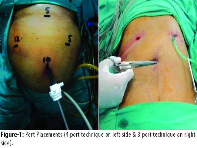
In terms of surgical procedures, the patients were positioned in modified lithotomy position and the surgeon stood between the legs with the first assistant (camera surgeon) and scrub nurse on the right and second assistant to the left of the patient. Pneumoperitoneum was achieved via closed technique and a suprabumbilical 10mm primary port for a 30 degree scope made. A right 5mm and a left 12mm mid-clavicular ports were used as working ports. In initial cases, a subxiphoid 5mm port was also used to retract the left lobe of liver, but as we gained experience we managed the same procedures with only 3 ports (Figure-1).
The pseudocyst was localised with the help of tactile and visual sensations. An anterior gastrostomy was then done using electro surgery (Figure-2).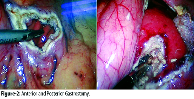
To confirm the exact location of the cyst, percutaneuos aspiration of the fluid using a long Veress needle or lumber puncture needle was performed.
A hole wide enough to insert an arm of the stapler was then created on the posterior gastric wall (Figure-2). The cyst fluid was aspirated and bleeding control was achieved by electro cautery and Ligature.
A laparoscopic roticulating linear stapler 45mm was used for creating anastomosis (Figure-3).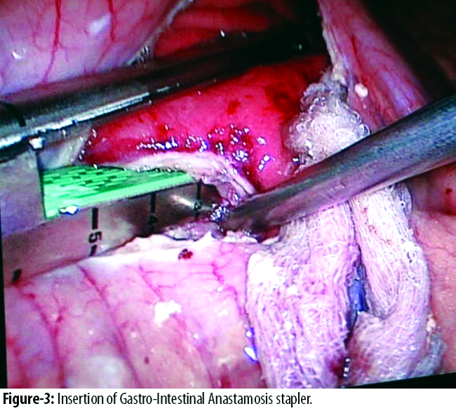
The cyst was then cleared of any necrotic material inside. Then a nasogastric catheter was placed through the stomach into the cyst and the anterior gastric wall was closed in two layers by intracorporeal sutures. Finally, a suction drain was placed near the anastomosis. The repair of the anterior wall was then checked by passing methylene blue through nasogastric (NG) tube and watching for any leak through the anterior suture line.
Results
The mean age of the 12 patients in the case series was 52±12 years (range: 35-65 years). There were 9(75%) males and 3(25%) females. All the patients had previous attacks of acute pancreatitits secondary to gall stones. Mean time to surgery was 14±3.44 weeks (range 12-20weeks) and the mean size of the cyst was 8±2.64cm (range:6-15cm).
All patients underwent a transgastric anterior cystgastrostomy with a stoma size of 4.5cm. Average operating time was 128±13.45min (range: 110-150 min). Neither technical trouble nor intraoperative complication was encountered in any patient and there was no intraoperative bleeding or leakage on anastamotic lines. No conversions to open were necessary and no patient required per-operative blood transfusion.
A standard laparoscopic cholecystectomy was added to the procedure in 6(50%) patients who had a history of acute biliary pancreatitis; no additional port was required if using 4-port technique, but an additional port was needed if using 3 ports. Three (25%) patients underwent a laparoscopic cholecystectomy after resolution of symptoms after 6 weeks.
Patients were put on oral fluid after 48 hours of surgery and received liquid diet on the third postoperative day depending on their clinical progress. Post-op pain score on the first post-op day was 4 (2-5) on the Visual Analogue Scale (VAS). Abdominal drain and NG tube were removed on the second postoperative day.
Patients were discharged from hospital when adequately mobile and tolerating liquid diet. Average hospital stay was 4.1±2.3 days. They were advised to continue with liquid diet for 5 days after surgery and to resume solid diet thereafter.
Patients were followed for an average of 6 months. All patients had complete resolution of symptoms on follow-up. Follow-up CT scans on 8(66.6%) patients showed complete resolution of the cyst.
Discussion
Even though the number of the patients was limited, the results demonstrate that laparoscopic cystogastrostomy was a safe and useful tool in the management of PP. Important thing, however, is to recognise which patient requires surgery.3-5 Timing and size are the two most important factors.12 In our series we waited for at least 12 weeks before opting for an intervention.
Indications for intervention in PPs include size >6cm in diameter, duration of more than 6 weeks, persistent symptoms (including epigastric pain, anorexia, nausea, vomiting, biliary obstruction, and duodenal obstruction), complications (including infection, haemorrhage and spontaneous rupture) and inability to differentiate it from a malignancy.9,13
Cystogastrostomy has emerged as a treatment of choice for non-resolving PP.14 Laparoscopic surgery is more frequently performed for the treatment of PP.15,16 It is minimally invasive and has well-established advantages with respect to postoperative pain, cosmesis, hospital stay and postoperative recovery period.17,18
A stoma size of at least 3cm has been recommended for drainage of cyst which in our study was 4.5cm.10,11 We preferred creating our stoma using an Endo Gastro-Intestinal Anastamosis (GIA) linear stapler of 45mm size. Stapled cystogastrostomy is fast becoming a standard for performing these enterotomies.14 Staplers can be used both with open technique and laparoscopic technique.19
We faced no difficulties in any of our patients while using staplers for creating ostomies. In fact, we found the stapler to be a quick, safe and effective technique for creating a communication between the cyst wall and the gastric wall. Staplers also prevented the ostomy from closing down spontaneously later on and maintained a continuous drainage pathway for the cyst even if it was found to be communicating with a major pancreatic duct.20 Laparoscopic surgery has the added advantage of performing a cyst wall biopsy if the diagnosis is in doubt.17,18
Another important use of laparoscopic drainage is for patients with gallstone pancreatitis. These patients can have a laparoscopic cholecystectomy performed in the same session. Six of our patients underwent a concomitant laparoscopic Cholecystectomy. No additional port was required if we were performing the surgery with 4 ports, but with a 3-port technique we needed to insert another port to do a Cholecystectomy.
Our operating time of 128min was considerably higher compared to other studies, but we are gradually improving on it. Post-op recovery was smooth for all of our patients. Mean hospital stay was 4.1 days which was also considerably higher compared to other studies,17-21 but since this was our first exposure to this procedure, we tended to keep our patients a bit longer to make sure no complications were encountered.
All patients were subsequently followed for a minimum period of 6 months and all had complete resolution of symptoms. Eight patients had a follow-up CT scan done after 6 months which showed resolution of cysts in all the 8 cases.
Another approach to PP is a posterior gastric approach.21 We still haven\'t had any experience with this approach, but a comparison of the two techniques needs to be done before a decision regarding the superiority of anterior or posterior approach can be made.
Conclusion
Laparoscopic cystogastrostomy was a safe and feasible method and provided efficient drainage of PP. Although our sample size was limited, but the safety profile of the procedure made it ideal for drainage of pseudocysts. The procedure may be considered standard treatment for non-resolving PPs. However, careful patient selection and necessary expertise is required.
References
1. Palanivelu C. Laparoscopic Pancreatic Surgery. Art of Laparoscopic Surgery-Textbook and Atlas. First ed: Jaypee; 2007.
2. Palanivelu C, Senthilkumar K, Madhankumar MV, Rajan PS, Shetty AR, Jani K, et al. Management of pancreatic pseudocyst in the era of laparoscopic surgery--experience from a tertiary centre. Surg Endos. 2007;21:2262-7.
3. Mehta R, Suvarna D, Sadasivan S, John A, Raj V, Nair P, et al. Natural course of asymptomatic pancreatic pseudocyst: a prospective study. Indian J Gastroenterol 2004;23:140-2.
4. Vitas GJ, Sarr MG. Selected management of pancreatic pseudocysts: operative versus expectant management. Surgery 1992;111:123-30.
5. Bradley EL CJJ, Gonzalez AC. The natural history of pancreatic pseudocysts: a unified concept of management. Am J Surg 1979;137:135-41.
6. Aljarabah M, Ammori BJ. Laparoscopic and endoscopic approaches for drainage of pancreatic pseudocysts: a systematic review of published series. Surg Endosc 2007;21:1936-44.
7. Way LW LP, Mori T. Laparoscopic pancreatic cystogastrostomy: the first operation in the new field of intraluminal laparoscopic surgery. Surg Endosc 1994;8:235.
8. Barragan B, Love L, Wachtel M, Griswold JA, Frezza EE. A comparison of anterior and posterior approaches for the surgical treatment of pancreatic pseudocyst using laparoscopic cystogastrostomy. J laparoendosc Adv Surg Tech A. 2005;15:596-600.
9. Singhal D, Kakodkar R, Sud R, Chaudhary A. Issues in management of pancreatic pseudocysts. JOP 2006;7:502-7.
10. Davila-Cervantes A, Gomez F, Chan C, Bezaury P, Robles-Diaz G, Uscanga LF, et al. Laparoscopic drainage of pancreatic pseudocysts. Surg Endosc 2004;18:1420-6.
11. Smadja C, Badawy A, Vons C, Giraud V, Franco D. Laparoscopic cystogastrostomy for pancreatic pseudocyst is safe and effective. J laparoendosc Adv Surg Techn A. 1999;9:401-3.
12. Warshaw AL, Rattner DW. Timing of surgical drainage for pancreatic pseudocyst. Clinical and chemical criteria. Ann Surg 1985;202:720-4.
13. Cooperman AM. Surgical treatment of pancreatic pseudocysts. Surg Clin North Am 2001;81:411-9.
14. Hindmarsh A, Lewis MP, Rhodes M. Stapled laparoscopic cystgastrostomy: a series with 15 cases. Surg Endosc 2005;19:143-7.
15. Park AE, Heniford BT. Therapeutic laparoscopy of the pancreas. Ann Surg 2002;236:149-58.
16. Chowbey PK, Soni V, Sharma A, Khullar R, Baijal M, Vashistha A. Laparoscopic intragastric stapled cystogastrostomy for pancreatic pseudocyst. J laparoendos Adv Surg Tech A. 2001;11:201-5.
17. Obermeyer RJ, Fisher WE, Salameh JR, Jeyapalan M, Sweeney JF, Brunicardi FC. Laparoscopic pancreatic cystogastrostomy. Surg Laparosc, Endosc Percutaneous Tech 2003;13:250-3.
18. Pekmezci S, Saribeyoglu K, Karahasanoglu T, Tasci H. Total laparoscopic cystogastrostomy for the treatment of pancreatic pseudocyst. J Laparoendosc Adv Surg Tech A. 2002;12:119-22.
19. Sheng QS, Chen DZ, Lang R, Jin ZK, Han DD, Li LX, et al. Laparoscopic cystogastrostomy for the treatment of pancreatic pseudocysts: a case report. World J Gastroenterol. 2008;14:4841-3.
20. Makris KI, St Peter SD, Tsao KJ, Ostlie DJ. Laparoscopic intragastric stapled cystgastrostomy of pancreatic pseudocyst in a child. J Laparoendosc Adv Surg Tech A. 2008;18:771-3.
21. Roth JS, Park AE. Laparoscopic pancreatic cystgastrostomy: the lesser sac technique. Surg laparosc, endosc percutaneous tech. 2001;11:201-3.
Journal of the Pakistan Medical Association has agreed to receive and publish manuscripts in accordance with the principles of the following committees:


