

Kiran Kamran ( Department of Anatomy, Foundation University Medical College, Rawalpindi. )
Muhammad Yunus Khan ( Department of Anatomy, CPSP Regional Center, Islamabad. )
Liaqat Ali Minhas ( Department of Anatomy, Rawal Medical College, Rawalpindi. )
March 2015, Volume 65, Issue 3
Original Article

Abstract
Objective: To study the effects of ethanol vapour exposure on development of atrial and ventricular septa of chick embryo.
Methods: The experimental study was conducted at the College of Physicians and Surgeons, Islamabad, from 2006 to 2007. The experimental and control groups were further divided into three subgroups based on the day of sacrifice. The experimental group was exposed to ethanol vapours produced in a specially-designed vapour chamber and then compared with age-matched controls.
Results: There were 90 eggs in each of the two groups. The development of inter-ventricular septum completed at day 7 of development in chick embryo. Ethanol vapour exposure produced a small discontinuity at day 10 of development in a chick embryo which may be labelled as ventricular septal defect since ventricular development is completed by day7. Interatrial septum formed till day 7 with small perforations which persisted till hatching.
Conclusion: Ethanol vapour exposure may lead to ventricular septal defect.
Keywords: Chick embryo, Ethanol vapour exposure, Ventricularseptal defect. (JPMA 65: 296; 2015)
Introduction
Alcohol has been a major teratogenic agent for centuries. Nowadays there is increasing awareness regarding the hazards of alcohol drinking during pregnancy, but the teratogenic effects of ethanol vapour inhalation are still not well known.1 Ethanol vapour is inhaled by a special device known as Alcohol Without Liquid Vaporizer (AWOL). The product manufacturers are of the opinion that ethanol vapour inhalation is not dangerous for health, but latest research on ethanol vapour exhibits its ill-effects on health.2
Ethanol intake during pregnancy causes severe developmental anomalies in the brain3 and heart.4 Prenatal ethanol vapour exposure would also predispose a foetus to congenital heart defects.5 Chick embryo is a valuable tool for studying the effects of a teratogen on the embryo. It is now widely being used as an animal model in several developmental studies.6
The objective of the current study was to evaluate the effects of ethanol vapour exposure on development of interatrial and interventricular septa in chick embryos and associated septal anomalies.
Materials and Methods
The experimental study was done at the College of Physicians and Surgeons, Pakistan (CPSP), Regional Centre, Islamabad, from 2006 to 2007. \'Desi\' chicken eggs were collected from the Poultry Research Institute Punjab, Rawalpindi. These eggs were divided into two groups — control group A and experimental group B. Group B was exposed to ethanol vapours. Each group was further subdivided into three equal subgroups 1, 2 and 3. The temperature inside the incubator was maintained at 102°F and the relative humidity was kept between 70% and 80%.7 The day when the eggs were placed in the incubator was taken as day 1. Cracked or refrigerated eggs were not excluded. Subgroup 1 was sacrifised at day 7; subgroup 2 at day 10 and subgroup 3 on hatching or day 22 whichever was earlier. Egg shells were opened up and hearts with great vessels were dissected out from embryos and were fixed, processed and then embedded in paraffin for spaced serial sections of 15 to 20 micrometre thickness. Serial sections were taken to observe any abnormality in the septa. Six µm thick sections were done for histological study of interatrial and interventricular septa. All the sections were stained with Haematoxylin & Eosin (H&E).
Group B was exposed to ethanol vapours in the range of 0.75mg/l to1.5mg/l. Ethanol vapours were produced in a specially designed apparatus and transmitted to the incubator. The level of vapours in the incubator was measured with the help of a breathalyser.7 Subgroup 1 was exposed to the ethanol vapours from day 1 to day 6 and then sacrifised on day 7. Subgroup 2 was exposed to ethanol vapours from day 1 to day 9 and sacrifised on day 10. Subgroup 3 was exposed to ethanol vapours from day 1 till day 9 and then sacrifised on hatching day or day 22 whichever was earlier.
Results
There were 90 eggs in each of the two groups, and 30(33.3%) in each of the three subgroups. In group A1, day 7 chick embryos had four heart chambers: right atrium, left atrium, right ventricle and left ventricle. Ventricular septation was complete (Figure-1).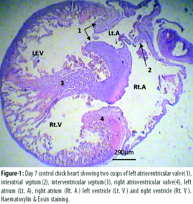
Interventricular septum had two parts: muscular and membranous. The lower three-fourth part of the interventricular septum consisted of rounded nuclei with no identifiable cell boundaries. Nuclei were loosely arranged in a meshwork of fibrous matrix. Some elongated nuclei could also be seen in between the rounded nuclei (probably representing intermyocardial fibroblast). The upper one-fourth part of the interventricular septum near the atrioventricular junction consisted of different shapes of nuclei ranging from oval, elongated, fusiform and flattened compactly packed in a meshwork of fibrous matrix. The ventricular septum was lined by flattened endothelial cells on both sides. This endothelial lining was continuous with the endothelial cells lining the trabeculated part of the ventricular wall.
Interatrial septum, which is formed from septum primum, was not completely closed and had small perforations in it. Interatrial septum was made up of rounded nuclei with no identifiable cell boundaries. It was lined on both sides with flattened endothelial cells. The day 7 heart in experimental group B1 also had comparable histology as that of control group A1.
Atrial septum had multiple perforations in the middle on day 10 (Figure-2).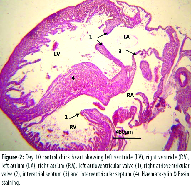
The ventricular septation was complete in day 10 heart of experimental group B2 and control group A2 with 1(3.3%) exception. Serial heart sections of specimen B2 (40\') showed that the interventricular septum was only made up of round to oval nuclei with no identifiable cell boundaries (representing the muscular part of the septum). This part of septum was lined with flattened endothelium on all sides. Above the free margin of the septum and just below the atrioventricular junction, both the ventricular cavities were in continuity (Figure-3).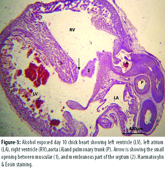
In later sections, part of the interventricular septum was seen developing just below the atrioventricular junction. It had nuclei ranging from oval, elongated; fusiform to flattened, compactly packed in a meshwork of eosinophilic matrix (representing membranous part of the septum). This membranous part of septum joined with the muscular part in later coming serial sections.
The chicks of group A3 had septal histology comparable to that of group B3.Atrial septation was still incomplete in both the subgroups.
Discussion
Interventricular septum of chick embryo has two parts: muscular and membranous. In the present study it was seen that ventricular septation had completed at day 7 with the fusion of both muscular and membranous parts of the septum. One study found that the primitive ventricle becomes uniformly trabeculated with highly structured sheets of myocytes lined by endocardial cells. These trabecular sheets combine and this process is finished by day 7,8 resulting in a muscular ventricular septum that divides the primitive ventricle into right and left ventricles. The interventricular septum grows towards the atrioventricular cushions and starts to fuse, leaving a small gap called a primary interventricular connection (interventricular canal) between the left and right ventricles. Further growth of the ventricular septum is by constant fusion of the adjoining trabecular sheets. One study found that ventricular septation completes between day 7 and 8.9 It was also seen in the study that the ventricle septation was complete on day 7. The portion of the interventricular septum that separates the inlet of the right ventricle from the left ventricular infundibulum is not formed by connective tissue as it is in humans, but it still corresponds to the membranous part of the human interventricular septum. It is also believed that this area is probably formed by different embryological components.10 In the current study it was seen that interatrial septum which formed from septum primum was not entirely closed and had small gaps in it at day 7 and these remained even till hatching. This is in accordance with the previous studies in which it was seen that interatrial septum of chick is formed by septum primum only and is completed postnatally; there is no contribution by septum secundum.11
The teratogenic effects of ethanol drinking have been well-known during the past few decades in humans as well as in animal models. Alcohol is a potential teratogen, and prenatal exposure to ethanol is a foremost avoidable reason of congenital defects known as Foetal Alcohol Syndrome (FAS).12 FAS produces defects in many organ systems, such as the central nervous system (CNS), gastrointestinal (GI) tract, and the cardiovascular system.13 Defects of cardiovascular system appear in approximately 50% of children diagnosed with FAS.13,14 The most frequently seen cardiac defects were that of the atrial septum. The incubation of the chick embryo with a small dose of ethanol (0.20 ml of 50% ethanol/egg) results in 43% ventricular septal defects; at a higher dose (0.4 ml of 50% ethanol/egg), 74% occurrence of aortic and ventricular septal defects were seen.15 In the present study, at day 10, in one of the alcoholic chick, it was seen that the interventricular septum was only made up of muscular part of the septum. This part of septum was lined with flattened endothelium on all sides. Above the free margin of the septum and just below the atrioventricular junction, both the ventricular cavities were in continuity. In later sections, membranous part of the interventricular septum was seen developing just below the atrioventricular junction. This membranous part of septum joined with the muscular part in later coming serial sections. Since ventricular septation is complete by day 7, this defect in the interventricular septum at day 10 in this study could be a ventricular septal defect. There could be several possible causes of this ventricular septal defect and other cardiac and foetal anomalies due to alcohol during development. Ethanol increases reactive-oxygen species that can be harmful to biomacromolecules resulting in cell injury and cell death.16 Oxidative stress can also result from decreased amounts of endogenous-antioxidant systems in the body like glutathione, ascorbic acid and catalase that causes increased production of reactive oxygen radicals. In vitro studies have shown that a known antioxidant resveratrol, decreases ethanol-induced damage.17
Low concentration of ethanol exacerbates processes leading to apoptosis18 and resulting in cellular death.19 Ethanol produces apoptosis of neural crest cells which play a key role in cardiac development. Death of neural crest cell produces foetal and other anomalies of the heart.20
The role of genetics in producing vulnerability towards alcohol toxicity is also a focus of research. A study found that different strain of mice given the similar amount of alcohol had different effects on different organs. These differential susceptibilities were more due to genetic influence, rather than maternal influence.21 In another study done on three strains of chick embryo it was seen that ethanol\'s effect on development of heart was under influence of foetal genome. One of the strains demonstrated pronounced cell death of cardiac neural crest cells but had normal development of the heart and aortic arches. Migration of neural crest cells and development of distal outflow tract were also normal in these embryos, which showed a capacity to repair initial losses. The second strain did not show cardiac defects. Hearts of the third strain had a distinctive phenotype with regards to ethanol exposure and displayed a thin compact layer of ventricle, dilatation, and reduced myosin/deoxyribonucleic acid and myosin/protein content, a phenotype that exhibits disturbed maturation of myocardium.22 The above-mentioned studies indicate that genetic makeup strongly affects the outcome of prenatal alcohol exposure.
In the present study the development of a ventricular septal defect in only one day 10 chick embryo could be due to genetic make-up. Chick embryos have different vulnerabilities towards the same dose of alcohol exposure due to the fact that ethanol\'s impact on cardiac development is governed by foetal genetics.
Conclusion
Ethanol vapour exposure during development may lead to ventricularseptal defects.
References
1. Caldwell KK, Goggin SL, Tyler CR, Allan AM. PrenatalAlcohol Exposure Is Associated with Altered Subcellular Distribution of Glucocorticoid and Mineralocorticoid Receptors in the Adolescent Mouse Hippocampal Formation. Alcohol Clin Exp Res 2014; 38: 392-400
2. Wikipedia The Free Encyclopedia: Alcohol Inhalation [online] updated 30/6/2014 [cited 2014 July 22]. Available from: URL: http://en.wikipedia.org/wiki/Alcohol_without_liquid
3. Pitel AL, Chanraud S, Müller-Oehring EM, Pfefferbaum A, Sullivan EV. Modulation of limbic-cerebellar functional connectivity enables alcoholics to recognize who is who. Brain Struct Funct 2013; 218: 683-95.
4. Hens L, Dambrink JH. Alcohol and drugs: twins or evil in a young heart. Acta Cardiol 2012; 67: 469-71.
5. O\'Leary CM, Nassar N, Kurinczuk JJ, de Klerk N, Geelhoed E, Elliott EJ, et al. Prenatal Alcohol Exposure and Risk of Birth Defects. Paediatric 2010; 126: e843-50.
6. Song G, Cui Y, Han ZJ, Xia HF, Ma X. Effects of choline on sodium arsenite-induced neural tube defects in chick embryos. Food Chem Toxicol 2012; 50: 4364-74.
7. Kamran K, Khan MY, Minhas LA. Effect of ethanol vapour exposure on survival of chick embryos. J Coll Physicians Surg Pak 2009; 19: 150-3.
8. Sedmera D, Pexieder T, Vuillemin M, Thompson RP, Anderson RH. Developmental patterning of the myocardium. Anat Rec 2000; 258: 319-37.
9. Waldo K, Miyagawa-Tomita S, Kumiski D, Kirby ML. Cardiac neural crest cells provide new insight into septation of the cardiac outflow tract: aortic sac to ventricular septal closure. Dev Biol 1998; 196: 129-44.
10. Martinsen BJ. Reference guide to the stages of chick heart embryology. Dev Dyn 2005; 233: 1217-37.
11. Morse DE, Rogers CS, McCann PS. Atrial septation in the chick and rat: a review. J Submicrosc Cytol 1984; 16: 259-72.
12. Serrano M, Han M, Brinez P, Linask KK. Fetal alcohol syndrome: cardiac birth defects in mice and prevention with folate. J Obstet Gynecol 2010; 203: 77-5.
13. Goh JM, Bensley JG, Kenna K, Sozo F, Bocking AD, Brien J, et al. Alcohol exposure during late gestation adversely affects myocardial development with implications for postnatal cardiac function. Am J Physiol Heart Circ Physiol 2011; 300: H645-51
14. Ren J, Wold LE, Natavio M, Ren BH, Hannigan JH, Brown RA. Influence of prenatal alcohol exposure on myocardial contractile function in adult rat hearts: role of intracellular calcium and apoptosis. Alcohol Alcohol 2002; 37: 30-7.
15. Mone SM, Gillman MW, Miller TL, Herman EH, Lipshultz SE. Effects of environmental exposures on the cardiovascular system: prenatal period through adolescence. Pediatrics 2004; 113: 1058-69.
16. Goodlett CR, Horn KH, Zhou FC. Alcohol teratogenesis: mechanisms of damage and strategies for intervention. Exp Biol Med (Maywood) 2005; 230: 394-406.
17. Hancock ML, Miller RR. Resveratrol can only partially attenuate ethanol-induced oxidative stress in embryonic chick brains. Nutr Neurosci 2006; 9: 121-9.
18. Castaneda F, Rosin-Steiner S. Low concentration of ethanol induced apoptosis in HepG2 cells: role of various signal transduction pathways. Int J Med Sci 2006; 3: 160-7.
19. McVicker BL, Tuma DJ, Kubik JL, Tuma PL, Casey CA. Ethanol-induced apoptosis in polarized hepatic cells possibly through regulation of the Fas pathway. Alcohol Clin Exp Res 2006; 30: 1906-15.
20. Garic-Stankovic A, Hernandez MR, Chiang PJ, Debelak-Kragtorp KA, Flentke GR, Armant DR, et al. Ethanol triggers neural crest apoptosis through the selective activation of a pertussis toxin-sensitive G protein and a phospholipase Cbeta-dependent Ca2+ transient. Alcohol Clin Exp Res 2005; 29: 1237-46
21. Ogawa T, Kuwagata M, Ruiz J, Zhou FC. Differential teratogenic effect of alcohol on embryonic development between C57BL/6 and DBA/2 mice: a new view. Alcohol Clin Exp Res 2005; 29: 855-63.
22. Jankala H, Eriksson PCJ, Eklund K, Sarviharju M, Harkonen M, Maki T. Effect of chronic ethanol ingestion and gender onheart left ventricular p53 gene expression. Alcoholism: Clin Exp Res 2005; 29: 1368-73.
Journal of the Pakistan Medical Association has agreed to receive and publish manuscripts in accordance with the principles of the following committees:


