

Ulku Kucuk ( Department of Pathology, Izmir Tepecik Education and Research Hospital, Izmir, Turkey. )
Emel Ebru Pala ( Department of Pathology, Izmir Tepecik Education and Research Hospital, Izmir, Turkey. )
Ebru Cakir ( Department of Pathology, Izmir Tepecik Education and Research Hospital, Izmir, Turkey. )
Ibrahim Cukurova ( Department of Otorhinolaryngology, Izmir Tepecik Education and Research Hospital, Izmir, Turkey. )
Murat Gumussoy ( Department of Otorhinolaryngology, Izmir Tepecik Education and Research Hospital, Izmir, Turkey. )
Umit Bayol ( Department of Pathology, Izmir Tepecik Education and Research Hospital, Izmir, Turkey. )
December 2014, Volume 64, Issue 12
Case Reports

Abstract
Haemangiomas, the most common type of benign vascular tumours, are rare in the oral cavity. Some of these lesions are congenital and show symptoms in late childhood or early adult life. A 32-years-old woman presented with a huge lesion on her tongue which caused dysphagia and dysphasia. She had first noticed the lesion when she was 6. Her obstructive symptoms started when she was 28 and, despite various medical treatments, the size of the lesion gradually increased. Magnetic resonance imaging revealed a 7x5x3cm mass on the right side of the tongue. Because of severe functional and cosmetic problems, the lesion was excised with partial haemiglossectomy. Histopathological examination was consistent with intramuscular haemangioma. Haemangiomas are benign tumours with a benign course and are rarely seen on the tongue. They have clinical importance when localised in the oral cavity. Different treatment modalities exist, but in cases of large tumours, surgery may be the mainstay treatment.
Keywords: Tongue, Haemangioma, Treatment.
Introduction
Haemangioma is the most common benign tumour of vascular origin, occurring most frequently in newborns, infants and children.1 The majority of haemangiomas involve the head and neck region and are rare in the oral cavity. They may occur on tongue, lips, buccal mucosa, gingiva, palatal mucosa, salivary glands, alveolar ridge and jaw bones.2 We describe a rare and unusual case of intramuscular (IM) haemangioma of the tongue in a 32-years-old female who had first noticed the lesion when she was 6.
Case Report
A 32-years-old woman presented with a huge lesion on her tongue which caused dysphagia and dysphasia. She had first noticed the lesion when she was 6. It had gradually increased in size, but she had not received any treatment till she was first admitted to our hospital with similar complaints in 2008. Sclerotheraphy was performed six times between 2008 and 2009. But the symptoms did not regress. A frequently preferred alternative technique, radiofrequency ablation, was thus performed between 2009 and 2010. Two years later, she was again admitted to our hospital and physical examination showed that the whole oral cavity was filled with the lesion; the tongue was diffusely enlarged; and showed purple colouration (Figure-1A). Physical examination showed that the lesion was in front of the circumvallat papilla of the tongue and there was no lesion at the hypopharynx side. Magnetic resonance imaging (MRI) revealed 7×5×3cm mass on the right side of the tongue. T1 weighted maxillofacial MRI revealed high pathological signals on the base of the tongue and multiple small dot-like signals in the hall of the tongue (Figure-1B).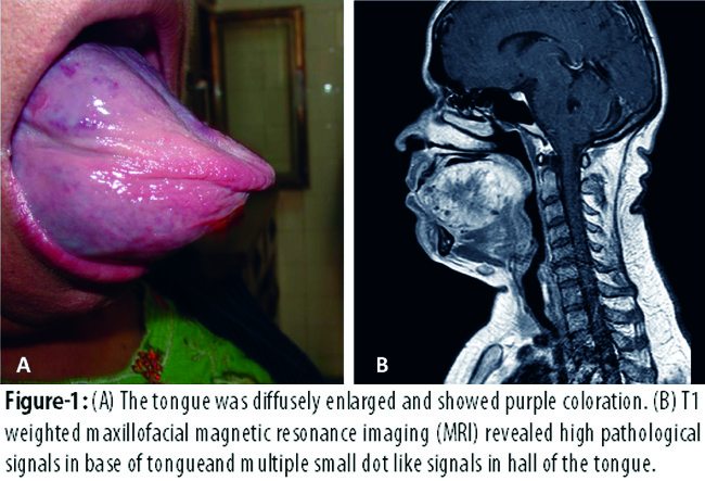
The lesion was partially excised with partial haemiglossectomy because of severe functional and cosmetic problems. For radiofrequency hemiglossectomy, Sutter BM-789 II Radiofrequency Unit with To-BiTE™ clamp was used. Thermal radiofrequency excision technique reduced intraoperative blood loss, haemorrhage and pain. Postoperatively, the patient did well with no complications. Ten months later, she presented with recurrence. Excision of the recurrent mass was performed. Histopathological examination of the specimens revealed different sized vessels dispersing muscle bundles, some of which were thrombosed and lined by bland endothelium. Occasional adipose tissue and cavernous vascular structures were also seen among fascicles of muscle (Figure-2).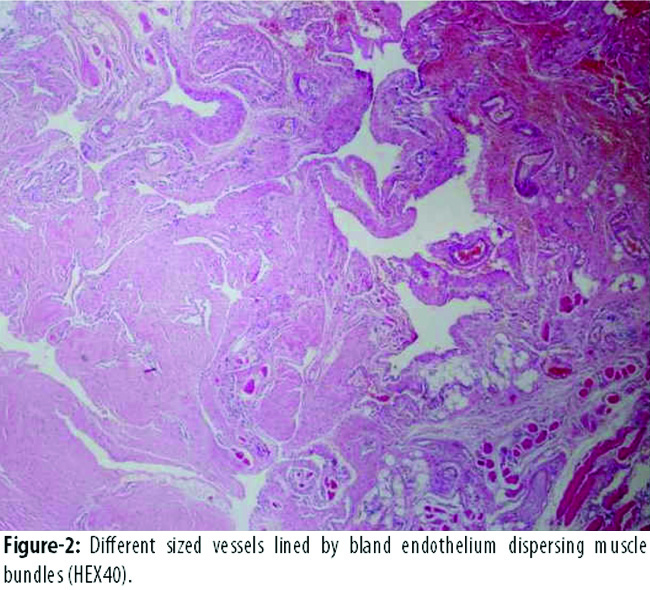
Six months after the last operation, the patient is well except dysphasia and has no recurrence.
Discussion
Haemangioma is one of the most common soft tissue tumours of infancy and childhood. Most haemangiomas are superficial lesions with a predilection to head and neck region. Vascular lesions of the tongue are rare. The young age of affected patients and the long duration of symptoms in some cases raise the possibility that many of these lesions are congenital tumours that slowly give rise to symptoms during late childhood or in early adult life.3
In 1987, a case was reported of mixed capillary and cavernous haemangioma of tongue in a 76-years-old woman, which was present since birth and was asymptomatic for more than 50 years. The patient had dislocated jaw presumably due to mass effect of haemangioma, which started growing after 56 years.4 In the present case, tongue enlargement was present since 22 years. When she had first noticed the lesion, she was six-years-old. The lesion had gradually increased in size and she was first admitted to our hospital when she was 28 and probably this was a congenital lesion.
Vascular lesions of the tongue may present with spontaneous haemorrhage in majority of cases. Other common complaints are pain, shortness of breath secondary to enlargement of the tongue, and disabilities of chewing and speaking.5 In the present case, major symptoms were dysphagia and dysphasia.
Most IM haemangiomas vary in their gross and microscopic appearances, depending on the histopathological subtype; capillary, cavernous or mixed type. In many cases it is not possible to classify these subtypes because they are all a part of histological spectrum.3
Microscopically, IM haemangiomas of the capillary type are composed of small capillary sized vessels, lined by plump endothelium, extending among muscle fibres. The cavernous form of IM haemangiomas are composed of large vessels lined by bland, markedly attenuated endothelium. The presence of adipose tissue is common in these tumours.3/sup> In the present case microscopic examination revealed different sized vessels some of which were thrombosed and lined by bland endothelium and dispersing muscle bundles.
A number of treatment options exist for these lesions including medical and surgical interventions. Medical management includes systemic and intralesional administration of corticosteroids.6 Conservative or further aggressive forms of treatment modalities may be applied for the haemangioma of the tongue. Embolisation, excision, cryotherapy, sclerotherapy, radiation, laser photocoagulation and chemotherapy methods has been used for the treatment of these lesions previously.5
Each treatment method has advantages and disadvantages. Conservative treatment frequentlygives rise to recurrences. On the other hand, aggressive treatment may cause lingual tissue loss and major functional disability.5 In the present case, despite the use of sclerotherapy and radiofrequency ablation techniques, the tumour did not regress and partial hemiglossectomy was performed.
Conclusion
Haemangioma is one of the most common soft tissue tumours of infancy and childhood. They are benign tumours with a benign course, but they have clinical significance in the oral cavity. They can give rise to clinical problems in speaking, deglutition and mastication when they are localised in tongue. Different medical treatment modalities exist but in cases of large tumours, surgery is the main option.
References
1. Kin HJ, Colombo M, Frieden IJ. Ulcerated hemangiomas: clinical characteristics and response to therapy. J Am Acad Dermatol 2001; 44: 962-72.
2. Gill JS, Gill S, Bhardwaj A, Grover HS. Oral Haemangioma. Case Rep Med 2012; 2012: 347939.
3. Weiss SW, Goldblum JR. Enzinger & Weiss\'s Soft Tissue Tumors. 5th ed. Philadelphia, USA: Mosby; 2008; 633-79.
4. Al-Khateeb TH. Benign oral masses in a Northern Jordanian Population-a retrospective study. Open Dent J 2009; 3: 147-53.
5. Kutluhan A, Bozdemir K, Ugras S. The treatment of tongue haemangioma by plasma knife surgery. Singapore Med J 2008; 49: 312-4.
6. Qureshi SS, Chaukar DA, Pathak KA, Sanghvi VD, Sheth T, Merchant NH, et al. Hemangioma of base of tongue. Indian J Cancer 2004; 41: 181-3.
Related Articles
Journal of the Pakistan Medical Association has agreed to receive and publish manuscripts in accordance with the principles of the following committees:


