

Ali A. Qazalbash ( Khyber Medical College, Peshawar. )
July 1982, Volume 32, Issue 7
Case Reports

Abstract
A patient with intra-abdominal haemangiopericytomas was seen, who presented with generalized abdominal pain and multiple masses in the abdomen, masses were of various sizes. Laparotomy revealed multiple tumours of various sizes scattered all over the abdomen, in line with the blood vessels of messentery, ornentum and abdominal wall. Multiple abdominal haemangiopericytonias were suspected. Diagnosis was confirmed on histopathological examination.
Introduction
Haemangioma are true blood vessel turnours, which may be present at birth or occur subsequently. Haemangiopericytoma is a rare vas-cular tumour first described by Stout and Murray (1942). It arises from the pericytes of Zimmerman of capillaries and arterioles of solid or hollow visera in the abdomen. It may also arise from capi laries and arterioles of subcutaneous tissues. Although uncommon, it has been reported in brain Kings, and parotid glands. It may be mulcieentric in origin and benign or malignant in nature.
Intra-abdominal haemangiopcricytoma may arise from retroperitoneal structures (Herdman, 1953; Hakala et al, 1970); from mesentery (Stout and Cassel, 1943) and from omentum (Stout 1949; Goldberger and Schein, 1968). In hollow visera it can arise from stomach (Marino, 1959) duodenum (Binder et at., 1973), jejunum (Olsen and Well wood, 1970), colon (Ault et al., 1951) and rectum (Slaterry et al., 1956). Black Winckel and Diddamas (1970) have reported 45% of haernangiopericytoma in capillaries of musculoskeletal system and 18% in the intra abdominal organs and retropcritoneal structures.
Case Report
A 50 year old man was admitted to the Medical Unit ‘A’ of Khyber Hospital, Peshawar, with the complaints of recurrent vomiting and attacks of abdominal pain for seven months. He also noticed masses in his abdomen which were progressively increasing in size. He further complained of lack of appetite and loss of weight.
A moderately built middle.aged man, slightly anaernic, pulse rate 80 per minute and blood pressure 120)70 mmHg.
Abdomen lookcd full. There were nontender masses of various sizes all over the abdomen, some of them were even mobile. Largest mass was in the right hypochondrium. Other systems were normal.
Inoestigations:
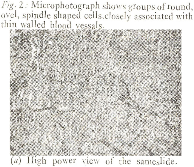
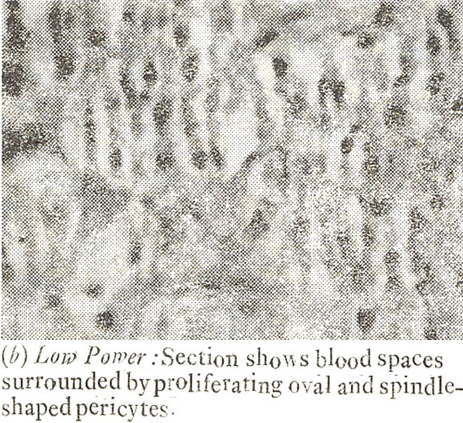
Haemoglobin was 12 gm% ,total and differential counts were normal, ESR was 36 mm 1st hour. Bleeding time, coagulation times, and urine examination were normal. Fasting blood sugar was 90 mg%. Casoni’s test was negative. Exploratory laparotomv revealed haemorrhagic fluid in peritoneal cavity; there were multiple masses of various sizes attached to the omentum mesentery and abdominal wall. These masses were highly vascular with big veins on the surfaces of these masses. Most of the masses were removed, the biggest one was 15 cm in size. No extra abdominal metastasis was seen. Histopathological studies of these masses revealed haemangiopericytoma (Figure 1 and 2).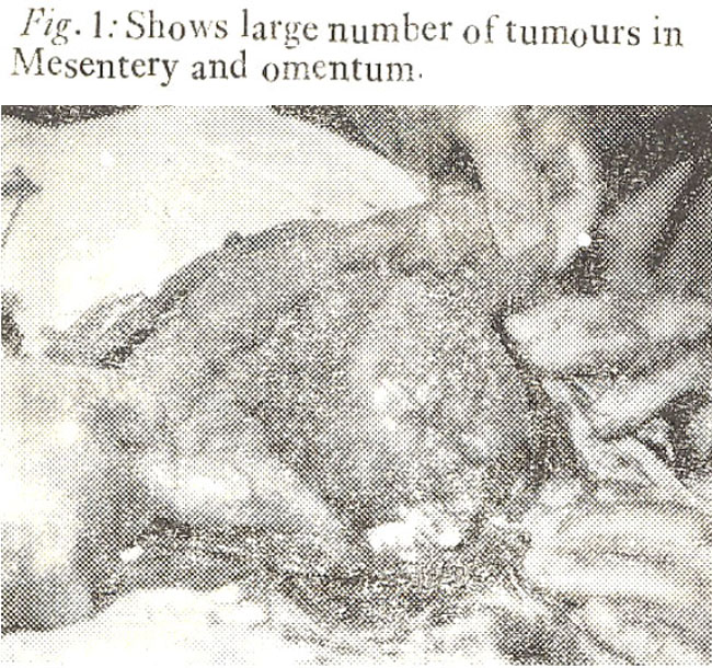
The patient died on 4th post-operative day.
Discussion
Haemangiopericytcma at first sight resem-. bles the haemangioeiidothelioma with numerous capillary channels surrounded and enclosed within nests and masses of round endotheijal cells; however, more careful examination discloses that the endothelial cells are some what organised and resemble the cells of glomangioma. Silver impregnation has shown that these are pericytes rather than endotheijal cells. These tumours were first thought to be benign, but recent studies have disclosed that as many as 50% are malignant (Robins, 1967), because of local invasiveness of the tumour or its spread into the adjoining structures. Twelve percent metastasize to the lung, bones, and liver through haematogenous spread. Regional lymph nodes are sometime affected (Stout, 1949).
Occasionally, the patient may have hypoglycaemia (Howard and David, 1959) which is attributable to the insulin-like substances, which are secreted by the tumour cells, or by the excessive glucose utilization by the primary or secondary tumour masses, because their excision has shown to abolish hypoglycaemia. Haenangiopericvtomas are usually less than 10 cm in size. A retroperitoneal 30 cm in diameter was reportcd by Binder et al. (1973) which was retro-peritoneal and was displacing the aorta and the ureter.
Treatment
The ideal treatment is surgical excision of the tumour masses, with wide excision of tissues, because of the involvement of ad jacent structures. Radical removal is apparently a permanent cure in 50% of cases (Fisher, 1960). Radiotherapy and Chemotherapy have also been recommended as palliative measures. The drugs recommended are Actinomycine and fluorouracil.
Acknowledgement
My thanks to Professor Dr. Feroz Shah for operating on this patient, to Dr. Fazal Ahmad for Histopathological studies andto Mr. Fazal-e-Wahid for typing.
References
1. Ault, G.W., Smith, R.S. and Castro, M.F. (1951) Hemangiopericytoma of the sigmoid colon; Surgery, Case report. 30:523.
2. Binder, S.C., Wolfe, H.J. and Deterling, R.A. Jr. (1973) Intra-abdominal hemangiopericytoma; report of four cases and review of the literature. Arch. Surgy., 107:536.
3. Black Winkel, K.D. and Diddamas, J.A. (1970) Hemangiopericytoma; report of a case and comprehensive review of the literature. Cancer, 25:896.
4. Fisher, J.H. (1960) Haemangiopericytoma; a review of twenty cases. Canad. Med. Ass. J., 83:1136.
5. Goldberger, R.E. and Schein, C.J. (1968) Hemangiopericytoma of omentum. Ann. Surg., 34:291.
6. Herdman, J.P. (1953) Primary retroperitoneal tumors. Br. J. Surg., 40:331.
7. Hakala, T.R., Page, D. and Fleischli, D.J. (1970) Paravesical Hemangiopericytoma. J. Urol., 103:436.
8. Howard, J.W. and David, P.L. (1959) Retroperitoneal heamangiopericytoma associated with hypoglycemia and masculinization. Del. Med. J., 31:29.
9. Marino, A.W. Jr. (1959) Hemangiopericytomas a review of literature and amplication. Dis. Colon Rectum, 2:438.
10. Olsen, E.G.J, and Well Wood, J.M. (1970) Hemangiopericytoma of the samll intestine. A review of three cases. Br. J. Surg., 57:66.
11. Robins, S.L. Blood vessels, in pathology. By Stanley L. Robbins. 3rd ed. Philadelphia, Saunders, 1967.
12. Stout, A.P. and Murray, M.R. (1942) Hemangiopericytoma, a vascular tumor featuring Zimmerman\'s pericytes. Ann. Surg., 116:26.
13. Stout, A.P. and Cassel, C.A. (1943) Hemangiopericytoma of omentum. Surgery, 13:578.
14. Stout, A.P. (1949) Hemangiopericytoma, a study of 25 new cases. Cancer, 2:1027.
15. Slaterry, L.R., Aronson, S.G. and Lowman, E.W. (1956) Hemangiopericytoma review of abdominal cases. Am. J. Surg., 91:985.
Related Articles
Journal of the Pakistan Medical Association has agreed to receive and publish manuscripts in accordance with the principles of the following committees:


