

S.M. Faruque ( Department of Biochemistry, Dhaka University, Dhaka-2, Bangladesh. )
Lamia Sharmeen ( Department of Biochemistry, Dhaka University, Dhaka-2, Bangladesh. )
S. A.M. Khairul Bashar ( Department of Biochemistry, Dhaka University, Dhaka-2, Bangladesh. )
September 1984, Volume 34, Issue 9
Original Article

Abstract
Post weaning rats were fed protein deficient dies for about 6 weeks until their body weight/brain weight ratio started to decrease. Effect of such malnutrition on the plaque forming response of antibody producing spleen cells against three bacterial antigens, Vibrio cholerae, Salmonella paratyphii and Staphylococcus aureus were studied. Mitogen stimulation of spleen cells was also studied using phytohemagglutinin and 3H-thymidine. Antibody plaque forming cell number and spleen cell stimulation response to phytohemagglutinin decreased significantly (P<001) in the malnourished rats as compared to the controls(JPMA 34 : 270, 1984).
Introduction
The association between malnutrition and infectious diseases1,2 and the role of protein as a nutrient against infection is well established.3 In malnutrition the immune status is impaired in various ways4-6 These include reduced antibody response’ altered distribution pattern of blood lymphocytes and impaired phagocytosis. The mechanism by which malnutrition causes reduced anti-body response is yet to be investigated. The present study attempts to investigate the effect of protein mal1putrition on the proliferation of spleen cells to produce antibody producing cells against injected antigens and on stimulation with phytohemagglutinin.
Material and Methods
Post weaning male rats of Long-Evans strain, weighing 75 ± 10 g were used. The animals were divided into two comparable groups according to body weight. One group was fed control diet; adequate in all essential nutrients while the other group was fed a diet deficient in protein (containing only 2% protein). On every 10th day, 3 rats from each gtoup were weighed and then killed to determine the body weight/brain weight ratios of the two groups. On the 40th day after initiation of the experiment, the body weight! brain weight ratio of the protein deficient rats was found to have decreased as compared to that found on the 30th day, and at this time the rats of both the groups were considered to be ready for immunological experiments. The animals were then divided into 4 batches, each batch consisting of S controls and 5 malnourished rats. One of these batches of rats were sacrificed and their spleens were removed aseptically for studying the proliferative response of spleen cells on stimulation with phytohemagglutinin. The rats of the remaining 3 batches were immunized against three bacterial ahtigens, Vibrio cholerae, Salmonella paratyphii and Staphylococcus aureus respectively. In each case, killed cells of the specific bacterium, washed with normal saline and resuspended- in a solution of 0.25 M’ sucrose, 1 M EDTA, and 5 M phosphate buffer, pH 7.4, was used as the antigen and a 0.3 ml portion of the bacterial suspension, containing about 5 X 10⁶ bacteria was injected intraperitonially. After 6 days of injecting the antigen all the rats were killed and their spleens were removed aseptically for studying the plaque forming response of antibody producing spleen cells against the specific antigens.
Plaque forming cells in the spleen were determined by the plaque assay7. In each case, the spleen cells were dispersed with a syringe in cold tissue culture medium (Eagles Modification of Minimal Essential Medium, Glasgow, U.K.) to prepare single cell suspension. To 2ml of 0.8% Difco agar dissolved in minimal essential medium (MEM) were added in rapid succession 0.1 ml of 100% DEAE dextran solution, 0.1 ml of the specific bacterial suspension (in MEM) containing approximately 106 active bacterial cells, and about 106 nucleated spleen cells. After rapid but gentle dispersion the mixture was poured on to petri dishes containing a basal layer of 1.5% Difco agar. The plates were incubated for three hours at 3 7°C, and to each plate 3 ml of guinea pig complement (diluted 1:10 times with normal saline) was added. The plates were further incubated for six hours at 37 C. Clear zones (plaques) on the confluent lawn of bacteçial growth appeared which represented where the specific bacteria-lysin had been released by the antibody forming cells. The plaques were counted by using low powered magnifying glass.
Mitogen stimulation response of spleen cells was evaluated by microculture technique8 using phytohemagglutinin (PHA) and 3H-thymi-dine. Results were expressed as stimulation index defined as the ratio between counts per minute of PHA containing culture divided by counts per minute of culture without PHA.
Results
Before immunization the average spleen weight in the malnourished rats was found to be less than that of the control rats. On immunization, no significant change in the average spleen weights áf either group was found (Table I).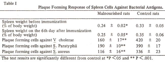
Spleen cell stimulation response to phytohemagglutinin decreased in the malnourished rats as compared to the controls. The mean stimulation index (Table II)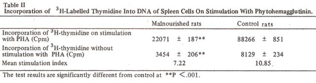
was found to have decreased in the malnourished rats, by about 33.6% as compared to the control rats. Plaque forming response of spleen cells was significantly lower in the malnourished rats. The average number of plaque forming cells in the control rats was 2 to 3 fold higher than that in the malnourished rats (Table I).
Discussion
Reduced plaque forming response of the spleen cells in the malnourished rats as compared to the normal rats sUggests that malnutrition causes impairment of immune status due to reduction in number or to faulty maturation of antibody producing cells in the spleen. Antibody formation in response to an antigen requires active collaboration between T and B lymphocytes and macrophages. T lymphocytes have a regulatory effect on the formation of antibody producing cells from B lymphocytes. The capacity of lymphocytes to synthesize DNA in response to mitogenic stimulus is an invitro correlate of proliferative response of lymphocytes against antigens. Since the spleen cells of the malnourished rats showed decreased stimulation response to the T-celmitogen, phytohemagglutinin (Table II), it is probable that in malnutrition either the formation of T lymphocytes in the spleen is reduced or their ability to respond is decreased. The present study thus suggests that in malnutrition the number of antibody producing ells in the spleen is reduced, probably due to a decrease in number or to impairment of function of T lymphocytes in the spleen.
References
1. Scrimshaw, N.S., Taylor, C.E. and Gordon ,J.E. Interactidns of nutrition and infection. WHO Monogr. Ser., 1968; 3 329.
2. Chandra, R.K. and Newberne, PM. Nutrition immunity and infection. New York Plenum Press, 1977.
3. Suskind, R.M. Malnutrition and immune response. New York, Raven Press, 1977.
4. McMurry, D.N., Loomis, S.A., Casazza, L.J., Rey, H. and Miranda, R. Development of impaired cell-mediated immunity in mild and moderate malnutrition. Am. J. Clin. Nutr., 1981; 34 : 68.
5. Samuel, A.M., Patel, B.D. and Mankodi, N. A study of immunoglobulins in protein-calorie malnutrition. Indian J. Med. Res., 1972; 60: 1278.
6. Chandra, R.K. Rosette-forming T lymphocyte and cell-mediated immunity in malnutrition. Br. J.Med., 1974;3 : 608.
7. Jerne, N.K. and Nordin, A.A. Plaque formation in agar by single antibody-producing cells. Science, 1963 140: 405.
8. Chandra, R.K. and Au, B. Single nutrient deficiency and cell mediated immune responses. III Vitamin A. Nutrition Research, 1981; .1 181.
Journal of the Pakistan Medical Association has agreed to receive and publish manuscripts in accordance with the principles of the following committees:


